Edited By: Jinming Yu

Precision Radiation Oncology, also known as the Blue Journal, presents an open access forum of collaboration and exchange for comprehensive and cutting-edge information on basic, translational, and clinical research in radiation oncology.
Precision Radiation Oncology is the first English Journal focusing on radiation oncology in China. It is officially endorsed by Chinese Society for Therapeutic Radiology and Oncology (CSTRO), Chinese Radiation Therapy Oncology Group (CRTOG), and the Radiation Oncology Physicians Brance of Chinese Medical Doctor Association.
Journal Metrics
- 2.3CiteScore
- 2.1Journal Impact Factor
- 56%Acceptance rate
- 37 days Submission to first decision
Precision Radiation Oncology is now recruiting Young Editorial Board Members. Please find more information here.

Why publish in Precision Radiation Oncology?
By publishing in the Blue Journal your research will enjoy:
- Your research will be open access and readily available to radiation oncologists, dosimetrists, radiation physicists, radiation biologists, radiation therapists, interventional radiologist, radiologists and more.
- Article publication charges are waived. There is no direct cost to you for publishing open access and increasing your downloads, shares and citations.
- Your work will be discoverable and widely distributed with indexing in Embase, Scopus and the Directory of Open Access Journals.
- Quality assured through a rigorous peer review process.
- Published in partnership with the Sino-American Network for Therapeutic Radiology and Oncology (SANTRO) and Chinese Anti-Cancer Association (CA-CA), with an experienced and globally represented Editorial Board.
Journal News


Create a FREE Wiley Online Library account and register to receive Content Alerts and instant notifications when research publishes in the Blue Journal.
- Receive personalized alerts at a pace that suits you.
- Keep track of a paper of interest with citation alerts.
- Save content to easily manage your reading list.
- Save searches for instant review later.
Articles
Feasibility study of a multi‐lesion cyberknife radiotherapy plan verification method using a 2D array with pre‐set roll angles
- 28 June 2025
Graphical Abstract
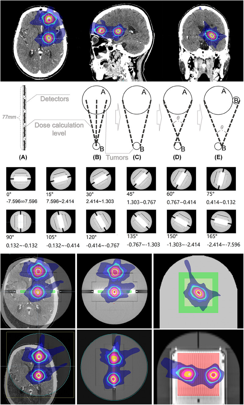
This study aims to solve the common problem in clinical practice of CyberknifeM6. In the verification of multi-lesion brain plans, when the 2D matrix is not set the roll Angle, only less than 1/3 of the cases can measure the dose of two targets.
The solution of using two-dimensional array and matching phantom design with relatively good resolution has better clinical applicability. In this study, a simplified model was designed to simplify the lesions with different location distribution, shape and size into a sphere, and calculate the roll angle required by the 2D array on the measurable limit distance. There are a large number of verification plans in this study, and the comparative analysis methods and conditions used are relatively perfect. The results obtained have high credibility.
Some specific suggestions for the improvement of the phantom for practical application are put forward.
Optimized fiducial marker placement using B‐spline surface modeling and graph theory for Cyberknife stereotactic body radiotherapy for superficial tumors
- 87-95
- 19 June 2025
Graphical Abstract
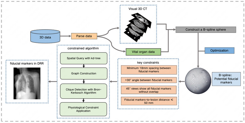
This study introduces a Fiducial marker placement planning algorithm, specifically for superficial tumors that are located 20-50 mm beneath the epidermis. The algorithm proposes numerous potential implantation sites by constructing and optimizing a B-spline surface. The time complexity of the algorithm proposed in this study is O(mlogm+m2+3(n/3)), significantly faster than the brute-force O(n3) approach. Experimental outcomes show that our algorithm can efficiently plan Fiducial marker placements, simplifying the planning process and providing valuable technical support for CyberKnife treatments.
Dosimetric and hematological toxicity analyses of bone marrow‐sparing intensity‐modulated radiation therapy for patients with cervical cancer treated with extended‐field radiation therapy
- 96-107
- 12 June 2025
Graphical Abstract
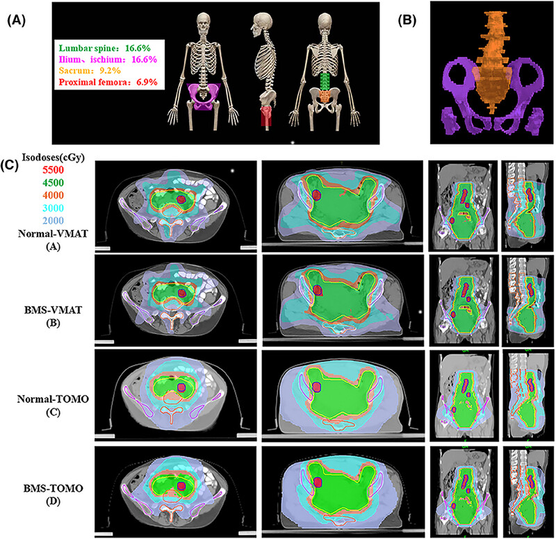
Bone Marrow Sparing Intensity Modulated Radiation Therapy is a feasible option for cervical cancer patients treated with extended-field radiation therapy, it may reduce hematologic toxicity and ensure the continuity of concurrent chemoradiotherapy.
Fast estimation of patient‐specific organ doses from abdomen and head CT examinations without segmenting internal organs using machine learning models
- 77-86
- 29 May 2025
Graphical Abstract
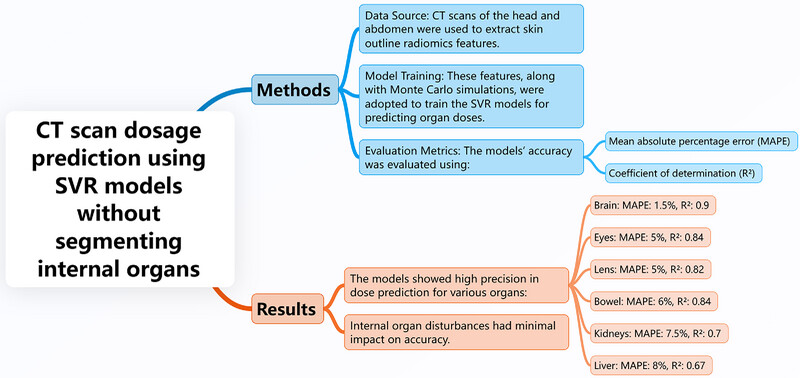
A new method using support vector regression (SVR) models predicts organ doses from CT scans based on skin radiomics, eliminating the need for organ segmentation. High accuracy was achieved for brain, eyes, lens, bowel, kidneys, and liver doses, improving clinical efficiency and accessibility in dose prediction.
Deep learning‐based dose prediction for low‐energy electron beam superficial radiotherapy
- 108-119
- 26 May 2025
Graphical Abstract
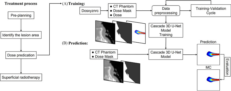
This study integrates Monte Carlo (MC) simulations with a cascaded 3D-UNet (C3D) deep learning model to achieve rapid and accurate dose predictions for low-energy electron radiotherapy. Utilizing CT phantoms of six body sites, the model demonstrates high prediction accuracy with a Gamma pass rate exceeding 92% within 1%/1mm tolerance and a computational speed approximately 140,000 times faster than traditional MC simulations. This approach enhances the efficiency and precision of treatment planning, addressing the challenges of surface contour variations in superficial electron beam therapy.
Deep learning (DL)-based advancements in prostate cancer imaging: Artificial intelligence (AI)-based segmentation of 68Ga-PSMSA PET for tumor volume assessment
- 120-132
- 3 May 2025
Graphical Abstract
This review highlights the use of 68Ga-PSMA PET in prostate cancer (PC) for tumor volume quantification, crucial for staging, treatment planning, and prognosis. Traditional manual segmentation methods are time-consuming and variable, while AI-based segmentation techniques, particularly deep learning models like convolutional neural networks, offer automated, accurate, and efficient solutions. AI-based segmentation improves reproducibility and processing times, aiding personalized medicine. However, challenges such as standardized protocols, extensive validation, and clinical integration need addressing for its widespread adoption in 68Ga-PSMA PET for PC, enhancing patient care.
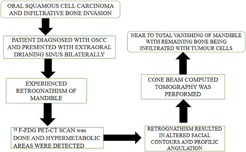
Massive Infiltrative Osteolysis of the Mandible in Oral Squamous Cell Carcinoma: A Case Report with a Review of the Literature
- 145-151
- 30 April 2025
Graphical Abstract
Radiotherapy after Neoadjuvant Immunochemotherapy in Unresectable Stage III Non‐Small Cell Lung Carcinoma: A Novel Therapeutic Approach?
- 143-144
- 30 April 2025
A safety study of ultra‐high dose rate FLASH radiotherapy in the treatment of superficial skin tumors: study protocol of a phase I trial (ChiCTR2400080935)
- 72-76
- 5 April 2025
Graphical Abstract
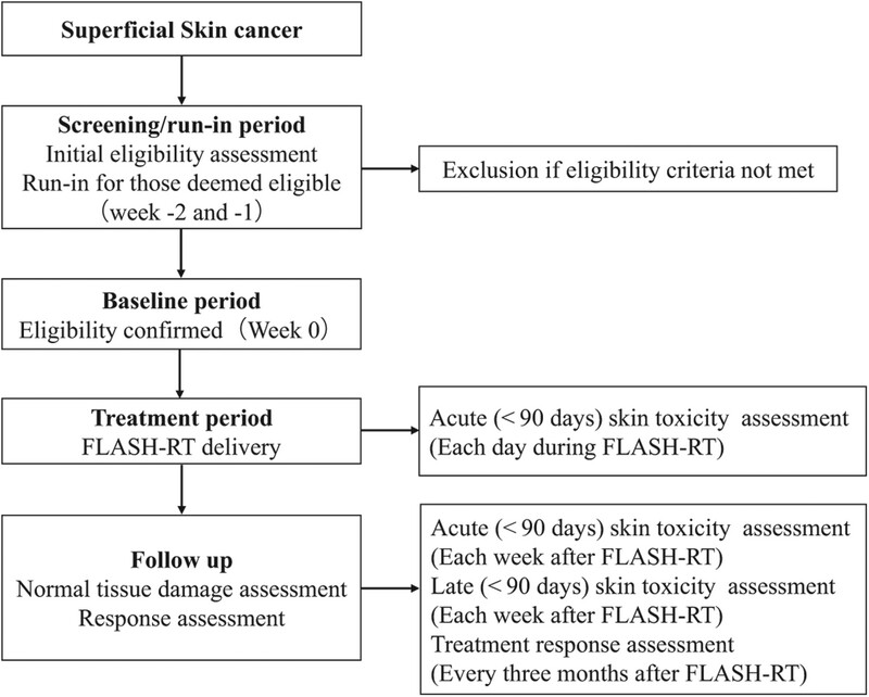
FLASH-RT has been shown to protect normal tissues, the clinical translation of FLASH-RT is ongoing.
We designed a phase I clinical trial to estimate the safety and healthy tissue spare by using a modified clinical linear accelerator
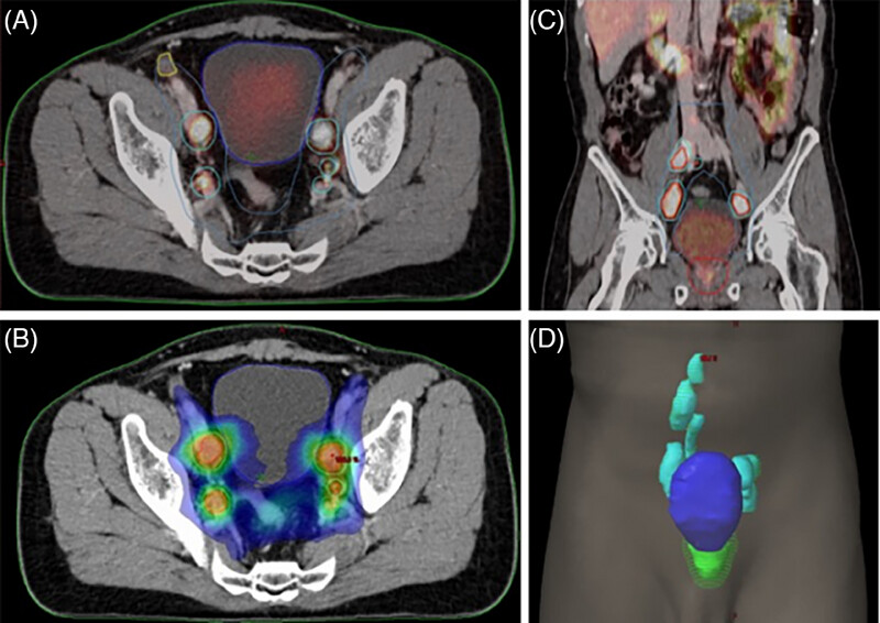
Application of high‐dose‐rate endorectal brachytherapy in the treatment of locally advanced rectal cancer
- 133-142
- 30 March 2025
Graphical Abstract

This image compares the studies included in this article with the meta-analysis from the Cochrane Library, indicating that the incorporation of brachytherapy in neoadjuvant treatment for rectal cancer can achieve better pathological response rates. Additionally, the use of brachytherapy can reduce the local recurrence rate in patients. However, there are no significant differences in long-term survival and distant metastasis.
Deep learning with attention modules and residual transformations improves hepatocellular carcinoma (HCC) differentiation using multiphase CT
- Precision Radiation Oncology
- 13-22
- 22 March 2025
Graphical Abstract
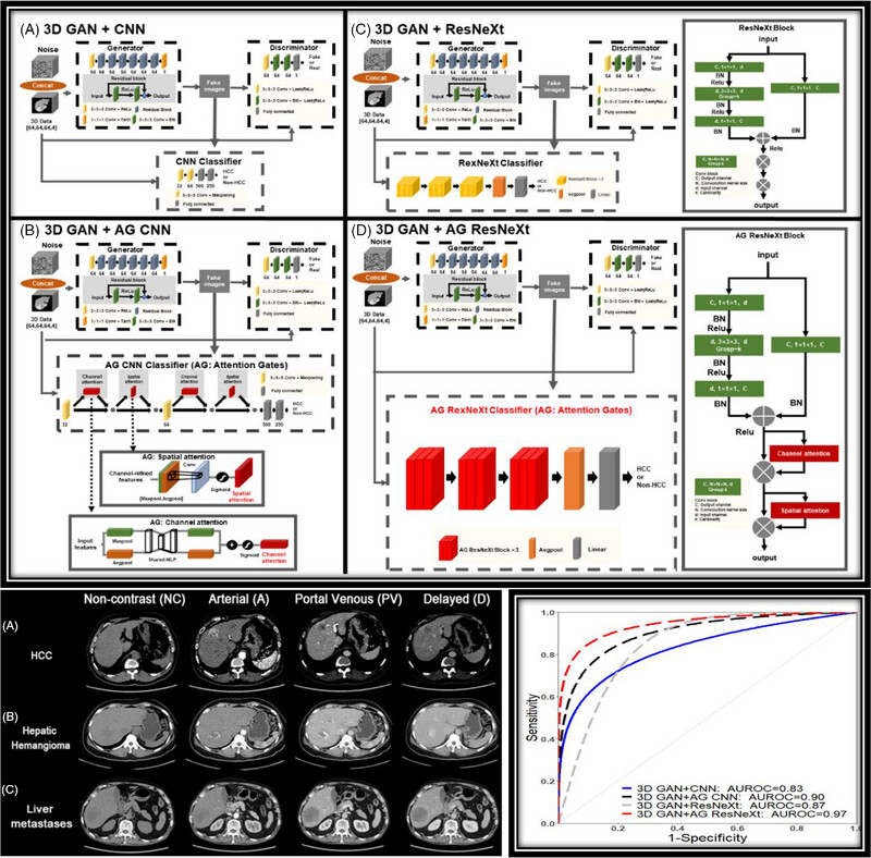
The proposed deep learning model with attentions and residual transformations (Top Figure (d) 3D GAN+AG ResNeXt) is applied to multi-phase CTs for HCC differentiation (Bottom Left). ROC curves (Bottom Right) showed the 3D deep GAN model with attentions and residual transformations (RED) had better performance.
Revolutionizing cancer treatment: The role of radiopharmaceuticals in modern cancer therapy
- Precision Radiation Oncology
- 145-152
- 11 September 2024
Graphical Abstract
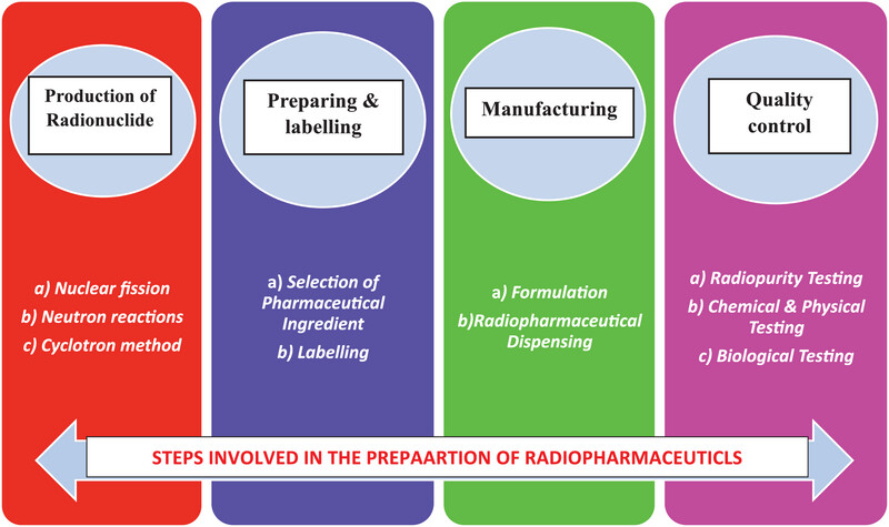
Steps involved in the preparation of radiopharmaceuticals
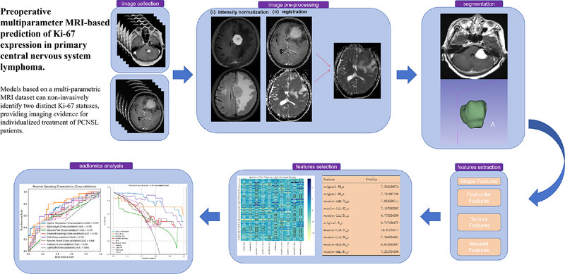
Preoperative multiparameter MRI‐based prediction of Ki‐67 expression in primary central nervous system lymphoma
- Precision Radiation Oncology
- 23-34
- 18 March 2025
Graphical Abstract
Stereotactic body radiation therapy for clinically diagnosed early‐stage non‐small cell lung cancer: Importance of accurate CT interpretation by experts
- Precision Radiation Oncology
- 30-36
- 10 March 2024
Graphical Abstract
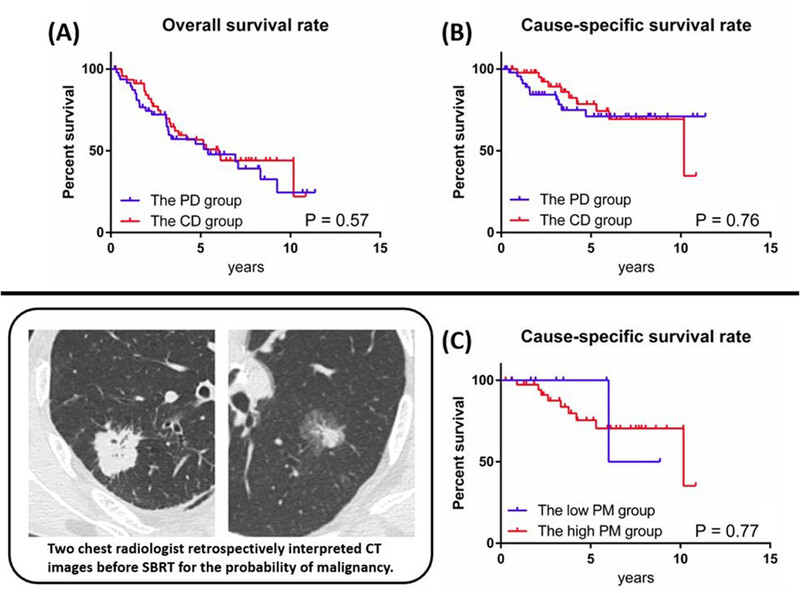
The present study investigated the clinical outcomes of stereotactic body radiotherapy (SBRT) for pathologically diagnosed (PD) and clinically diagnosed (CD) early-stage non-small cell lung cancer and examined the importance of accurate CT interpretations by experts. Overall survival and cause-specific survival curves showed no significant difference between the PD and CD groups. (a, b). The CD group was divided into the low probability of malignant (PM) group and the high PM group by CT interpretation. Although there was no significant difference in the OS and CSS curves of the high and low PM groups (c), lung cancer mortality was lower in the low PM group (25% [1 of 4]) that in the high PM group (47.4% [9 of 19]).
Clinical results of definitive radiotherapy for local recurrent kimura disease in the head and neck after surgery: A retrospective study
- Precision Radiation Oncology
- 119-125
- 21 August 2024
Graphical Abstract
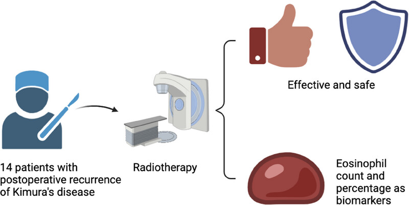
The graphic on the left represents definitive radiotherapy for selected patients with local recurrence of kimura disease in the head and neck after surgery.
The figure on the right is divided into upper and lower parts. The upper part
The graph is divided into upper and lower parts. The upper part
- Use a shield (for safety) and a thumbs-up icon (for efficacy).
- Text below the icon: “Radiation therapy is effective and safe for recurrent Kimura disease.”
- The lower half includes blood drop icons representing biomarkers.
- Add text: “Peripheral blood eosinophil count and percentage are reliable biomarkers.”
Research progress of cardiotoxicity caused by radiotherapy in breast cancer
- Precision Radiation Oncology
- 153-158
- 21 September 2024
Graphical Abstract
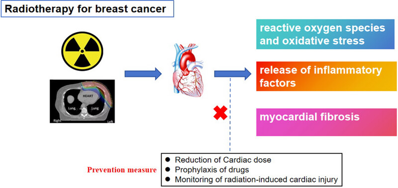
Breast cancer has surpassed lung cancer as the most common type of malignancy worldwide. Treatments for breast cancer include surgery, chemotherapy, radiotherapy, targeted therapy, endocrine therapy, immunotherapy, and hyperthermia. Radiotherapy plays an important role in breast cancer treatment. Patients with early breast cancer can have longer survival after combined treatment, but cardiotoxicity caused by radiotherapy may affect long-term prognosis. This article reviews cardiac damage caused by radiotherapy in breast cancer.
Enhanced Electroacoustic Tomography with Supervised Learning for Real‐time Electroporation Monitoring
- Precision Radiation Oncology
- 110-118
- 22 September 2024
Graphical Abstract
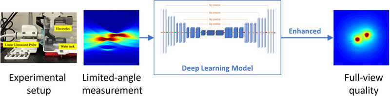
This study proposed a supervised learning-based workflow to address the ill-conditioning in EAT reconstruction. Electroacoustic signals were detected by a linear array and initially reconstructed into EAT images, which were then fed into a deep learning model for distortion correction.
Consensus statement on the exploration of clinical translation and application of electron ultra‐high dose rate FLASH radiotherapy
- Precision Radiation Oncology
- 4-12
- 3 March 2025
Graphical Abstract
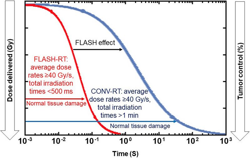
• FLASH-RT as a novel irradiation technique shows a protective effect on normal tissues while maintaining comparable tumor killing effect as CONV-RT, also known as the FLASH effect.
• We develop a consensus statement on the exploration of clinical translation of FLASH-RT, and provide insights for the further application of this technology in clinical practice.
Progress in radiotherapy for small‐cell lung cancer
- Precision Radiation Oncology
- 207-217
- 31 July 2023
Graphical Abstract
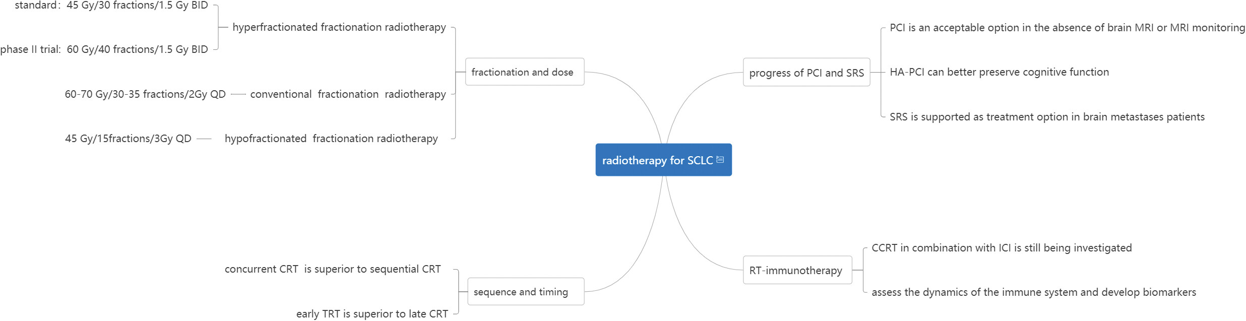
Small cell lung cancer (SCLC) is a highly malignant neuroendocrine tumor that is prone to extensive metastasis. Compared to non-small cell lung cancer (NSCLC), the overall treatment of SCLC is slow to progress.Therefore, we summarize the new evolving therapeutic strategies (fractionation and dose, sequencing and timing) , improved radiotherapy techniques (progress of PCI and SRS) and discuss the possibilities and prospects of radiotherapy combined with immunotherapy for SCLC.
Auto‐segmentation of the clinical target volume using a domain‐adversarial neural network in patients with gynaecological cancer undergoing postoperative vaginal brachytherapy
- Precision Radiation Oncology
- 189-196
- 7 August 2023
Graphical Abstract
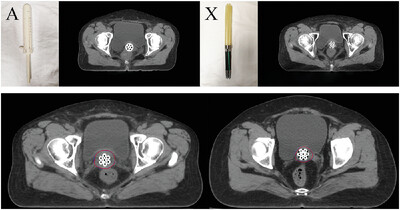
An auto-segmentation DANN model is constructed using 60 patients treated with applicator A as well as 10 patients treated with applicator X. More than 80% of CT slices contoured by the DANN model are acceptable.
Revolutionizing cancer treatment: The role of radiopharmaceuticals in modern cancer therapy
- Precision Radiation Oncology
- 145-152
- 11 September 2024
Stereotactic radiotherapy: An educational narrative review
- Precision Radiation Oncology
- 47-58
- 21 March 2024
Chinese clinical practice guidelines for the prevention and treatment of radiation‐induced dermatitis
- Precision Radiation Oncology
- 160-172
- 10 September 2023
Chinese clinical practice guidelines for the prevention and treatment of radiation‐induced esophagitis
- Precision Radiation Oncology
- 225-236
- 20 September 2023
Chinese clinical practice guidelines for the prevention and treatment of radiation‐induced rectal injury
- Precision Radiation Oncology
- 237-255
- 25 December 2023
A new formula for calculating normal tissue complication probability
- Precision Radiation Oncology
- 126-131
- 19 September 2024
Stereotactic body radiotherapy takes on Lung Oligometastases: Latest breakthroughs
- Precision Radiation Oncology
- 85-91
- 9 May 2024
Feasibility planning study of lattice radiotherapy for palliation in bulky tumors
- Precision Radiation Oncology
- 209-217
- 28 November 2024
Research progress of cardiotoxicity caused by radiotherapy in breast cancer
- Precision Radiation Oncology
- 153-158
- 21 September 2024
Recent issues


Latest news
Sign up for email alerts
Tools
More from this journal
- Open Access License and Copyright
- Institutional and Funder Payments
- Call for Papers: Special Issue Radiation Therapy for Rare Tumor of Head and Neck
- Wiley Open Access
- Publishing with Precision Radiation Oncology
- Call for Papers: Special Issue
- Top downloaded articles published in 2022
- Themed Collection: Guidelines for radiotherapy
- Themed Collection: MRI guided precision radiotherapy
- Themed Collection: Proton and carbon ion therapy
- Themed Collection: Innovations, Advances, and Challenges in Precision Radiation Oncology Physics
- Themed Collection: Stereotactic Body Radiation Oncology
Why publish with Precision Radiation Oncology?
- Your research will be open access
- Article publication charges are waived
- Indexed by Embase, Scopus, DOAJ
- Quality guaranteed with rigorous peer review
Submit your manuscript now