Journal list menu

 Issue
IssuePrecision Radiation Oncology: Volume 8, Issue 4
165-231December 2024
Export Citations
Download PDFs
ISSUE INFORMATION
ORIGINAL ARTICLE
Mimicking large spot-scanning radiation fields for proton FLASH preclinical studies with a robotic motion platform
- Pages: 168-181
- First Published: 24 October 2024
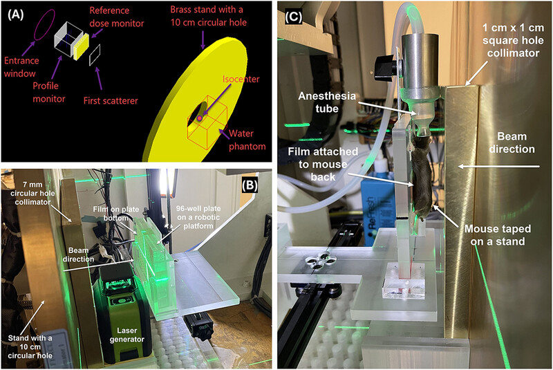
(A) Schematic of Monte Carlo modeling of the proton FLASH beamline.
(B) Image of the robotic motion platform and a self-leveling laser positioning system for high-throughput irradiation of cells or organoids in culture.
(C) Lateral view of the mouse setup with the robotic platform and a custom holder for the mouse and the anesthesia device.
Radiation induced liver injury (RILI) evaluation using longitudinal computed tomography (CT) in image-guided precision murine radiotherapy
- Pages: 182-190
- First Published: 09 November 2024
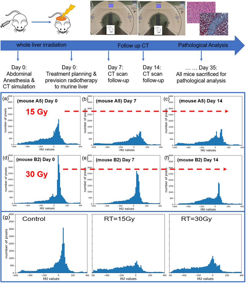
Image-guided, dose-escalated, precision radiation experiments to murine liver were performed, followed by longitudinal CT studies. In addition to body weight and mean HU reductions, HU histogram of liver parenchyma in the irradiated groups showed longitudinal changes in tissue density and different HU distribution compared to the non-irradiated livers.
3D gamma analysis between treatment plans for nominally beam-matched medical linear accelerators using PyMedPhys
- Pages: 191-199
- First Published: 27 November 2024
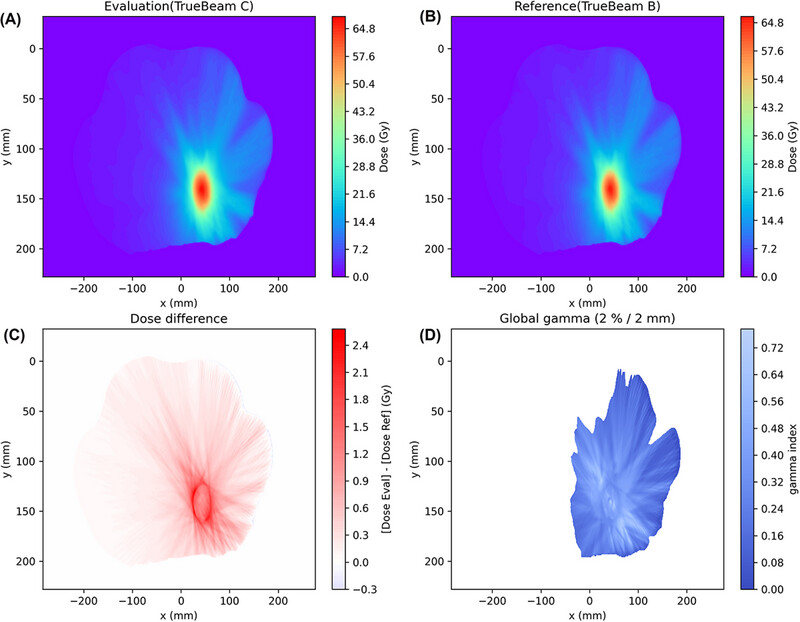
Illustration of 3D gamma analysis results using PyMedPhys. (A). Dose map on a selected slice of the evaluation plan. (B). Dose map of a selected slice of the reference plan. (C). Dose difference map on a selected slice. (D). Global gamma index on a selected slice. This example was from patient No. 40 (lung SBRT).
Heart ultrasound and biomechanical evaluation of radiation-induced heart toxicity using transthoracic echocardiogram (TTE) and dynamic mechanical analysis (DMA)
- Pages: 200-208
- First Published: 12 November 2024
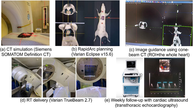
Precision radiation to murine heart is performed with CT simulation, VMAT treatment planning, CBCT image guidance, and dose delivery, followed by weekly transthoracic echocardiography (TTE). Results show RIHD evaluation with ultrasound imaging and elastic modulus measurement is feasible. Increased myocardial stiffness, abnormal diastolic relaxation, more collagen deposition, and reduced tissue elasticity are observed in irradiated cardiac tissue, which may deepen our understanding of RIHD and facilitate to improve patients’ quality of life.
Feasibility planning study of lattice radiotherapy for palliation in bulky tumors
- Pages: 209-217
- First Published: 28 November 2024
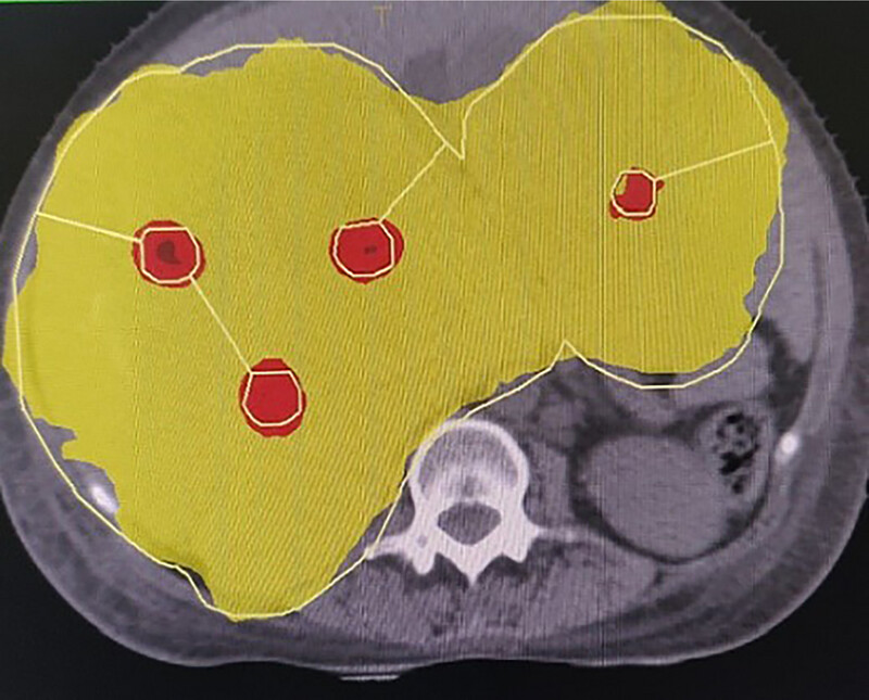
Lattice radiotherapy is dosimetrically feasible and can deliver high doses to small volumes called vertices within a low dose prescribed to the entire tumour volume while sparing organs at risk adequately. It could potentially improve outcomes in patients with large tumours in terms of palliation of symptoms and tumour shrinkage by the high dose peaks distributed within tumour volume, which is said to trigger immune mechanisms and bystander effects. The surrounding conventional doses ensure adequate organs at risk sparing. Here, we present a dosimetric feasibility study of ten patients and a comparison with conventional palliative radiotherapy. Lattice radiotherapy is a feasible option with VMAT and spares surrounding normal structures. Further prospective clinical trials are required to confirm its efficacy. .
Exploration of individualized neoadjuvant therapy model for operable esophageal cancer: A Surveillance, Epidemiology, and End Results database analysis
- Pages: 218-226
- First Published: 08 December 2024
Neoadjuvant therapy has been widely used and achieved considerable clinical efficacy in locally advanced esophageal cancer. For stage II esophageal cancer, its stage is still early, and the degree of malignancy is between stage I and stage III. This study compared the efficacy of nCRT, nCT and surgery alone, and found that nCRT is still recommended for stage II esophageal cancer.
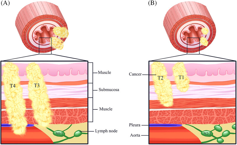
In addition, this study proposed that the tumor biological characteristics may vary according to the T and N tumor burden in esophageal cancer with the same clinical stage. Therefore, stage III operable esophageal cancer was divided into two tumor invasion patterns based on T and N tumor burden: “Local invasive type” (T3N1M0, T4N0-1M0) and “Regional metastatic type” (T1-2N2-3M0, Figure 1).
The study explored the neoadjuvant therapy model for esophageal cancer based on different tumor invasion patterns. This study found that among stage III operable esophageal cancer, the “Local invasive type” benefited more from nCRT, while the “Regional metastatic type” tended to benefit from nCT. With the increasingly diversified models of neoadjuvant therapy for esophageal cancer, this finding may have guiding significance for the selection of neoadjuvant therapy model for operable esophageal cancer .
CASE REPORT
[18F]-NOTA-FAPI-04 PET/CT downgraded the staging of a breast cancer patient and changed their treatment management
- Pages: 227-231
- First Published: 27 November 2024
![[18F]-NOTA-FAPI-04 PET/CT downgraded the staging of a breast cancer patient and changed their treatment management](/cms/asset/a07ba4c5-43bc-4bb3-bf86-90a728ab9cb2/pro61245-gra-0001-m.jpg)
In Brief: Qin et al. find an interesting case of a 47-year-old woman 41 days after breast-conserving surgery and sentinel lymph node biopsy for right breast cancer. [18F]-FDG PET/CT scan in the lymph nodes of right axillary fossa, right intrathoracic muscle, and right clavicle was considered as metastatic. However, no intense [18F]-NOTA-FAPI-04 uptake was found on [18F]-NOTA-FAPI-04 PET/CT scan. These [18F]-NOTA-FAPI-04-negative lymph nodes had been later confirmed by histopathology. Tumor staging was downgraded from cT1N3M0 to stage cT1N0M0.This interesting case suggested that [18F]-NOTA-FAPI-04 PET may have the potential for differential diagnosis of benign and malignant lymph nodes and improving the accuracy of breast cancer staging.
Sign up for email alerts
Tools
More from this journal
- Open Access License and Copyright
- Institutional and Funder Payments
- Call for Papers: Special Issue Radiation Therapy for Rare Tumor of Head and Neck
- Wiley Open Access
- Publishing with Precision Radiation Oncology
- Call for Papers: Special Issue
- Top downloaded articles published in 2022
- Themed Collection: Guidelines for radiotherapy
- Themed Collection: MRI guided precision radiotherapy
- Themed Collection: Proton and carbon ion therapy
- Themed Collection: Innovations, Advances, and Challenges in Precision Radiation Oncology Physics
- Themed Collection: Stereotactic Body Radiation Oncology

Relevant Open – Access Health Sciences journals

Journal of Clinical Laboratory Analysis (1987-Present)
Thoracic Cancer (2010-Present)
Why publish with Precision Radiation Oncology?
- All articles are fully open access and immediately freely available to read, download and share
- Easily comply with funder and institutional mandates
- High standard, rigorous peer review process
- All articles are published under a Creative Commons license, so you’ll retain copyright
Article Publication Charges are currently waived for all articles submitted to the journal!
Submit your manuscript now