Editor-in-Chief: Dr Philippe Vielh

Cytopathology is the affiliated title of over 20 national cytology societies. We publish articles relating to aspects of cytology that will increase our knowledge and understanding of the aetiology, diagnosis and management of human disease.
We make a major contribution to Continuing Medical Education in clinical cytology, and are essential reading for every practicing cytopathologist and cytotechnologist who wishes to keep abreast of new developments in this rapidly developing field.
Journal Metrics
- 2.3CiteScore
- 1.1Journal Impact Factor
- 33%Acceptance rate
- 16 days Submission to first decision
Editor's Choice Volume 36, Issue 4 (July 2025)
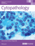
The Diagnostic Role of TFF1, TFF3, FOXA1, CA XII and TRPS1 in Serous Effusions
- Cytopathology
- 408-417
- 10.1111/cyt.13489
Graphical Abstract
TFF1, TFF3, FOXA1, CA XII and TRPS1 are sensitive breast carcinoma markers. FOXA1 is the most sensitive, and TRPS1 is the most specific of these 5 markers. The expression of TFF1 in breast carcinoma effusions may be informative of survival.
On the Cover
Articles
Primary Cutaneous Rosai‐Dorfman Disease: A Cyto‐Histo Correlate
- 21 July 2025
Graphical Abstract
Primary cutaneous Rosai Dorfman disease (RDD) is a rare proliferative disorder of histiocytes involving skin exclusively. Cytomorphological features of two such cases affecting a 20-year-old male (multicentric) and an 8-year-old male are presented here. The diagnosis is challenging and a high index of suspicion and systemic correlation are necessary for early diagnosis and treatment.
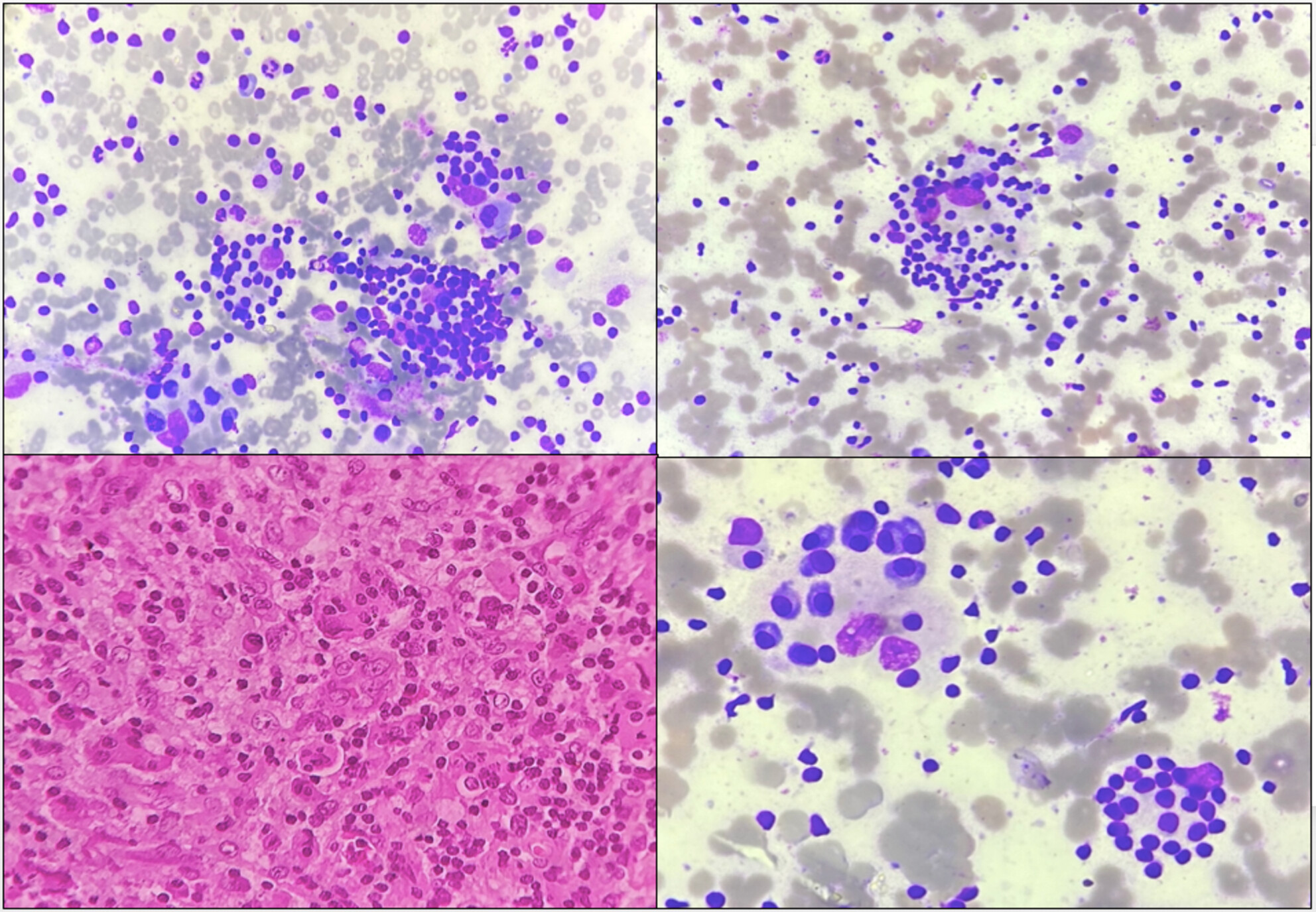
Cytomorphology of Renal Hydatid Cyst Mimicking as Simple Renal Cyst
- 10 July 2025
Graphical Abstract
Mixed inflammatory cell infiltrate along with the presence of numerous Hooklets.
From the Daily Peer Review of Abnormal Pap Test Slides to the Monitoring of Individual and Laboratory Performances: 5 Years of Data Collection and New Potential (Key) Performance Indicators
- 5 July 2025
Graphical Abstract
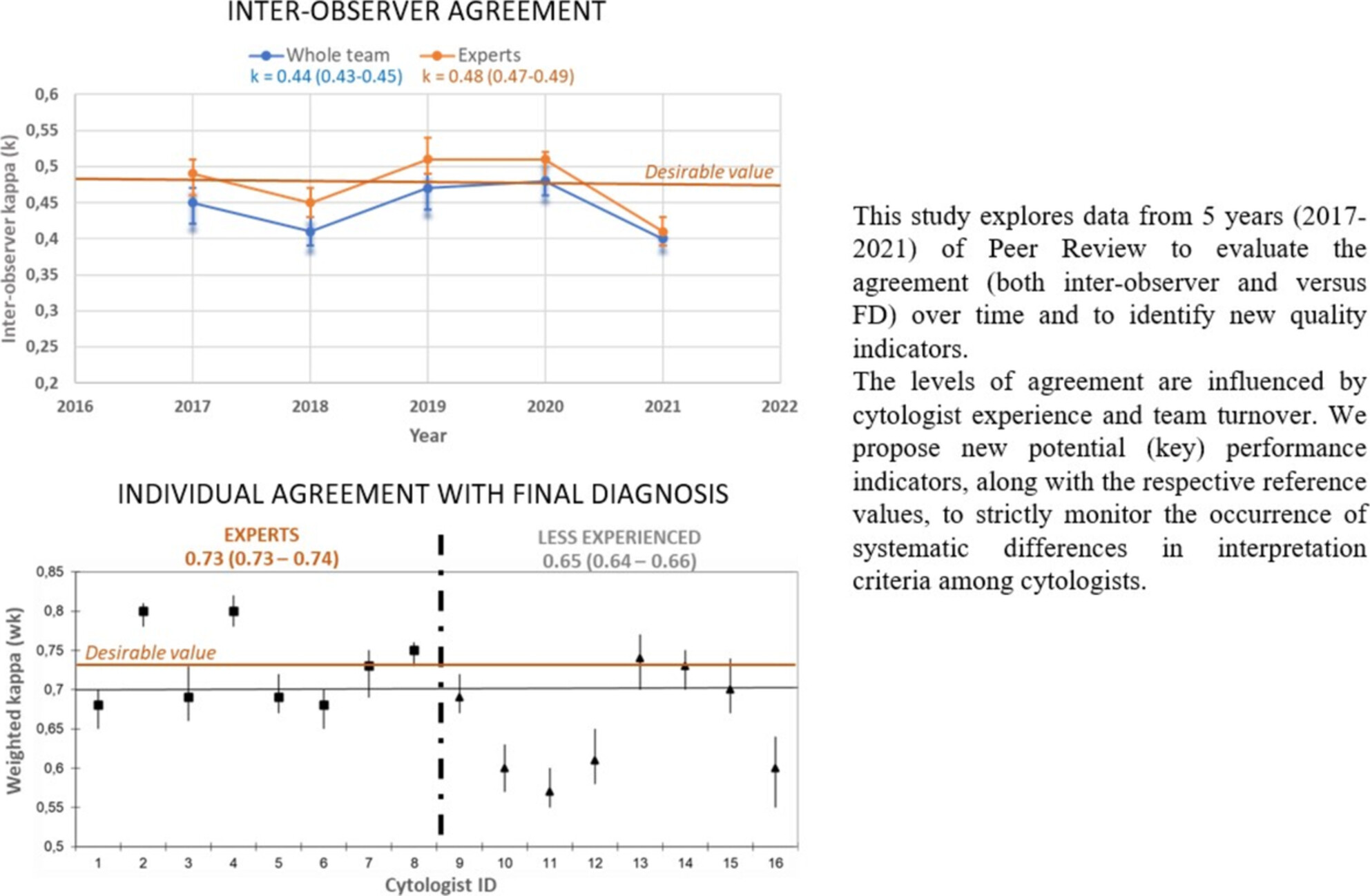
The study explores data from 5 years of Peer Review to evaluate the trend of agreement level (inter-observer and versus final diagnosis) over time and proposes new potential key performance indicators (KPIs), along with the relative reference values. These KPIs should be used to strictly monitor the occurrence of systematic differences in interpretation criteria among cytologists and to implement prompt corrective actions in case of significant deviations in diagnostic accuracy.
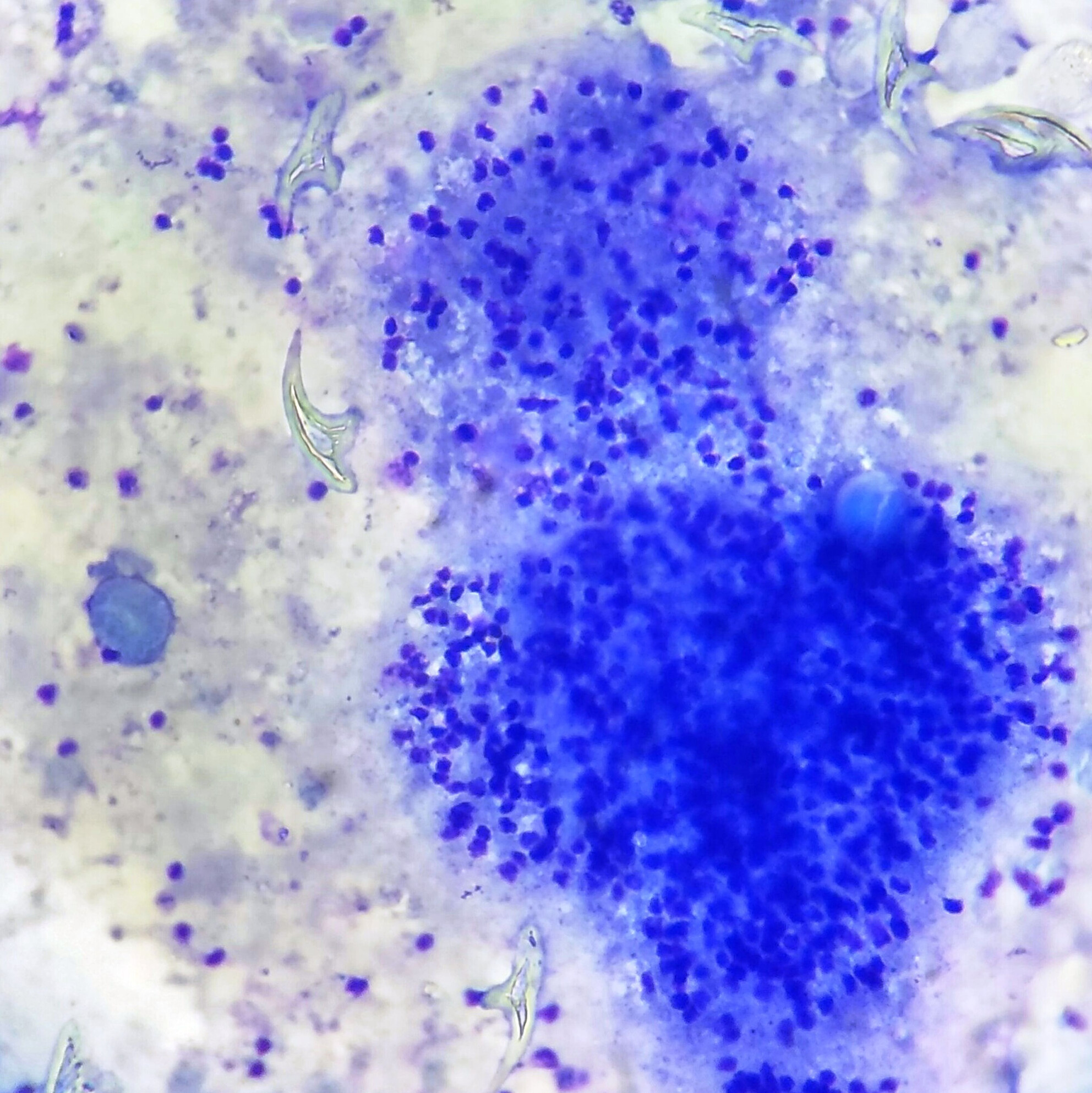
Cytological Diagnosis of Amyloidosis From Orbital Papules
- 1 July 2025
Graphical Abstract

We report a case of systemic amyloidosis with eyelid papules, diagnosed on FNAC. A 43-year-old male presented with periocular papules for 2 months. FNAC revealed acellular glassy pale eosinophilic material with apple green birefringence on Congo Red stain under polarised light. Periorbital papules are an uncommon presentation of amyloidosis. Any periorbital infiltrative dermatosis should raise suspicion of local or systemic amyloidosis.
Metastatic Malignant Phaeochromocytoma in Ascitic Fluid: Cytological Diagnosis of a Rare Entity
- 30 June 2025
Graphical Abstract
In summary, a review of the literature showed that there are only isolated case reports of ruptured pheochromocytoma and to the best of our knowledge this report is the first to document metastatic phaeochromocytoma in peritoneal fluid within the cytopathology literature. Our case report emphasises the importance of clinical history in the context of the cytopathologic evaluation, and good cell block preparation, which allows the use of IHCs in challenging cases as it may lead to the discovery of unusual metastatic sources.
The following is a list of the most cited articles based on citations published in the last three years, according to CrossRef.
The presence of a cytopathologist increases the diagnostic accuracy of endoscopic ultrasound‐guided fine needle aspiration cytology for pancreatic adenocarcinoma: a meta‐analysis
- 159-171
- 27 May 2013
Cell blocks in cytopathology: a review of preparative methods, utility in diagnosis and role in ancillary studies
- 356-371
- 11 August 2014
Impact of image analysis and artificial intelligence in thyroid pathology, with particular reference to cytological aspects
- 432-444
- 5 April 2020
Abstract
Thyroid pathology has great potential for automated image analysis/artificial intelligence algorithm application on whole-slide images. Studies to date mainly deal with the assessment of immunohistochemical staining, quantification of cellular and nuclear parameters and discrimination between benign and malignant nodules. They show that correlation of automated assessment of immunohistochemical staining with manual pathologist's assessment is high and diagnostic performance of automated models is comparable with expert pathologist diagnosis
Thyroid FNA cytology in Asian practice—Active surveillance for indeterminate thyroid nodules reduces overtreatment of thyroid carcinomas
- 455-466
- 2 November 2017
Abstract
Although Asian thyroid practices have implemented the American Thyroid Association guidelines and the Bethesda system for reporting thyroid cytopathology, significant deviations in actual risk of malignancy have been reported. With review of the literature and analyses of thyroid practices in Asia, the author examines the underlining reasons why actual ROMs reported in Asia are so different from those in Western practice based on the author's perspective.
Current state of whole slide imaging use in cytopathology: Pros and pitfalls
- 372-378
- 5 February 2020
Abstract
This review summarizes the advantages and the disadvantages of the use of whole slide imaging (WSI) in cytopathology, starting from the cytological specimen to the entire laboratory and istitution. Whole Slide Imaging provides high-quality standardized slides, but there is still a need for validation studies before full adoption for primary diagnosis. Whole Slide Imaging in cytology retains most of the advantages of surgical pathology, and in the near future barriers to adoption will be overcome.
Recent issues
 Volume 36, Issue 4
Special Issue: Interventional Cytopathology
i, 295-429July 2025Guest Editor(s):
Karen Villar Zarra, Ronald Balassanian