

Journal list menu

Export Citations
Download PDFs
2024: Volume 35
Morphology quiz: Large inguinal lymphadenopathy in a young man
- First Published: 06 April 2024
A 25-year-old male with fever, joint pain and inguinal masses. Lab tests reveal polyadenopathy. Negative serology. Ultrasound shows a 42 mm hypoechoic node. FNAC and core-needle biopsy show… what? Read the article to find out.
A sub-hepatic nodule in a young female: Chase the case
- First Published: 17 March 2024
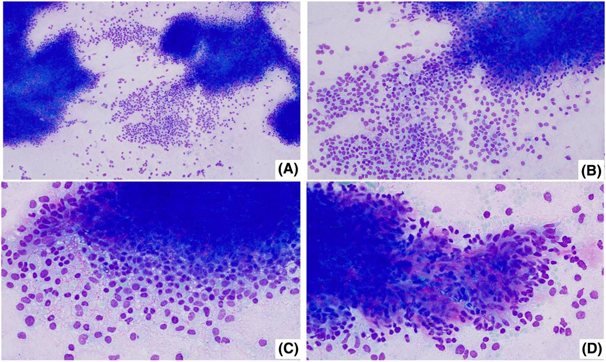
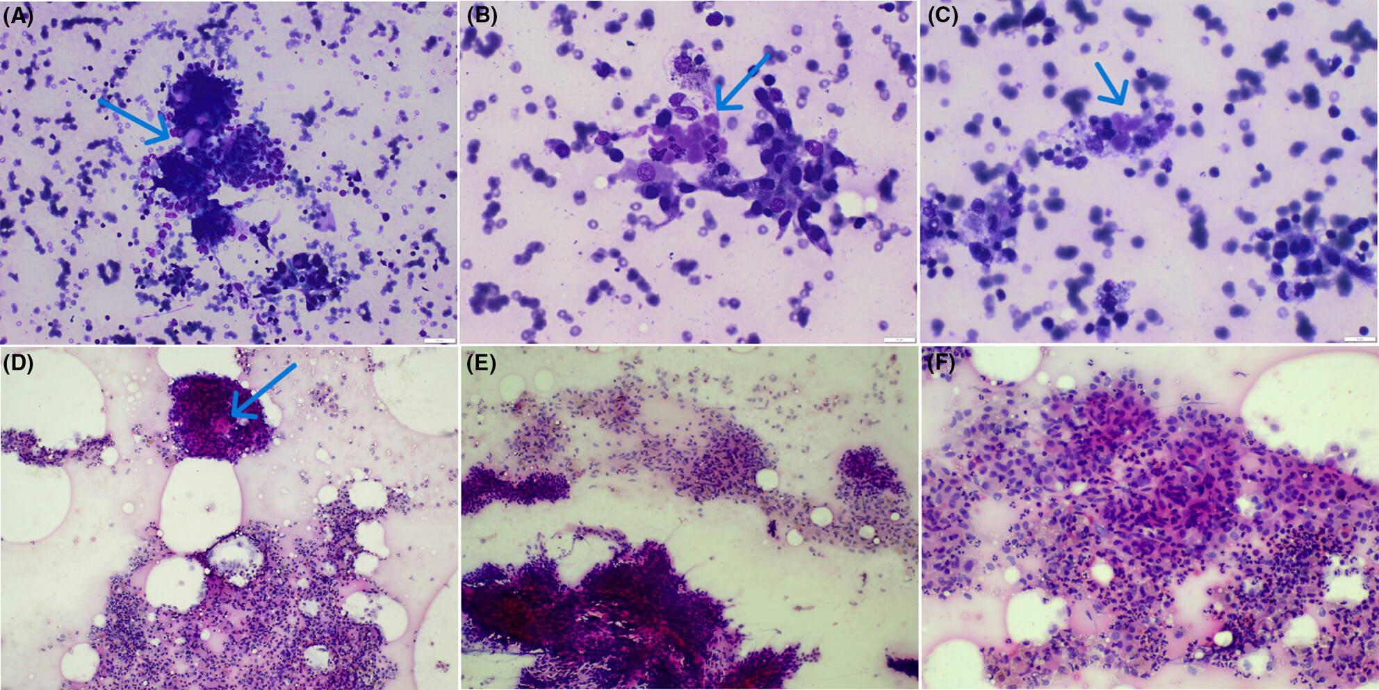
Sertoli–Leydig cell tumours (SLCTs) are rare, mixed sex-cord stromal tumours composed of varying proportions of both Sertoli and Leydig cells, which account for <0.5% of all ovarian tumours. The cytomorphologic features of SLCTs are not well described in literature. Herein, we describe the cytomorphologic features of an SLCT at an uncommon metastatic site in a young female.
Sertoli–Leydig cell tumours (SLCTs) are rare, mixed sex-cord stromal tumours composed of varying proportions of both Sertoli and Leydig cells, which account for <0.5% of all ovarian tumours. The cytomorphologic features of SLCTs are not well described in literature. Herein, we describe the cytomorphologic features of an SLCT at an uncommon metastatic site in a young female.
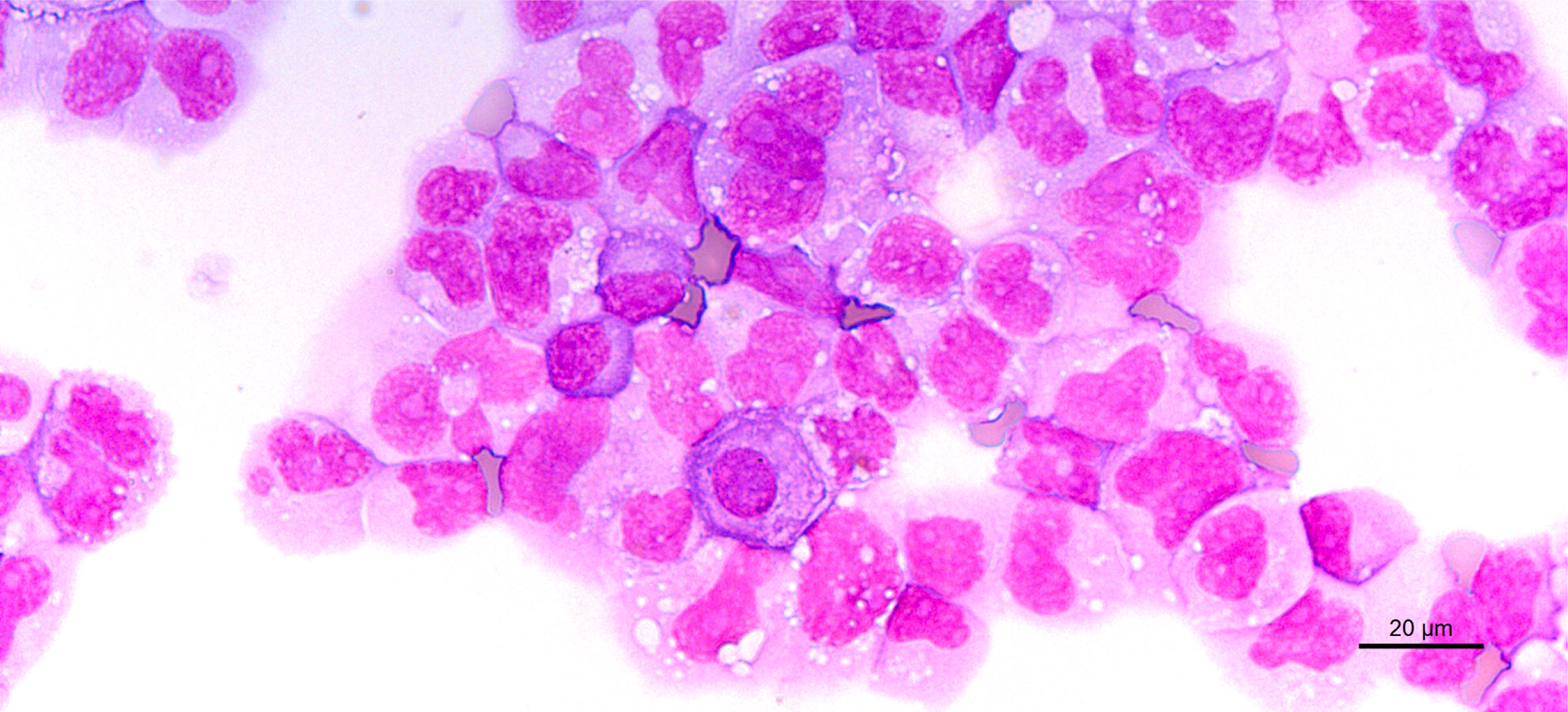
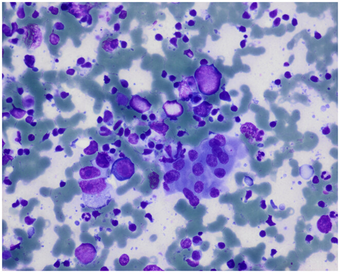
Vacuoles in a bone marrow smear: Not always reactive
- First Published: 09 July 2024
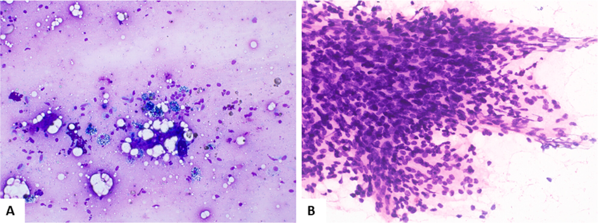
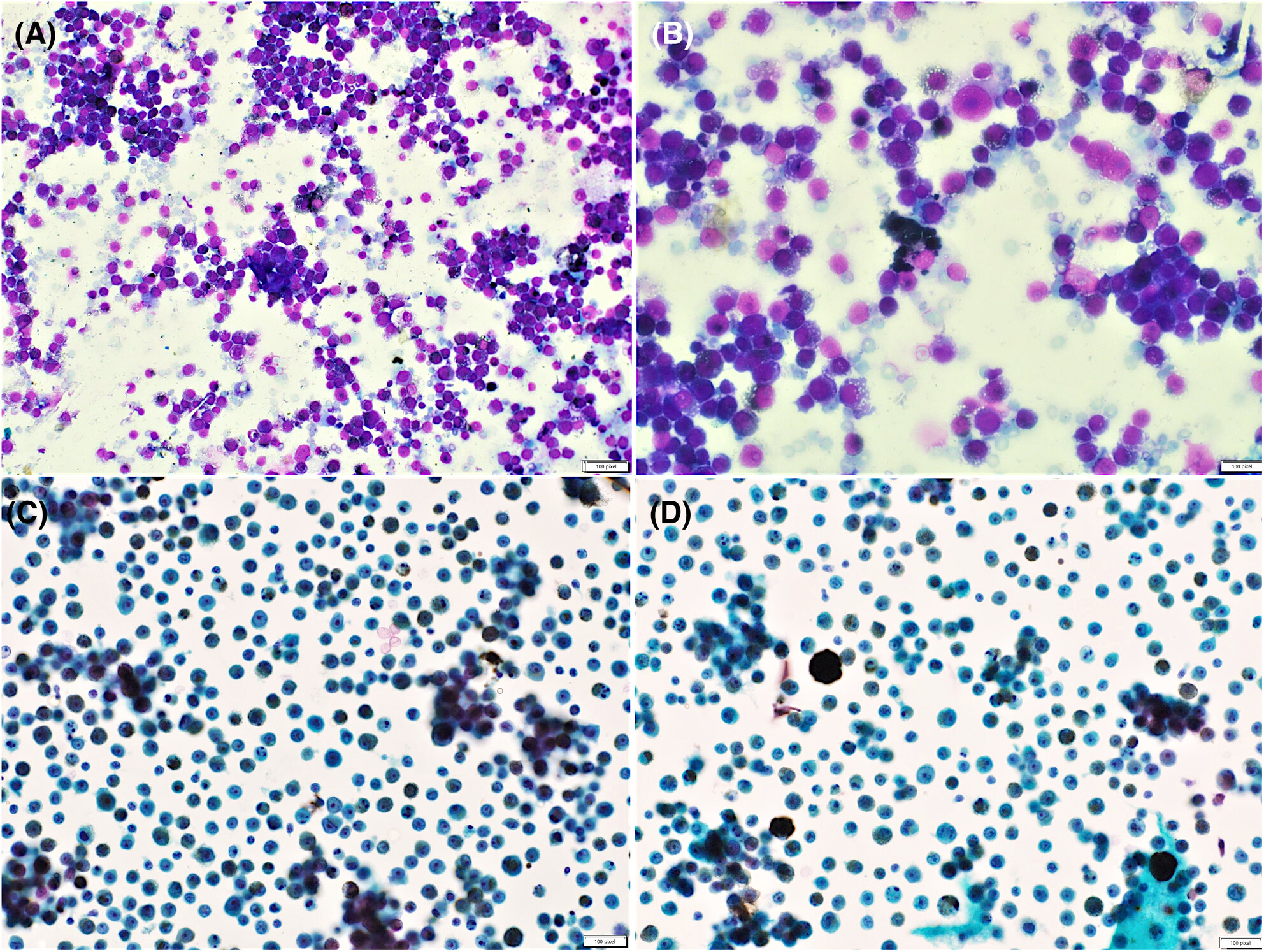
Vacuoles are a common observation in bone marrow smears, often related to reactive causes. The authors report a clinical case with this cytology finding in a 67-year-old male patient with systemic symptoms.
Pericardial effusion: Unusual immunohistochemical expression
- First Published: 31 July 2024
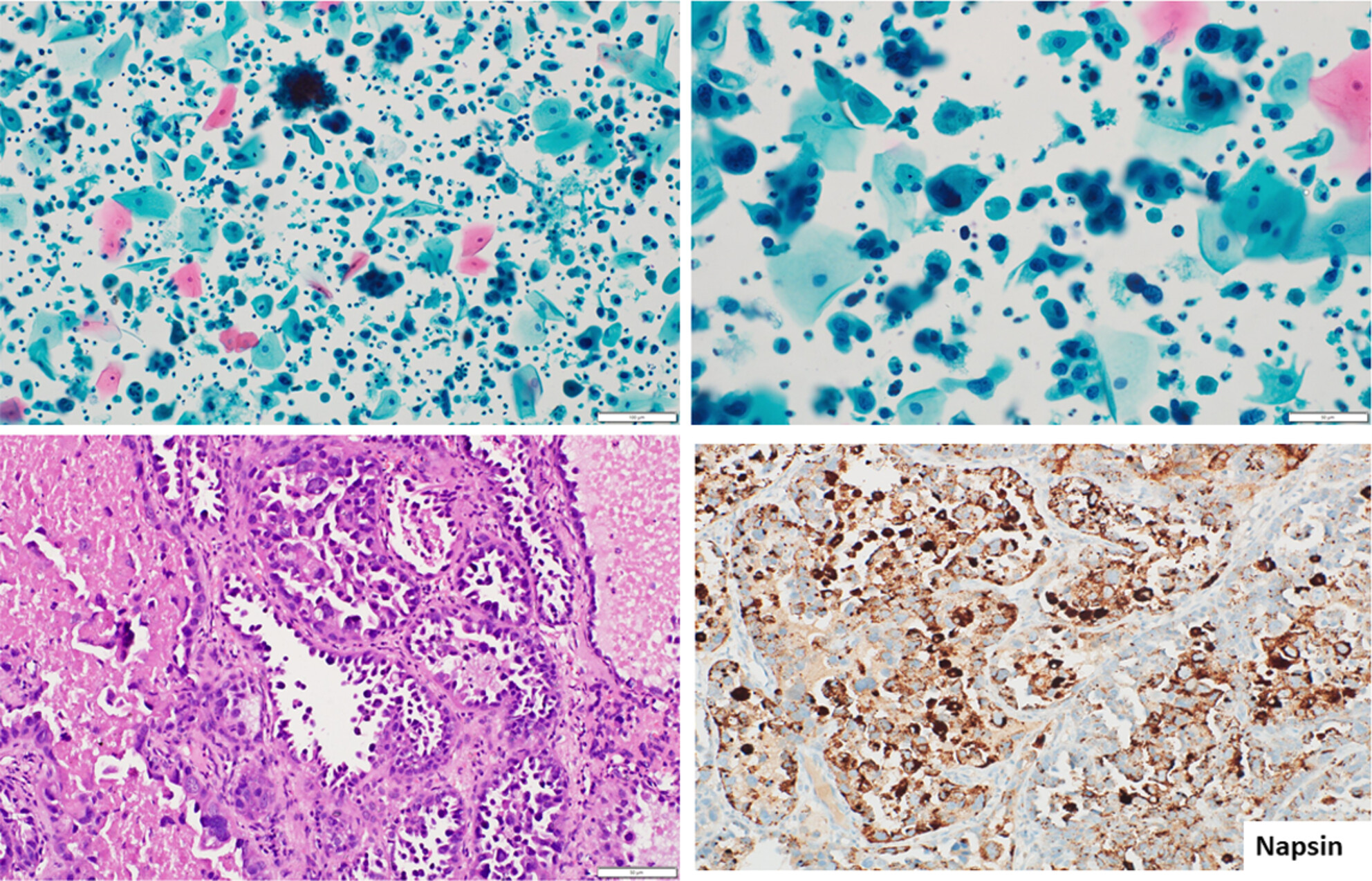
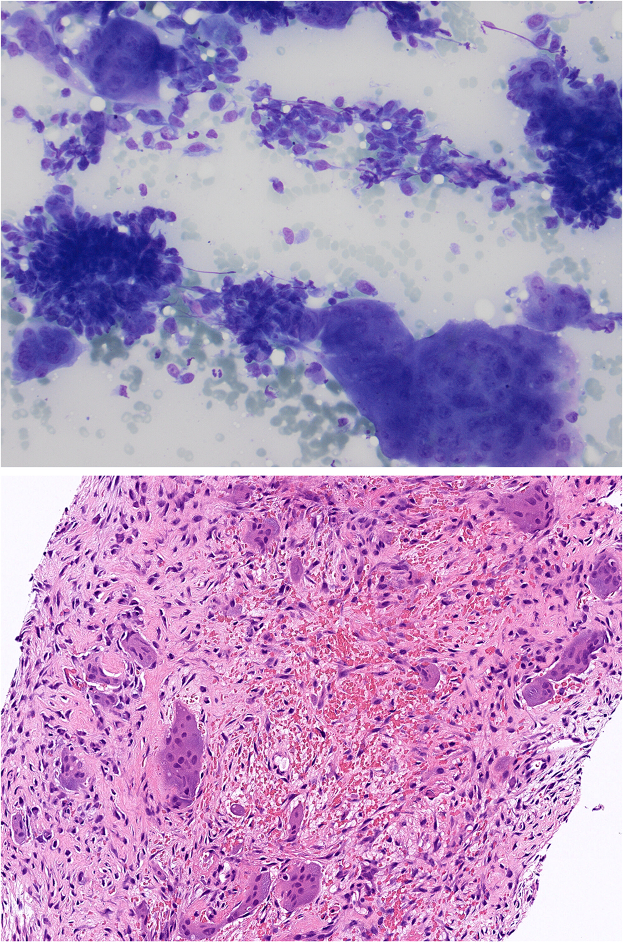
Endometrial Gastric-type Carcinoma is a rare disease, especially in cytology. A 65-year-old woman was admitted to the hospital due to intermittent chest tightness with dyspnoea. The chest CT showed a soft tissue shadow of the left lung with bronchial occlusion and pericardial effusion. What was her diagnosis?
The diagnostic conundrum of hyper eosinophilia—Sheer tenacity of a parasite
- First Published: 28 August 2024
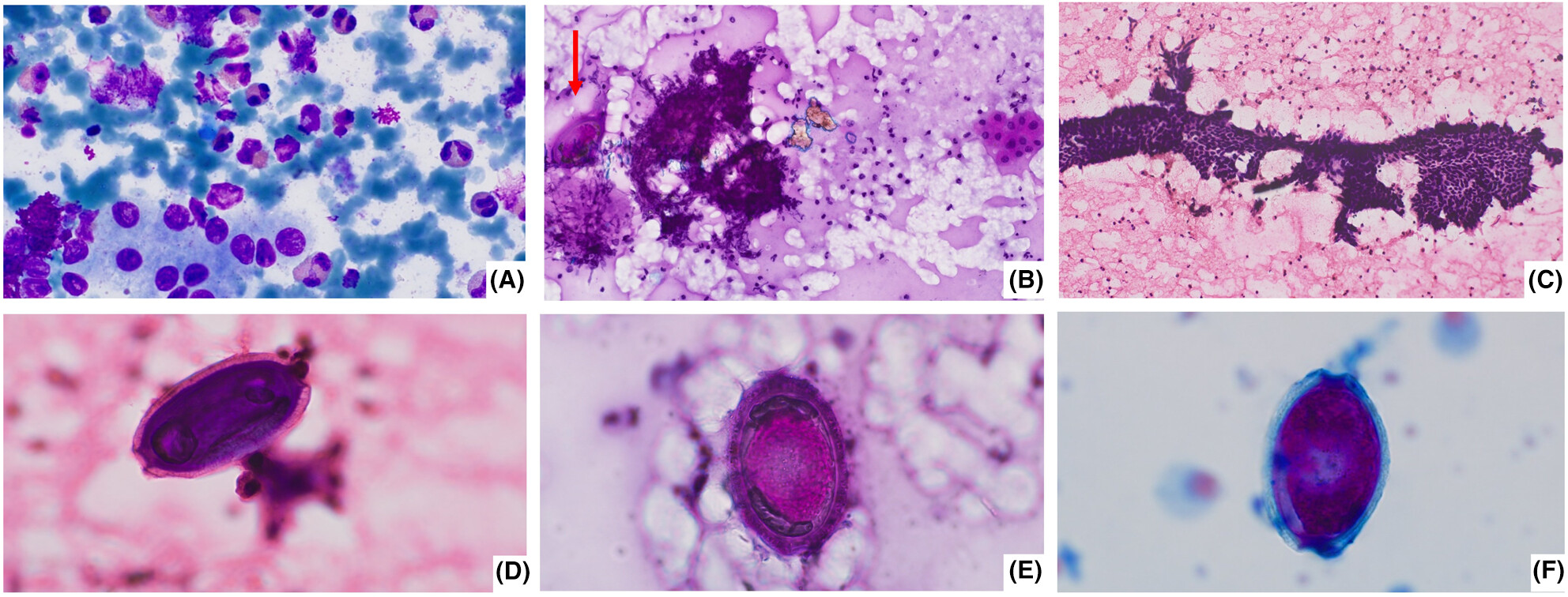
We present an interesting and rare case of Capillaria hepatica infection in a 2-year-old boy, who presented with fever, rash, hepatomegaly and peripheral eosinophilia. FNAC of hepatic lesion showed parasitic eggs and PCR from the aspirate confirmed the diagnosis. We describe the cytomorphological features and provide educational multiple-choice questions related to the topic.
All groin swellings are not lipoma
- First Published: 22 August 2023
An interesting case of an abdominal wall swelling near an old operative scar showing epithelial and stromal cells.
In this present paper, fine needle aspiration cytology of an inguinal swelling is discussed which may often create diagnostic confusion.
Bugs' eyes and black monsters: Ascitic fluid cytology in an elderly male with hematochezia
- First Published: 12 October 2023
Anorectal malignant melanomas are rare, accounting for less than 2% of all melanomas. Malignant effusions developing secondary to malignant melanoma are highly uncommon. Herein, we present the cytomorphological features of a metastatic anorectal malignant melanoma presenting with ascites at the initial clinical presentation.
Usual breast lump with unusual features
- First Published: 24 October 2023
The co-existence of granulomatous mastitis and collagenous spherulosis in a breast lump is an uncommon finding. The awareness of cytomorphological features can help corroborate a cytological diagnosis.
A palpable breast lump in an elderly female warrants urgent attention and fine needle aspiration is a rapid, reliable method of evaluation. An elderly female with a firm breast lump mimicking malignancy was subjected to fine needle aspiration cytology (FNAC). Smears showed ill-formed granulomas, inflammatory cells and homogeneous hyaline stromal globular elements intermingled with the benign ductal epithelial and myoepithelial cells.
Cytology of a parietal swelling in a 52-year-old man
- First Published: 30 October 2023
Primary FNA diagnosis of brown tumour is challenging because overlapping of cytomorphologic features with other giant cell lesions. Clinical information, imaging and laboratory tests benefits the correct diagnosis.
Fine-needle aspiration cytology of presacral myelolipoma: Cytomorphological features of a rare entity and review of the literature
- First Published: 04 December 2023
Presacral myelolipoma is an uncommon benign tumor, and its diagnosis can be challenging oncytology specimens. This case emphasizes the importance of fine needle aspiration cytology as an initial and valuable diagnostic tool for evaluating presacral masses. The identification of a combination of mature adipose tissue and hematopoietic elements in varying proportions is a crucial feature in FNA cytology. This underscores the role of FNA cytology in providing an accurate diagnosis and guiding subsequent management decisions.
Cutaneous balloon-cell melanoma metastases to the axillary lymph node: Exploring cytomorphologic features and differential diagnoses on fine needle aspiration biopsy
- First Published: 09 January 2024
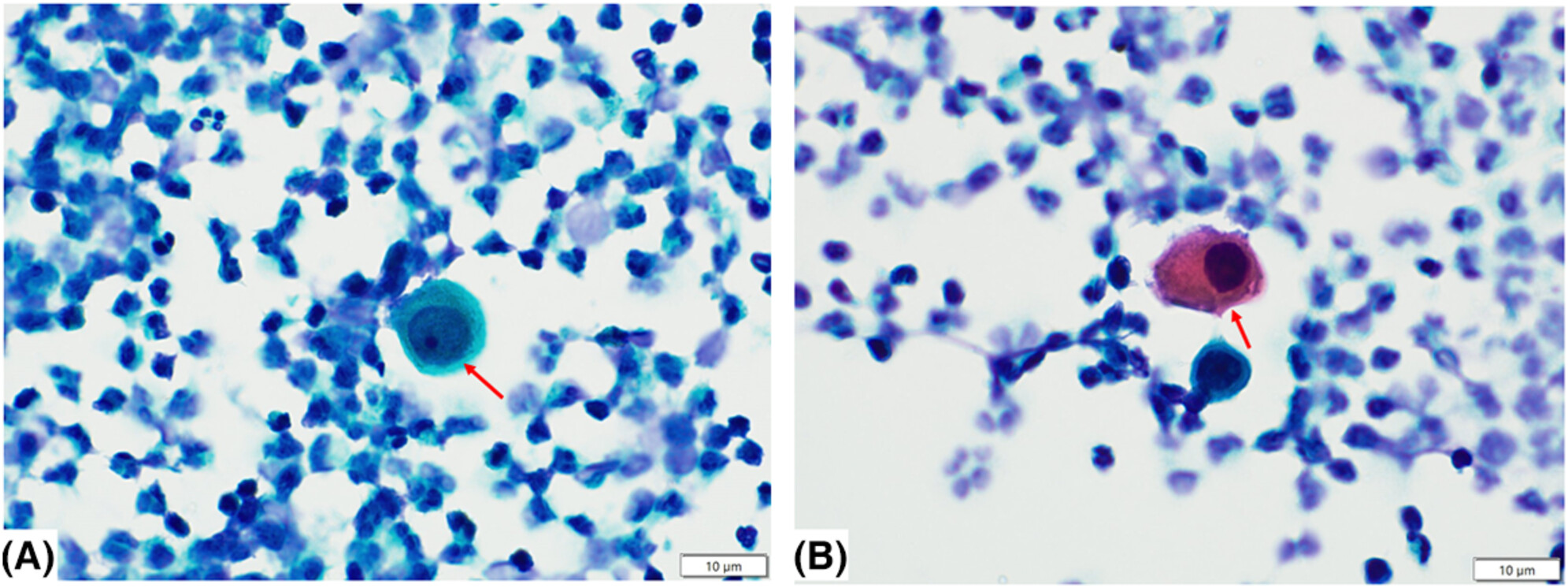
Balloon cell melanoma (BCM) is an exceptionally uncommon histological variant, making up <1% of all malignant melanomas. Diagnosing balloon cell melanoma on cytological specimens can be challenging due to its scarcity and its similarity to other medical conditions. A comprehensive clinical assessment and histological analysis, coupled with immunohistochemical staining, play a crucial role in distinguishing balloon cell melanoma from various benign and malignant skin conditions. The differential diagnoses encompass spitz nevus, balloon cell nevus, clear cell sarcoma of tendons and aponeuroses (melanoma of soft tissues), metastatic clear cell renal cell carcinoma, sebaceous carcinoma, and benign adnexal tumours like clear cell hidradenoma.
Malignant melanoma encompasses a spectrum of histopathological subtypes, each with unique clinical and cytological characteristics. Notably, balloon-cell melanoma (BCM) emerges as an exceptionally rare and diagnostically challenging variant. Marked by the presence of distinct balloon-like, clear cytoplasmic vacuoles within melanoma cells, BCM stands apart from other melanoma subtypes. Despite its rarity, the distinctive cytological features of BCM make it a compelling subject of investigation, emphasizing the crucial role of Fine Needle Aspiration (FNA) cytology in ensuring accurate diagnosis and guiding subsequent management decisions.
2023: Volume 34
CSF cytology in a patient with vertigo and earache
- First Published: 16 July 2023
Large atypical cells in cerebrospinal fluid in a patient with earache and vertigo.
In this Enigma Portal case, we described uncommon cerebrospinal fluid findings in a case of vertigo and earache in a 40-year-old man.
Intracranial squamous papillary lesion
- First Published: 02 August 2023
Intracranial lesions with squamous differentiation raise a diagnostic challenge, which can include benign or malignant, primary and metastatic lesions. Here is the case of a 49-year-old woman, in which intraoperative cytology helped the differential and final diagnosis.
Quiz case: Abnormal haematopoietic cells in pleural effusion
- First Published: 18 August 2023
This case was presented because of the number of plasmablasts in a patient with a medical history of multiple myeloma. Flow cytometry is a “gold standard” technique for the diagnosis of haematological malignancies. This technique works for all fluids and should be performed in effusions (pleural, pericardial, ascites) in cases of suspected haematological malignancy. Alternatively, immunohistochemistry using appropriate markers could be performed if flow cytometry is not available.
This case illustrates a pleural infiltration by plasmablasts. Myelomatous cells were characterised by immunocytochemistry and flow cytometry.
Granulomatous thyroiditis—A rare case and the diagnostic approach
- First Published: 21 May 2023
The presence of well-formed granulomas, multinucleated giant cells containing colloid, and degenerative changes in thyroid follicular epithelium are the key diagnostic features of granulomatous (subacute, de Quervain's) thyroiditis. Fine needle aspiration cytology is a confirmatory test that is helpful in cases without any clinical suspicion and to exclude differential diagnoses.
Cytological assessment of a thyroid mass
- First Published: 28 June 2023
This case highlights an uncommon diagnosis encountered in thyroid fine needle aspiration, highlighting differentials based on the clinical features and how cytomorphology can distinguish these.
Enigma portal: Undifferentiated thyroid malignant neoplasm in a patient with Hashimoto's thyroiditis
- First Published: 25 April 2023
Undifferentiated anaplastic thyroid tumours are uncommon and constitute a diagnostic challenge on fine needle aspiration. A case showing large, single neoplastic cells in a background of Hashimoto's disease is presented.
Spindle-nucleated cells in cervical liquid-based cytology: Difficulties in distinguishing between epithelial and non-epithelial tumours based on morphology
- First Published: 30 January 2023
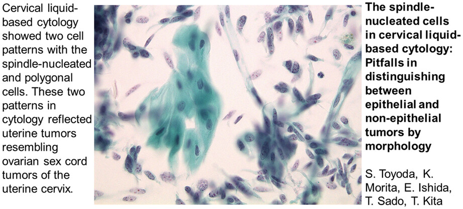
Uterine tumours resembling ovarian sex cord tumours of the uterine cervix are highly sporadic. Cervical liquid-based cytology revealed two cell patterns: spindle-nucleated cells and polygonal cells.
Not all black fungus is mucormycosis: A challenging case of Medicopsis romeroi
- First Published: 17 January 2023
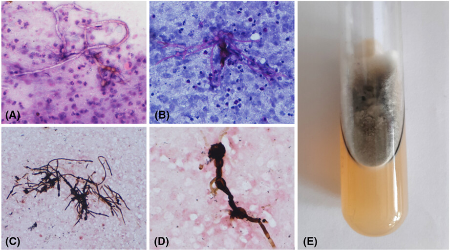
In this case of phaeohyphomycosis, fine needle aspiration cytology enabled a rapid diagnosis and prompt treatment. This infection is quite prevalent in immunocompromised individuals; however, the Medicopsis romeroi species is a rare causative agent. These cases are associated with inadequate response to standard antifungal therapy and require discussion.