Fine-needle aspiration cytology of presacral myelolipoma: Cytomorphological features of a rare entity and review of the literature
Abstract
Presacral myelolipoma is an uncommon benign tumor, and its diagnosis can be challenging oncytology specimens. This case emphasizes the importance of fine needle aspiration cytology as an initial and valuable diagnostic tool for evaluating presacral masses. The identification of a combination of mature adipose tissue and hematopoietic elements in varying proportions is a crucial feature in FNA cytology. This underscores the role of FNA cytology in providing an accurate diagnosis and guiding subsequent management decisions.
1 CASE PRESENTATION
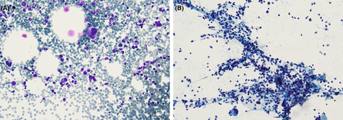
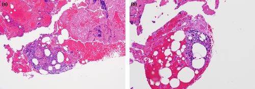
A 68-year-old female presented with no significant past medical history presented with lower back pain localized to the left side. MRI revealed a 4.0 cm presacral mass centred on the midline and directly abuts the anterior sacrum without infiltrative features. The patient underwent fine-needle aspiration (FNA) for further evaluation. Diff-Quik and Papanicolaou stained smear showed abundant haematopoietic elements intermixed with mature fat droplets. The haematopoietic cells were composed of immature myeloid and erythroid precursor cells admixed with neutrophils, lymphocytes and eosinophils. Few megakaryocytes were seen. Figure 1A,B. Cellblock of FNA showed mature adipocytes with no significant atypia and trilineage haematopoiesis with markedly increased megakaryocytes. Figure 2A,B.


2 QUIZ
- What is your diagnosis?
- Myelolipoma
- Extramedullary haematopoiesis
- Lipoma
- Liposarcoma
- Myeloid sarcoma
- The above lesion is most encountered in:
- Retroperitoneum
- Presacral region
- Adrenal gland
- Mediastinum
- Which of the following is true about the above lesion?
- Most encountered in males
- No malignant transformation or recurrence
- Often bilateral in the adrenal gland
- Adrenal lesions are hormonally active, complementary laboratory testing is recommended.
ANSWERS TO THE QUIZ
- a. The illustrated lesion depicts a presacral myelolipoma featuring dispersed haematopoietic cells intermingled with mature fat cells. The potential differential diagnosis encompasses extramedullary haematopoiesis, lipoma, liposarcoma and myeloid sarcoma. It is crucial to distinguish that lipoma, liposarcoma and myeloid sarcoma consist solely of mature fat cells without the presence of haematopoietic elements.
- c. Myelolipoma is most commonly found in the adrenal glands; however, it can also occur in extra-adrenal locations such as the retroperitoneum, presacral region and mediastinum.
- b. Myelolipoma typically impacts individuals aged 50–70 years, with no specific gender preference, and tends to occur unilaterally. Malignant transformation or recurrence is uncommon. Adrenal myelolipomas are generally hormonally inactive, and routine laboratory tests are not necessary.
3 EXPLANATION
Myelolipoma is a rare benign neoplasm composed of a variable proportion of a mixture of mature adipocytes and haematopoietic elements. While myelolipoma is predominantly encountered in the adrenal gland, the occurrence in extra-adrenal sites such as the presacral region, retroperitoneum, liver and mediastinum has been reported in the literature.1 The pathogenesis of myelolipoma is not well understood and is currently undergoing ongoing research to further characterize the unique cytogenetic and molecular alterations that contribute to the neoplastic process in myelolipoma.
Presacral myelolipoma commonly presents as an incidental finding on radiologic imaging; however, large lesions may be symptomatic and present as encapsulated, heterotopic fat-containing masses supported by a network of reticulum fibres. Fine-needle aspiration (FNA) cytology offers a minimally invasive and effective means of obtaining cytological samples from deep-located lesions.2
In the illustrated case, the differential diagnosis included myelolipoma and extramedullary haematopoiesis. Surgical resection and microscopic examination revealed a combination of fully mature adipocytes and extramedullary trilineage haematopoietic cells, resembling hypercellular bone marrow, accompanied by a significant elevation in megakaryocyte count. The overall findings with a correlation with histopathologic examination revealed a definitive diagnosis of presacral myelolipoma. The cellular composition of presacral myelolipoma in the cytological sample is usually heterogeneous, reflecting the coexistence of mature adipocytes and haematopoietic elements. The relative proportions of adipocytes and haematopoietic cells may vary. The cytological appearance of adipocytes is typically bland and unremarkable without significant nuclear atypia or pleomorphism. The adipocytes are large with abundant clear or vacuolated cytoplasm. The nuclei of adipocytes are small, round to oval and eccentrically located due to the abundant cytoplasm. The trilineage haematopoietic elements include myeloid and erythroid cell precursors. The myeloid elements reveal the presence of individual cells or clusters with varying degrees of maturation of granulocytes and monocytes. Multidisciplinary diagnostic approaches combining radiologic imaging features, FNA and core needle biopsy of a presacral myelolipoma aid in accurately diagnosing this unique entity.
Fine-needle aspiration cytology of several presacral lesions reveals mature adipocytes that may overlap with the cytopathologic features of myelolipoma; however, the trilineage haematopoietic elements are key features of presacral myelolipoma and are not typically seen in other entities such as lipoma, angiomyolipoma and liposarcoma. Table 1 summarized cases of presacral myelolipoma diagnosed via FNA cytology.3, 4
| Case Study | Age | Gender | Diagnostic procedure | Clinical history | Cytology finding | Immunohistochemical staining |
|---|---|---|---|---|---|---|
| Yang et al.5 | 40 | M | Computed tomography (CT)-guided percutaneous fine needle aspiration (FNA) biopsy | Prolonged steroid therapy for bronchial asthma | In an air-dried smear stained with Diff-Quik: hematopoietic cells of various lineages and fragments of collapsed stroma within a backdrop of fat droplets | Megakaryocytic, myelocytic, and eosinophilic lineages were highlighted by Factor VIII immunostain, chloroacetate esterase, and LUNA-E histochemical stain, respectively |
| Skorpil et al.4 | NA | NA | Magnetic resonance imaging and aspiration cytology | Rectal cancer | Various elements of hematopoietic cells | NA |
| Gill et al.6 | 71 | Female | EUS-FNA and Trucut biopsy | NA | Various elements of hematopoietic cells | Myeloperoxidase stain confirmed the diagnosis of myelolipoma |
| Varone3 | 55 | Female | Fine needle aspiration, core needle biopsy | Breast cancer | mature adipose tissue admixed with trilineage hematopoietic cells (red blood cells, white blood cells and megakaryocytes) | NA |
| Our case | 68 | Female | MRI and fine needle aspiration cytology | No significant past medical history | mature adipose tissue admixed with trilineage hematopoietic cells | NA |
Presacral myelolipoma and presacral extramedullary haematopoiesis are two benign mature adipocyte lesions that can present as discrete, encapsulated, lipomatous masses with variable amounts of non-lipomatous trilineage haematopoietic components and should be included in the differential diagnosis of a well-circumscribed fat-containing presacral lesions.
Extra-adrenal presacral myelolipoma often raises a concern for liposarcoma due to the frequent resemblance of radiologic features. Aspirate cytology shows hypercellular cell groups with atypical spindle cells. Lipoblasts can be seen with vesicular mature adipocytes, while lipoma is usually composed of mature adipocytes demonstrating nuclear vacuolization with no significant atypia. The lack of abundant trilineage haematopoiesis helps in differentiating this entity.
Angiomyolipoma should be considered in aspirates cytology of extrarenal masses when an admixture of three components, including smooth muscle spindle cells, mature adipocytes and thick-walled hyalinized blood vessels is observed. Notably, trilineage haematopoietic elements are not typically seen.
Myeloid sarcoma, commonly associated with acute myeloid leukaemia and encountered in the skin, gastrointestinal tract, bone and lymph node, presents with predominant myeloid blasts composed of single cells with large, round eccentrically located nuclei, coarse chromatin occasional binucleation and prominent nucleoli.
In many cases, surgical excision is pursued followed by histopathologic examination to confirm the diagnosis. The specimen typically exhibits a mix of fully mature adipocytes and extramedullary trilineage haematopoietic cells, simulating hypercellular bone marrow, often associated with a notable increase in megakaryocyte count.
4 AUTHOR CONTRIBUTIONS
NS was involved in conceptualization, data curation, writing—original draft, and writing—review and editing; PV was involved in conceptualization, writing—review and editing; ZL was involved in conceptualization and writing—review and editing; OS was involved in conceptualization, writing—original draft, and writing—review and editing. AA was involved in conceptualization, data curation, writing—original draft, and writing—review and editing
5 ACKNOWLEDGEMENTS
Authors acknowledged the submission of the manuscript.
CONFLICT OF INTEREST STATEMENT
The authors declare no conflicts of interest.
Open Research
DATA AVAILABILITY STATEMENT
Data are available upon request from the corresponding author.


