Cytomorphology of Renal Hydatid Cyst Mimicking as Simple Renal Cyst
Funding: The authors received no specific funding for this work.
ABSTRACT
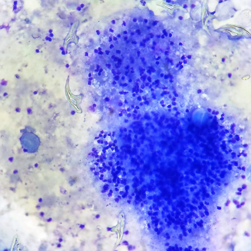
Echinococcosis is one of the zoonotic illnesses. Echinococcus granulosus larvae are the causative agents. Renal hydatid cysts are extremely rare and are typically unilateral. If they evolve slowly, they may not show any symptoms for years. Only 2%–3% of cases of hydatidosis have isolated renal hydatid cysts, making them rare. In this case, the radiological and clinical results suggested only a simple renal cyst, and cytopathology clinched the final diagnosis. A 20 mL turbid cyst fluid sample was sent for cytological analysis. Many scolices with radially arranged hooklets, scattered hooklets, macrophages, foreign body giant cells, and necro-inflammatory cell infiltrate were visible under the microscope.
Inside This Month's Cytopathology
In this study, the author highlights the cytology features of renal hydatidosis clinically and radiologically, mimicking a simple renal cyst. This case demonstrates the relevance of cytopathological screening in the early detection of unusual illnesses at rare locations.
Tweeting (~280 Characters) Through Cytopathology Twitter Handle
A study of 24 #renal lesions found #FNA #fluid cytology on laparoscopic deroofing of the left renal cyst. Smears show singly scattered hooklets, intact scolices with radially arranged hooklets in a background of acellular debris and foreign body giant cells. #CytoJ #CytoPath #hydatid cyst #renal cyst #cytology.
Graphical Abstract
Conflicts of Interest
The authors declare no conflicts of interest.
Open Research
Data Availability Statement
The data that support the findings of this study are available from the corresponding author upon reasonable request.


