Journal list menu

 Issue
IssueiRADIOLOGY: Volume 3, Issue 3
Themed Section: Fetal imaging, maternal and children imaging
183-252June 2025Guest Editor(s):
Export Citations
Download PDFs
ISSUE INFORMATION
FETAL IMAGING, MATERNAL AND CHILDREN IMAGING
A Clearer Picture: MRI's Expanding Role in High-Risk Pregnancy Care
- Pages: 188-190
- First Published: 13 June 2025
Utilization of MRI in Fetal Surgery
- Pages: 191-202
- First Published: 19 June 2025
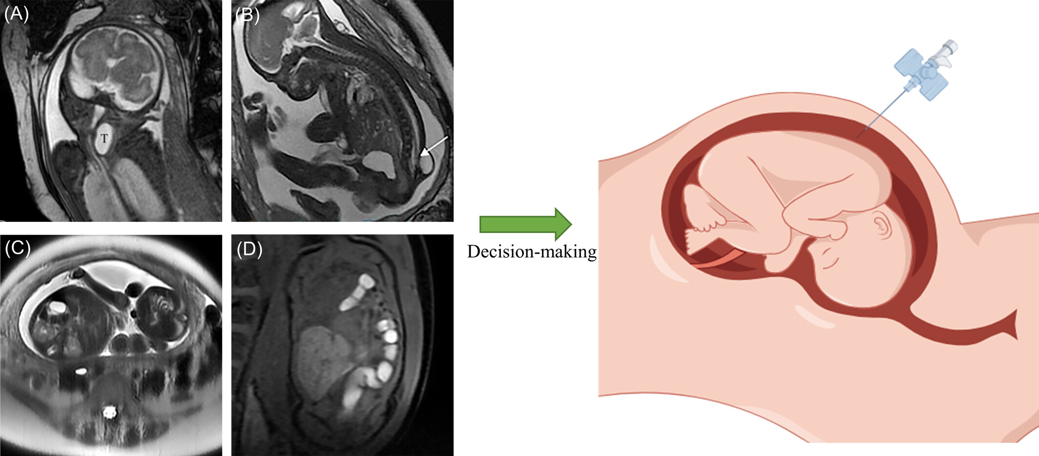
The ex utero intrapartum treatment (EXIT) procedures, open fetal surgery, fetoscopic surgery, and percutaneous interventions are critical approaches in fetal medicine, particularly for managing complex congenital anomalies. Fetal MRI plays a pivotal role in pre-procedural planning and real-time monitoring, ensuring optimal outcomes by providing detailed anatomical and functional assessments. These procedures collectively enhance survival rates and long-term health for affected fetuses. Fetal MRI: (a) thyroglossal duct cyst, (b) myelomeningocele, (c) twin-twin transfusion syndrome, and (d) diaphragmatic hernia.
Congenital Intracranial Tumors: Prenatal Diagnosis by Fetal Magnetic Resonance Imaging
- Pages: 203-208
- First Published: 16 June 2025

Fetal intracranial tumors are rare, comprising 0.5%–1.9% of all childhood tumors, with their true incidence potentially underestimated. This review highlights the role of fetal MRI, particularly with advanced sequences, in diagnosing common fetal brain tumors such as teratomas, astrocytomas, and choroid plexus papillomas, often presenting unique histological and imaging characteristics.
Value of Magnetic Resonance Spectroscopy for Examining Fetal Brain Development in Mid- to Late Pregnancy
- Pages: 209-213
- First Published: 19 May 2025
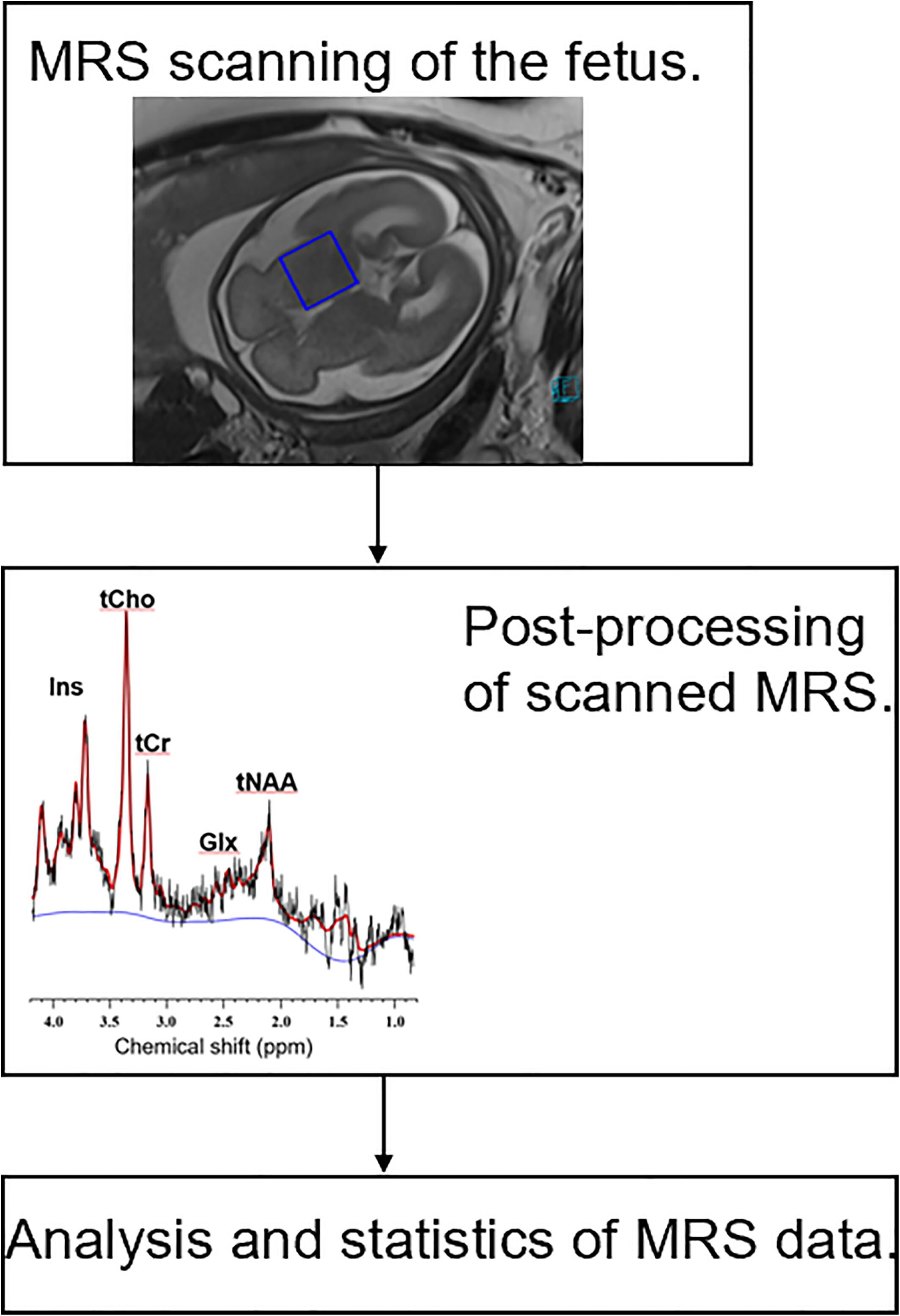
This study demonstrates that the ratio of tNAA to tCr increases during gestation, which correlates with an increase in the number of neurons and cellular energy metabolism in the fetal brain as gestational weeks progress. Additionally, tCho levels exhibit a progressive decrease with advancing gestational weeks, indicating the ongoing development of the fetal brain during myelin formation in late gestation.
Prenatal Ultrasound and Magnetic Resonance Imaging Features and Postnatal Outcomes of Congenital Hepatic Hemangioma: A Retrospective Analysis
- Pages: 214-221
- First Published: 13 June 2025
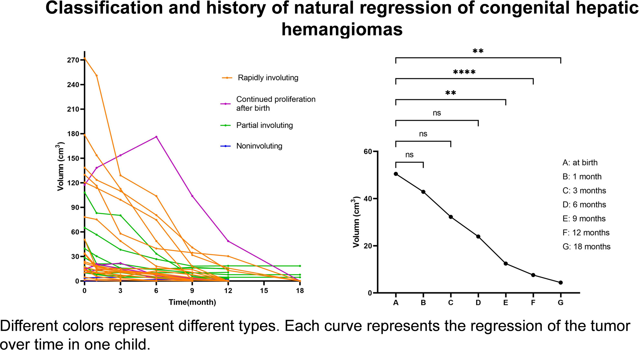
This study explored the imaging features of congenital hepatic hemangiomas of different sizes. A history of spontaneous regression of congenital hepatic hemangioma was identified at follow-up. A new type of congenital hepatic hemangioma—the spontaneous regression type after continued proliferation after birth—was also found.
Association Between Fetal Myocardial Alterations and Congenital Heart Disease Based on Post-Mortem Myocardial MRI
- Pages: 222-230
- First Published: 09 May 2025
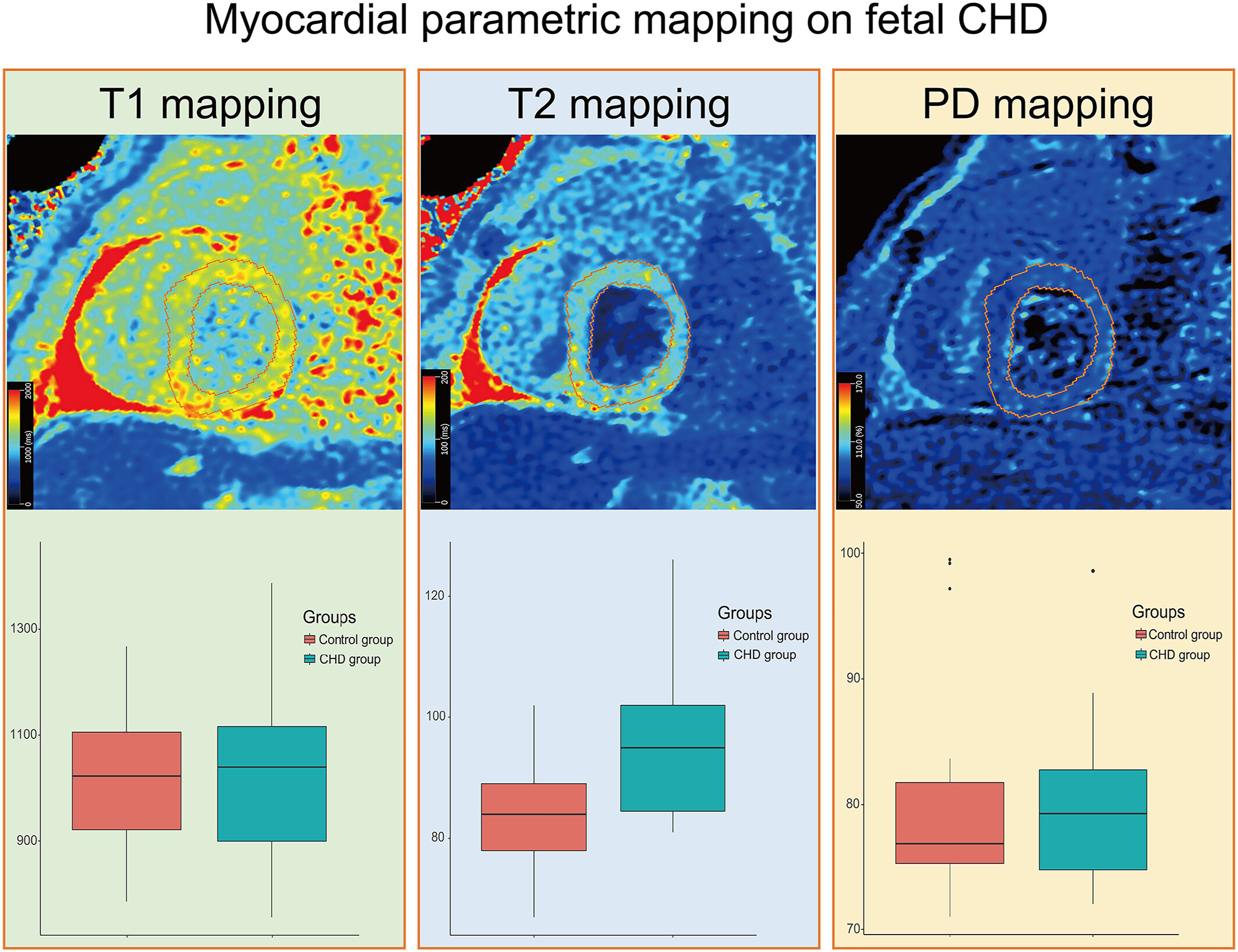
Applying myocardial parametric mapping based on post-mortem MRI is feasible for non-invasive tissue characterization. We speculated that the elevated T2 relaxation may be an essential myocardial tissue characterization of cyanotic congenital heart disease, potentially suggesting myocardial edema, which may provide evidence to guide appropriate therapeutic intervention in fetuses with congenital heart disease.
Prenatal Diagnosis and Management of Congenital Tracheal Stenosis
- Pages: 234-236
- First Published: 27 March 2025
REGULAR ARTICLES
Use of Prenasal Thickness, Nasal Bone Length and Their Ratio in Diagnosing Down Syndrome at 16-25 weeks' of gestation in India: A Retrospective, Observational, Case Control Study
- Pages: 239-247
- First Published: 14 June 2025
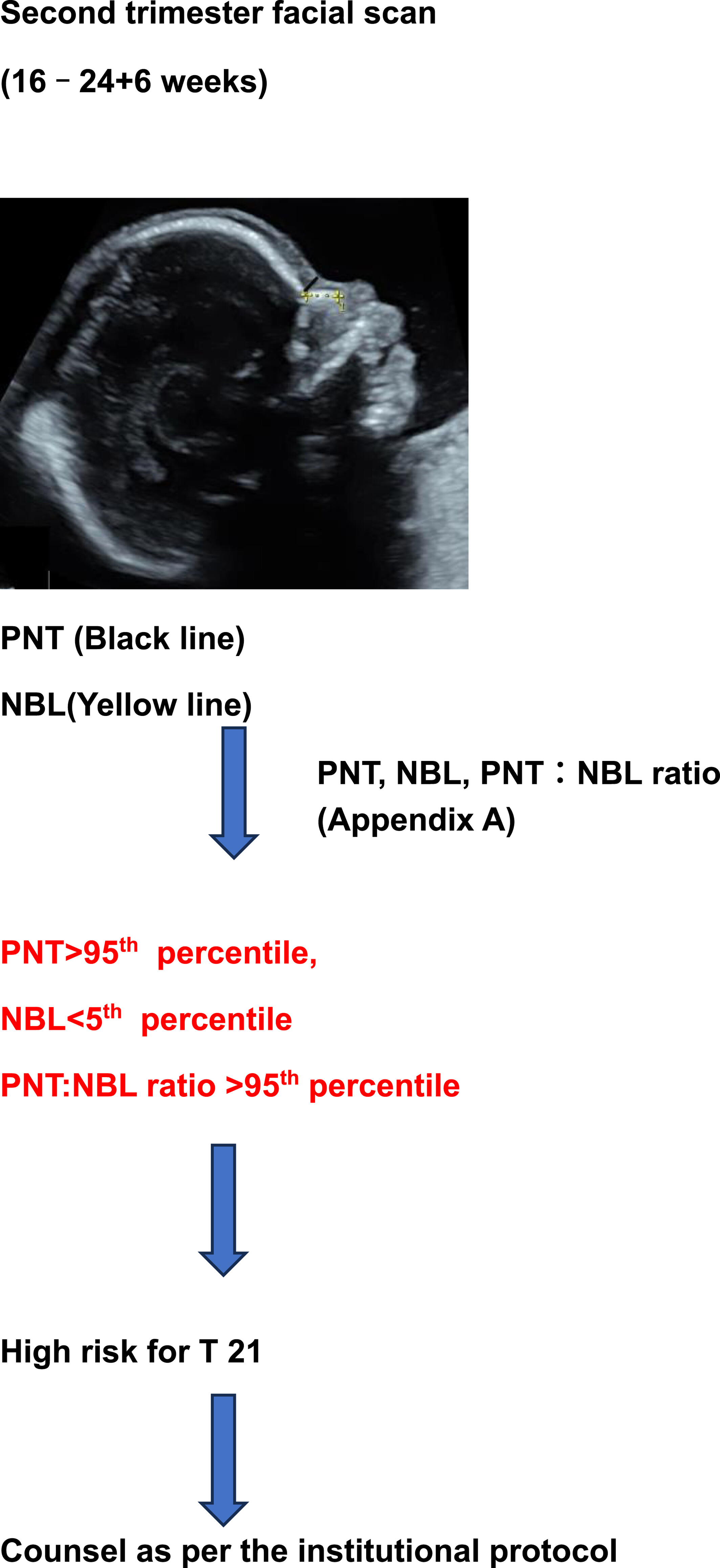
Proposed algorithm for Diagnosing T 21 at 16-25 weeks' gestation in India using two dimensional Prenasal thickness, nasal bone length and their ratio.
Automated Color Coding in Musculoskeletal MR Imaging
- Pages: 248-252
- First Published: 13 June 2025
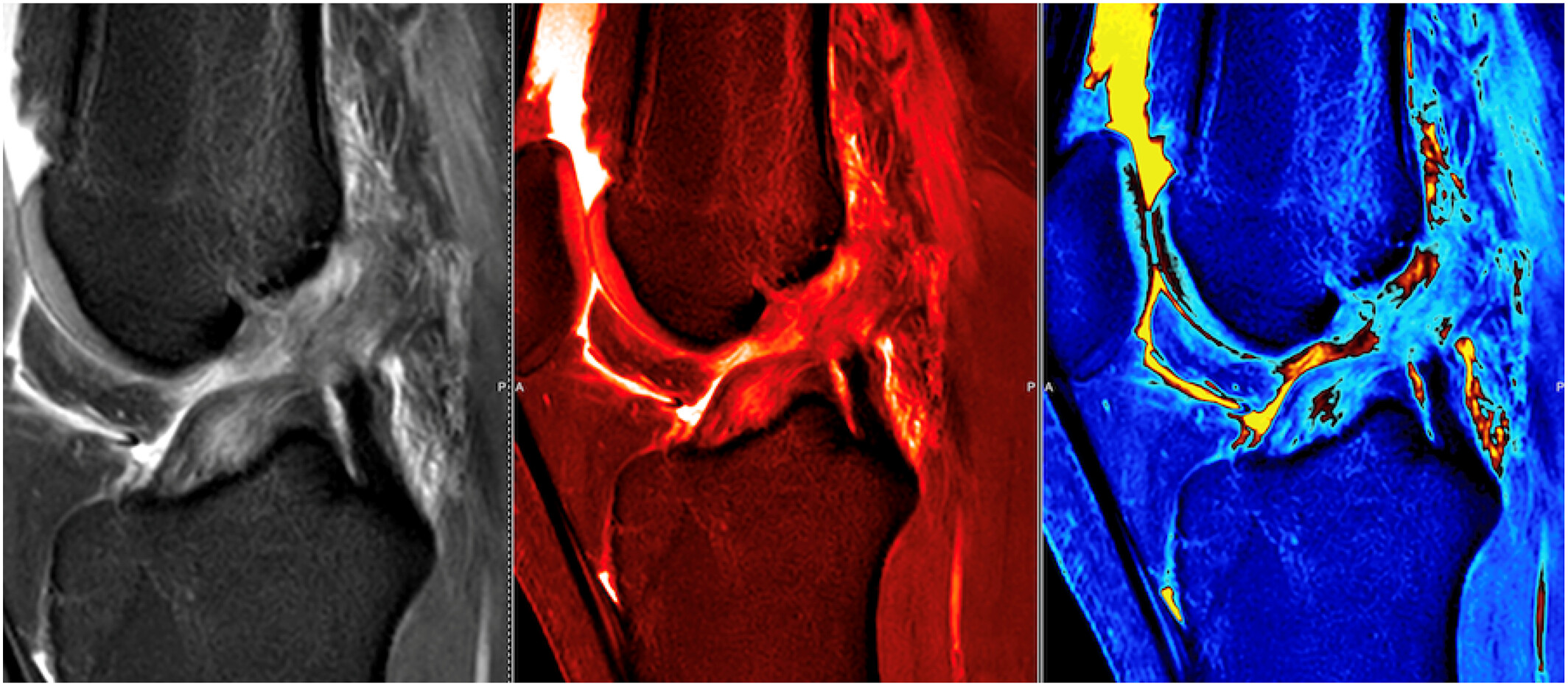
Automated color coding of the MRI signal intensities may enhance the visualization of various pathologies, potentially leading to improved diagnostic accuracy and image quality. Incorporating color-coded MR image reconstruction into routine practice could ultimately lead to more accurate diagnoses, faster patient care, and better overall outcomes, highlighting the future potential of this innovation in advancing medical imaging technology. ACL tear-gray scale, hot rod and parathyroid color-coded MRI.
Sign up for email alerts
Tools
Peer-Review Metrics
- Submission to first decision: 18
- Submission to final decision: 28
- Submission to acceptance: 33
Published on behalf of Tsinghua University Press and The Affiliated Hospital of Guizhou Medical University. An official publication of Chinese Society of Molecular Imaging in Chinese Biophysical Society.


More from this journal
Understanding Open Access
As an open access journal, all articles in iRadiology are freely available to read, download and share immediately on publication. Learn more about open access: