Retroperitoneal Ectopic Pregnancy
Funding: The authors received no specific funding for this work.
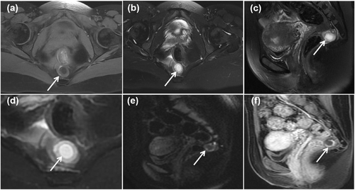
A 35-year-old woman with a history of regular menstruation presented with a positive urine pregnancy test and elevated blood human chorionic gonadotropin concentrations. Color Doppler ultrasound showed multiple slightly hyperechoic areas within the uterine cavity. She was admitted to the hospital with a preliminary outpatient diagnosis of “suspected molar pregnancy, pending further evaluation.” After electrocution and curettage, no villous tissue was identified, and postoperative human chorionic gonadotropin concentrations failed to decline. Pelvic MRI showed a round, thick-walled cystic mass in the anterior sacral region (Figure 1). Color Doppler ultrasound showed that the cystic mass contained a yolk sac, fetal bud, and fetal cardiac activity (Figure 2). Surgical pathology subsequently confirmed the presence of villous tissue, consistent with a diagnosis of ectopic pregnancy.

The MRI showing the location and signal characteristics of ectopic pregnancy. (a) Axial T1WI shows a thick-walled cystic mass with a slightly low signal in the sacral anterior space. (b) Axial FS-T2WI shows a slightly high-signal “middle ring” in lesions. (c) FS-T2WI in the sagittal position. (d) A low-signal “inner ring” can be seen in the focal capsule on axial FS-T2WI. (e) Axial DWI shows a high signal in the focal “middle ring.” (f) The capsular wall was enhanced in the sagittal view, but no enhancement was found in the capsular cavity.

Ultrasound images showing the fluctuation of the fetal bud and heart. (a) Gestational sac structures (yellow arrow) and the fetal bud (red arrow) in the presacral space, and (b) the yolk sac (green arrow) and the fetal cardiac activity (red arrow).
Author Contributions
Qingying Song: writing – original draft preparation (lead), writing – review and editing (lead). Dan Li: writing – original draft preparation (supporting), writing – review and editing (supporting).
Acknowledgments
The authors have nothing to report.
Ethics Statement
The study was reviewed and approved by the Ethics Committee of Linyi Maternal and Child Health Hospital (QTL-YXLL-2023080).
Consent
Informed consent was waived because the patient’s information has been anonymized, which was approved by the ethics committee.
Conflicts of Interest
The authors declare no conflicts of interest.
Citing Literature
Open Research
Data Availability Statement
The data is sourced from the big data and artificial intelligence research platform of Linyi Maternal and Child Health Hospital. The data that support the findings of this study are available from the corresponding author upon reasonable request.


