Journal list menu

 Issue
IssueiRADIOLOGY: Volume 2, Issue 6
THEMED SECTION: Innovation and Application of Artificial Intelligence in Radiology
i-v, 525-608December 2024Guest Editor(s):
Export Citations
Download PDFs
ISSUE INFORMATION
INNOVATION AND APPLICATION OF ARTIFICIAL INTELLIGENCE IN RADIOLOGY
Artificial intelligence in medical imaging
- Pages: 525-526
- First Published: 15 December 2024
Artificial intelligence and radiomics applied to prostate cancer bone metastasis imaging: A review
- Pages: 527-538
- First Published: 26 September 2024
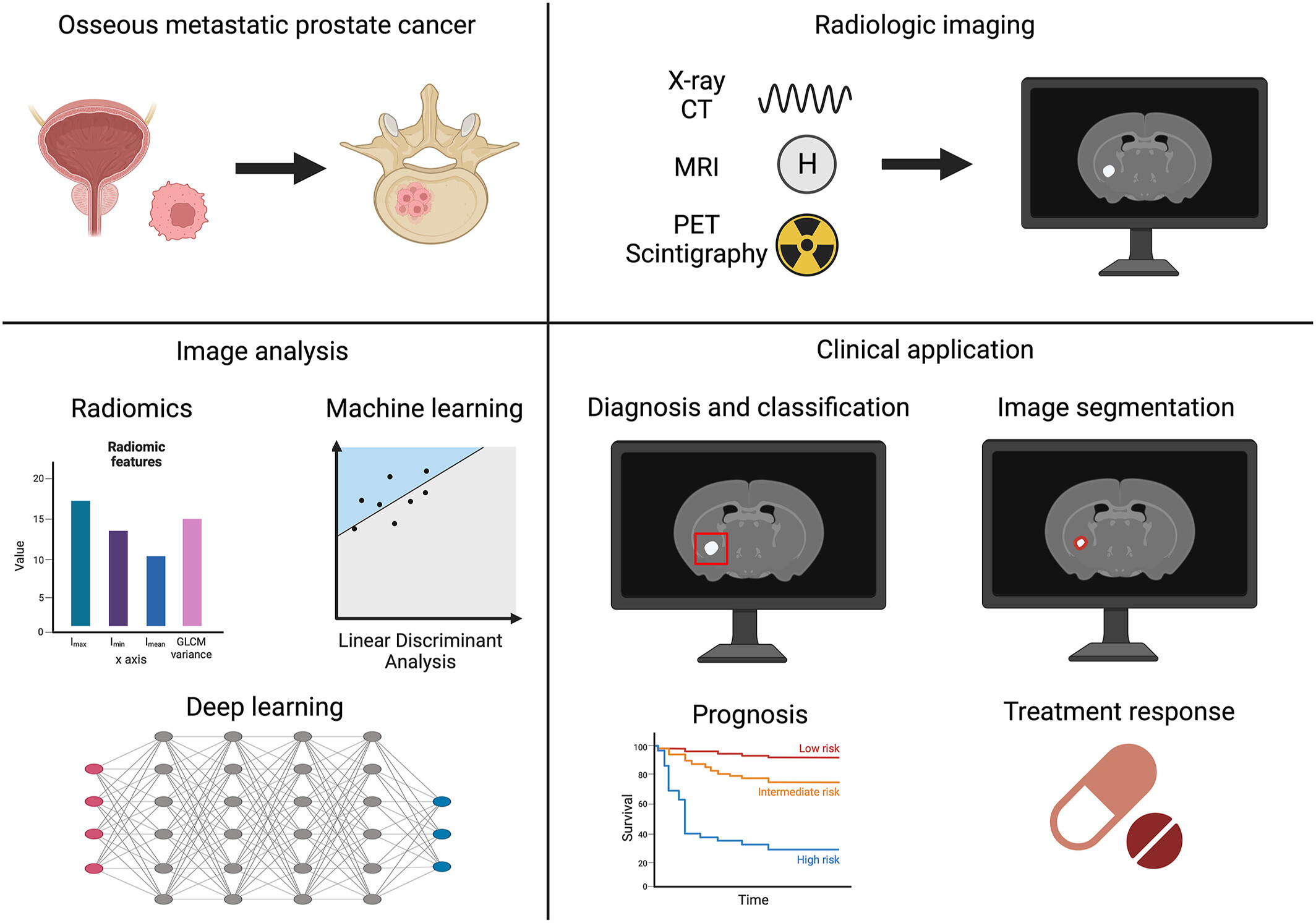
Overview of inclusion criteria for this review. The medical domain is metastatic prostate cancer to the bone. The imaging modalities are radiologic and nuclear imaging (X-ray, CT, MRI, positron emission tomography, positron emission tomography-computer tomography, scintigraphy). The area of emphasis is quantitative imaging analysis (radiomics, machine learning). The clinical applications include diagnosis/classification, segmentation, prognosis, and treatment response.
Fairness in artificial intelligence-driven multi-organ image segmentation
- Pages: 539-556
- First Published: 23 October 2024
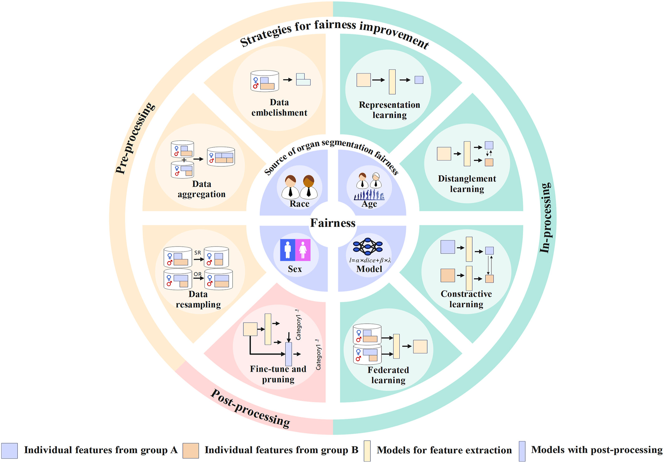
This review first discusses the fairness in multi-organ segmentation, including the sources of fairness issues and strategies for fairness improvement with the comments on the researches on these topics. Besides, we analyze the limitations of current researches and give suggestions for fairness assessment and improvement in organ segmentation.
Artificial intelligence in the diagnosis of cerebrovascular diseases using magnetic resonance imaging: A scoping review
- Pages: 557-570
- First Published: 11 November 2024
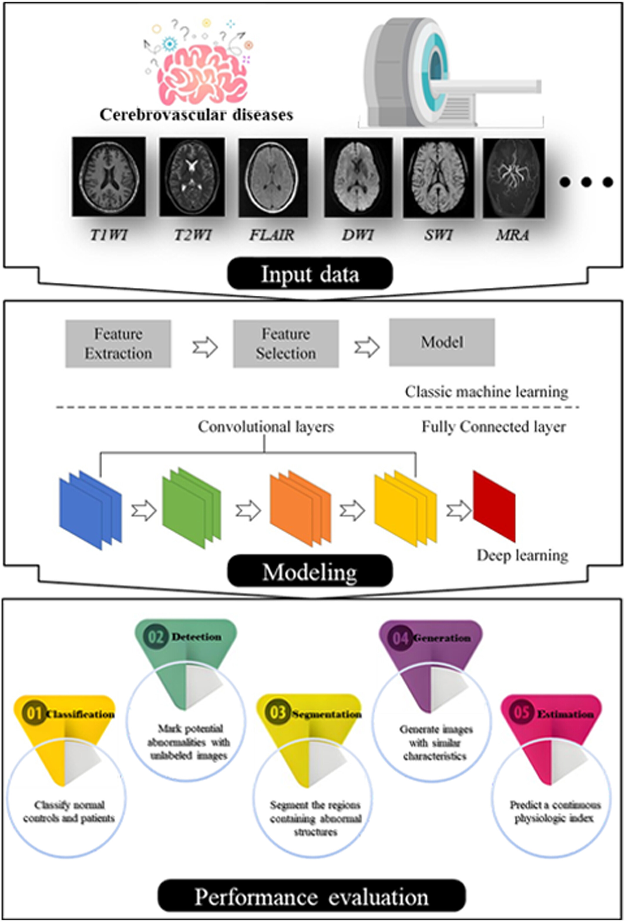
This scoping review identifies and summarizes the technical methods and related clinical applications of artificial intelligence (AI) in cerebrovascular diseases using MR imaging. We obtained a general overview of the field, modeling and evaluation metrics, and also illustrated the key questions of the cerebrovascular diseases research and graded the clinical utility of their AI solutions. Although most attention is devoted to improving the performance of AI models, this scoping review suggests that the availability of algorithms, reliability of external validations, and consistency of evaluation metrics may facilitate an improved clinical applicability and acceptance.
Deep learning-based reconstruction on intensity-inhomogeneous diffusion magnetic resonance imaging
- Pages: 571-583
- First Published: 01 November 2024
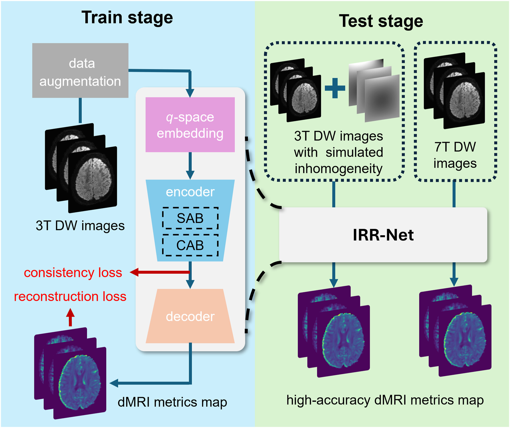
An attention-based reconstruction model and a training framework incorporating data augmentation and consistency loss have been proposed to achieve inhomogeneity-resistant dMRI metric reconstruction. The proposed method can be trained on 3T dMRI data and directly applied to 7T data without fine-tuning, eliminating the need for homogeneous ultrahigh field dMRI data. The effectiveness of the model has been validated on 7T datasets and synthetic homogeneous 3T datasets.
An investigation into the applicability of rapid artificial intelligence-assisted compressed sensing in brain magnetic resonance imaging performed at 5 Tesla field strength
- Pages: 584-593
- First Published: 08 December 2024
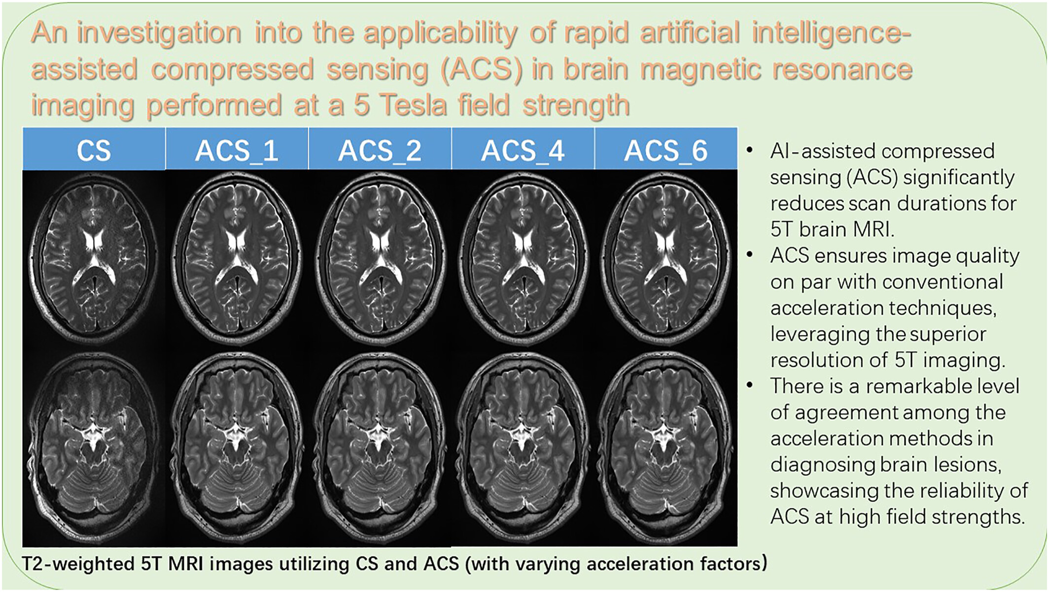
Our findings revealed that artificial intelligence-assisted compressed sensing acceleration significantly reduced the acquisition times of T1-, and T2-weighted sequences at most 43% and 53% respectively, compared to traditional compressed sensing at 5 T. Importantly, this acceleration was achieved while maintaining excellent image quality, demonstrated by higher or comparable SNR and CNR values.
REGULAR ARTICLES
Key imaging perspectives on Achilles tendon tears—A radiological roadmap: Pictorial essay
- Pages: 594-602
- First Published: 12 November 2024
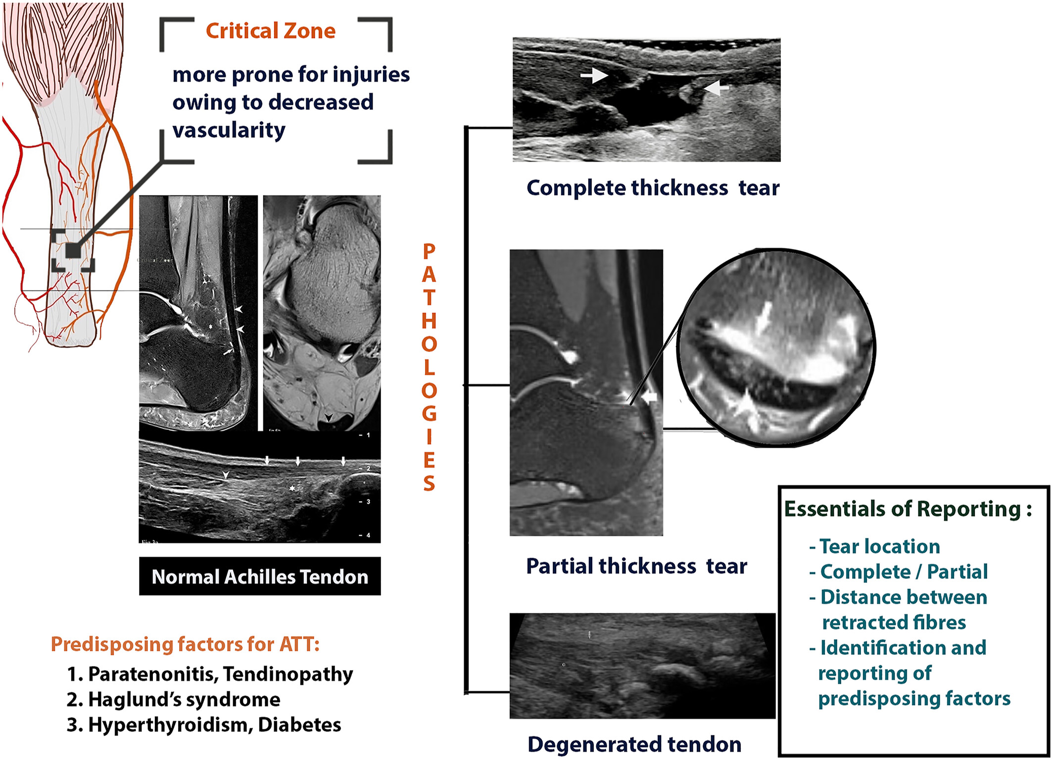
Essentials of reporting Achilles tendon tears: Accurate description of tear location and extent (partial vs. complete). Measurement of tendon gap or retraction. Identification and reporting predisposing factors influencing treatment decisions. Collaboration with orthopedic surgeons for optimal patient management.
Foot pain as first presenting symptom of renal cell carcinoma, due to metastatic lesion in medial cuneiform
- Pages: 603-608
- First Published: 02 December 2024
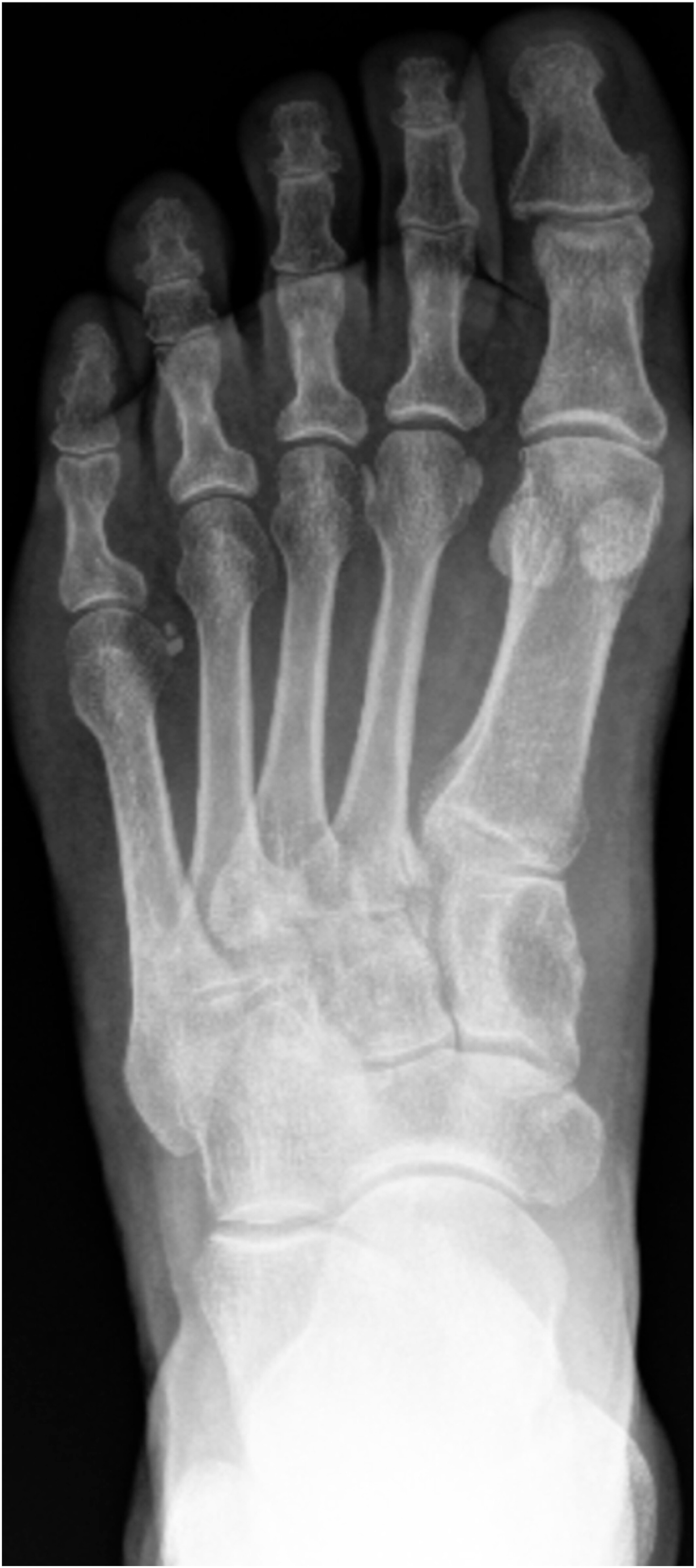
Metastatic renal cell carcinoma (RCC) lesions in the foot are a rare entity and uncommon finding in a series of foot radiographs ordered to investigate foot pain. We report the case of a 72 year old male who experienced left foot pain for a year, before developing intermittent haematuria and right flank pain, and subsequently being found to have right RCC with an osseous metastatic lesion in the left medial cuneiform.
Sign up for email alerts
Tools
Peer-Review Metrics
- Submission to first decision: 18
- Submission to final decision: 28
- Submission to acceptance: 33
Published on behalf of Tsinghua University Press and The Affiliated Hospital of Guizhou Medical University. An official publication of Chinese Society of Molecular Imaging in Chinese Biophysical Society.


More from this journal
Understanding Open Access
As an open access journal, all articles in iRadiology are freely available to read, download and share immediately on publication. Learn more about open access: