Journal list menu

 Issue
IssueJournal of Biophotonics: Volume 10, Issue 8
943-1094August 2017
Export Citations
Download PDFs
Cover Pictures
Front Cover: Imaging of fatty tumors: appearance of subcutaneous clipomas in optoacoustic images (J. Biophotonics 8/2017)
- Page: 943
- First Published: 11 August 2017
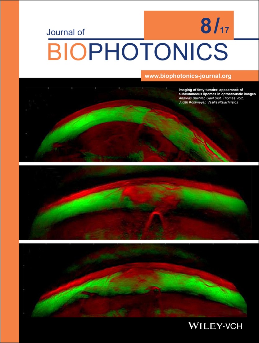
A wide variety of subcutaneous soft-tissue masses may be seen in clinical practice. Handheld multispectral optoacoustic imaging technology (MSOT) was used in a proof-of-principle study to image superficial fatty tumors. Findings indicated that MSOT offers highly complementary contrast to sonography. The images show a pseudo-color representation of three different multi-spectral dataset obtained from subcutaneous lipomas. Signals from blood and fat obtained by multi-spectral unmixing are displayed in red and fat in green, respectively. Further details can be found in the article by A. Buehler et al. on pp. 983–989.
Issue Information
Contents
Review Article
Editor's Choice
Mueller polarimetric imaging for surgical and diagnostic applications: a review
- Pages: 950-982
- First Published: 02 May 2017
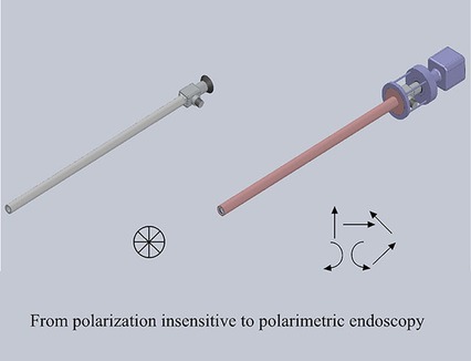
Polarization is a fundamental property of light that is able to provide an interesting and unique insight into tissue. Tissue polarization properties convey morphological, micro-structural and compositional information of tissue with great potential for label free characterization of tissue pathological changes. Recent progress in tissue polarimetric imaging and polarization resolved endoscopy paved the way for translation of polarimetric imaging to surgery and tissue diagnosis.
Letters
Editor's Choice
Imaging of fatty tumors: appearance of subcutaneous lipomas in optoacoustic images
- Pages: 983-989
- First Published: 09 May 2017
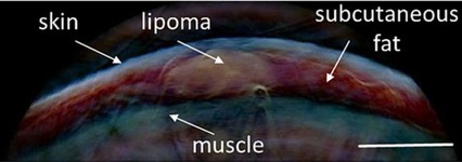
A wide variety of superficial soft tissue masses may be seen in clinical practice. To identify the nature and exact origin of the mass, imaging may be necessary. Novel hand-held multispectral optoacoustic imaging technology (MSOT) was used in a pilot study to image superficial fatty tumors, and it was compared qualitatively to ultrasonography. MSOT was found to offer highly complementary contrast to sonography, suggesting that MSOT may help improve the evaluation of subcutaneous soft tissue masses.
Sensitivity of spatially offset Raman spectroscopy (SORS) to subcortical bone tissue
- Pages: 990-996
- First Published: 02 May 2017
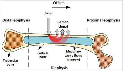
The development of spatially offset Raman spectroscopy (SORS) has enabled deep, non-invasive chemical characterization of turbid media. This study uses SORS to investigate a murine model of the genetic bone disorder osteogenesis imperfecta. The results suggest that SORS is more sensitive to disease-related biochemical differences in subcortical trabecular bone and marrow than conventional Raman measurements.
Full Articles
Endoluminal application of glass-capped diffuser for ex vivo endovenous photocoagulation
- Pages: 997-1007
- First Published: 10 August 2016
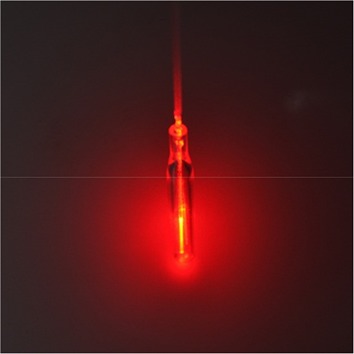
Endovenous laser ablation (EVLA) is a minimally invasive method to thermally treat varicose veins. This paper introduces the new design of glass-capped diffusing applicators and validates the feasibility of endoluminal applications for EVLA. Due to circumferential light emission with lower irradiance, the proposed applicator maintained the maximum temperature of 70-80°C and achieved uniform thermal denaturation in venous tissue. The results indicate that the glass-capped diffuser can be a feasible delivery device to treat varicose veins in an effective manner.
Autofluorescence hyperspectral imaging of radiofrequency ablation lesions in porcine cardiac tissue
- Pages: 1008-1017
- First Published: 22 August 2016
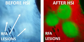
Effective treatment of atrial fibrillation, the most prevalent cardiac arrhythmia, is highly dependent on the correct placement of radiofrequency ablation lesions. However, few options are currently available to guide lesion placement. Autofluorescence hyperspectral imaging relies on subtle changes in the autofluorescence emission spectrum to successfully delineate radiofrequency ablation lesions placed on the endocardial surface of left atrium. Our results support the application of autofluorescence hyperspectral imaging for improved radiofrequency ablation guidance.
Changes in endogenous UV fluorescence and biomechanical stiffness of bovine articular cartilage after collagenase digestion are strongly correlated
- Pages: 1018-1025
- First Published: 07 October 2016
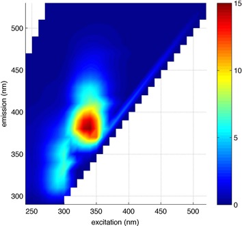
Articular cartilage contains a variety of fluorescent molecules, including constituents of collagen and other structural proteins. Ultraviolet fluorescence spectroscopy was carried out on bovine cartilage samples before and after digestion by collagenase, a model of osteoarthritis. Changes in the stiffness of the cartilage were found to correlate with changes in the UV fluorescence spectrum of the cartilage.
Autofluorescence flow sorting of breast cancer cell metabolism
- Pages: 1026-1033
- First Published: 12 October 2016
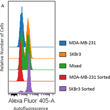
Clinical cancer treatment aims to target all cell subpopulations within a tumor. Autofluorescence microscopy of the metabolic cofactors NAD(P)H and FAD has shown sensitivity to anti-cancer treatment response. Alternatively, flow cytometry is attractive for high throughput analysis and flow sorting. This study measures cellular autofluorescence in three flow cytometry channels and applies cellular autofluorescence to sort a heterogeneous mixture of breast cancer cells into subpopulations enriched for each phenotype.
Fabricating upconversion fluorescent nanoparticles modified substrate for dynamical control of cancer cells and pathogenic bacteria
- Pages: 1034-1042
- First Published: 07 September 2016
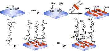
A short descriptive and popular text on the general aim and value of my paper is that "we developed a novel UCNP-based substrate for dynamic capture and release of cancer cells and pathogenic bacteria under NIR-control. The UCNPs harvest NIR light and convert it to ultraviolet light, which subsequently result in the cleavage of photoresponsive linker (PR linker) from the substrate, and on demand allows the release of a captured cell"
Structural damage of Bacillus subtilis biofilms using pulsed laser interaction with gold thin films
- Pages: 1043-1052
- First Published: 07 October 2016
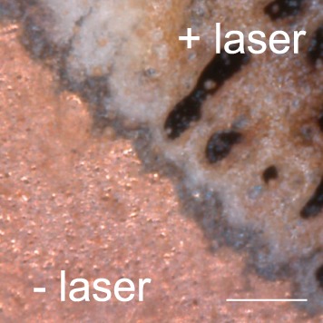
Biofilms can have a negative impact in medical and industrial settings. Distinct changes on the surface structure and overall morphology are induced on two day-old B. subtilis biofilms using the interaction of 532 nm pulsed laser with gold thin films. After irradiation of biofilms in the presence of gold film, an increased number of dead bacterial cells was detected. Thus, this technique may be utilized in targeting and eradicating matured biofilms.
Biocompatible astaxanthin as novel contrast agent for biomedical imaging
- Pages: 1053-1061
- First Published: 12 September 2016
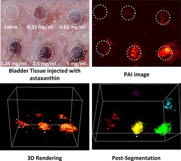
This study investigates the feasibility of marine-oriented astaxanthin as a biocompatible photoacoustic image (PAI) contrast agent for mapping urinary bladder tumors. Ex vivo imaging experiments show that astaxanthin can enhance PA image contrast due to strong light absorption of astaxanthin at the visible spectrum. Astaxanthin-assisted PAI is able to distinguish the margins of the artificial tumor as well as to visualize them in 2D and 3D images. Astaxanthin can be a potential biocompatible contrast agent for PAI-assisted bladder cancer detection.
Optical redox ratio and endogenous porphyrins in the detection of urinary bladder cancer: A patient biopsy analysis
- Pages: 1062-1073
- First Published: 07 October 2016
New avenues are sought for the early detection of urinary bladder cancer, a common cancer which is often missed during routine examination. One suggested technique is autofluorescence spectroscopy, however much is still unknown about autofluorescence properties in health and disease. We present new findings regarding differences in autofluorescence profiles between healthy and diseased tissue – reflecting alterations to biochemical processes – which may be of clinical worth for disease diagnosis.
Raman spectroscopy and PCA-SVM as a non-invasive diagnostic tool to identify and classify qualitatively glycated hemoglobin levels in vivo
- Pages: 1074-1079
- First Published: 23 December 2016
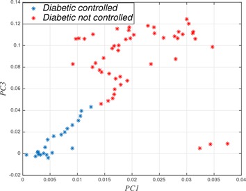
Glycated hemoglobin is a gold standard recommended test by the World Health Organization as a diagnostic and control took in diabetic patients. Unfortunately is a high-cost test. Raman spectroscopy combined with multivariate methods improves significantly the accuracy of this diagnostic tool (specificity 99% & sensitivity 99%). In this work we show the first study of Raman spectroscopy in vivo in diabetic and healthy patients, to classify qualitatively high and low levels of glycated hemoglobin.
Remission spectrometry for blood vessel detection during stereotactic biopsy of brain tumors
- Pages: 1080-1094
- First Published: 07 October 2016
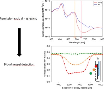
To increase the safety of stereotactic brain biopsies, it is crucial to avoid hemorrhages due to blood vessel rupture. By illuminating the tissue that is to be sampled and comparing the remitted intensities at two wavelengths with strongly differing hemoglobin absorption, blood vessels can be recognized before biopsy sampling. The method is easily implementable into a conventional biopsy needle and clearly discriminates between vessels within and outside the needle's coverage.
Sign up for email alerts
Tools
More from this journal
Related Titles
 Volume 19, Issue 14July 22, 2025
 Volume 5, Issue 3-4August-November 2023
 Volume 13, Issue 21July 25, 2025