Journal list menu

 Issue
IssueJournal of Biophotonics: Volume 9, Issue 5
Special Issue:Image Processing in Biomedical Diagnosis
427-559May 2016
Export Citations
Download PDFs
Cover Picture
Cover Picture: J. Biophotonics 5/2016
- Page: 427
- First Published: 09 May 2016

Modern methods of image analysis and processing are one of the cornerstones of research for biophotonic diagnostics. Rapid progress in methods for the analysis of medical images require continuous knowledge updates. This special issue shows new trends in analysis and processing with particular emphasis on imaging in visible light and infrared radiation used in an air-puff tonometer, OCT, IR spectroscopy and Raman spectroscopy.
Issue Information
Contents
Editorial
Image Processing in Biomedical Diagnosis
- Pages: 434-435
- First Published: 09 May 2016
Full Articles
Numerical analysis of corneal curvature dynamics based on Corvis tonometer images
- Pages: 436-444
- First Published: 21 March 2016
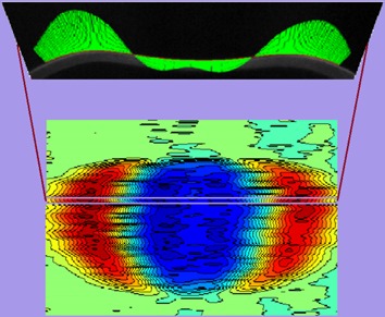
Curvature distribution is the most important optical and biomechanical property of the cornea. Investigation of very fast variations of these distributions during corneal deformations can give a lot of important information. Processing of images of a very fast deforming cornea from the Corvis ST tonometer enables a new description and definition of new geometrical curvature parameters of individual cornea. They could be used in a modern approach to the examination of corneal biomechanics.
Application of corneal tomography before keratorefractive procedure for laser vision correction
- Pages: 445-453
- First Published: 15 April 2016
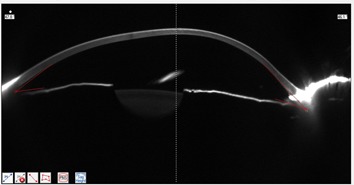
Ectasia after refractive surgery is among the complications most feared by surgeons. The presence of ectatic disease in the preoperative state is the main risk factor for ectasia. A ‘traditional' evaluation is insufficient because there have been several cases of ectasia post- laser-assisted in situ keratomilusis (LASIK) surgery without any identified risk factor. A comprehensive corneal structural analysis based on corneal tomography can detect susceptible corneas with very mild, subtle disease. The Ectasia Susceptibility Score (ESS) is new tool for preoperative evaluation, with significantly improved accuracy for detecting susceptible cases.
Determining in vivo elasticity and viscosity with dynamic Scheimpflug imaging analysis in keratoconic and healthy eyes
- Pages: 454-463
- First Published: 11 January 2016
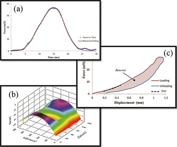
The paper shows a novel method of determining the corneal biomechanical properties. Combined with air puff force of CorVis ST and corneal displacement, which extracted from CorVis ST video using proposed imaging analysis, the STSC and Aabsorbed were generated to represent the corneal elasticity and viscosity properties, respectively. In clinical studies, the STSC and Aabsorbed metrics were statistically different in patients with keratoconus compared with those in healthy controls.
Review Articles
In vivo human corneal deformation analysis with a Scheimpflug camera, a critical review
- Pages: 464-477
- First Published: 12 February 2016
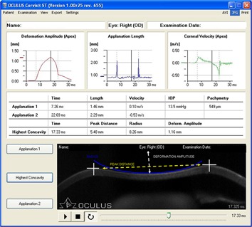
This review provides a complete and deep analysis of the most recent papers evaluating the new device for corneal deformation detection: Corvis ST (Oculus Optikgeräte GmbH, Wetzlar, Germany). Particular attention has been paid in comparison of results of research about the same group of patients (healthy eyes, keratoconus ones, glaucoma ones, refractive surgery ones etc), in order to let the reader better understand the innovation and the utilities that this device is able to provide. Section about new, experimental, prospectives has been added.
Performance evaluation of automated segmentation software on optical coherence tomography volume data
- Pages: 478-489
- First Published: 11 March 2016

Reliable data analysis and proper diagnosis of various retinal diseases requires an appropriate ground truth dataset that reflects the realities of everyday retinal features observed in clinical settings. The major intent of this review is to show a number of important issues surrounding the lack of a representative and practical dataset that could be used as a common ground truth for the evaluation of the performance of OCT quantitative methods.
Full Articles
Prostate cancer diagnosis by optical coherence tomography: First results from a needle based optical platform for tissue sampling
- Pages: 490-498
- First Published: 09 February 2016
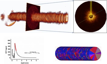
Functional needle based OCT of the prostate demonstrates a potential for differentiation between healthy and malignant prostate tissue. The optical attenuation coefficient was significantly higher in malignant tissue compared to benign prostate tissue. By performing quantitative analysis of OCT data, it can be expected to avoid diagnostic boundaries that exist in conventional pathology, such as delay in diagnosis and subjectivity. Quantitative OCT has the potential to aid in swift diagnosis which enables faster and better treatment.
Hybrid method of strain estimation in optical coherence elastography using combined sub-wavelength phase measurements and supra-pixel displacement tracking
- Pages: 499-509
- First Published: 13 November 2015
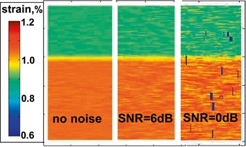
A novel hybrid method which combines sub-wavelength-scale phase measurements and pixel-scale displacement tracking for robust strain mapping in compressional optical coherence elastography is proposed. Its robustness is enabled by direct fitting of local phase gradients obviating the necessity of phase unwrapping and error-prone numerical differentiation. The significantly extended range of measurable strains strongly reduces errors in phase-gradient estimation and ensures high robustness with respect to both additive and decorrelation noises.
Some selected quantitative methods of thermal image analysis in Matlab
- Pages: 510-520
- First Published: 10 November 2015

A new algorithm for automated quantitative thermal image analysis method has been presented. The practical implementation of the discussed image analysis methods has been done in Matlab. It enables to perform fully automated, quantitative and reproducible measurements of selected parameters in thermal images. The paper also shows two examples of the use of the proposed image analysis methods for the area of the skin of a human foot and face. In addition, the full source code of the developed application that can be used without licensing restrictions has been attached to the paper.
Fully unsupervised inter-individual IR spectral histology of paraffinized tissue sections of normal colon
- Pages: 521-532
- First Published: 12 February 2016
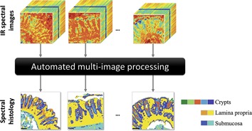
An automatic multi-image IR spectral histology for the simultaneous analysis of a set of spectral images is presented. This approach is based on a joint extended multiplicative signal correction to numerically deparaffinize the tissue sections, followed by an automated joint K-Means clustering using the hierarchical double application of a validity index. Thus, murine and human colon histological structures were correctly identified at both the intra- and the inter-individual levels.
Systematic evaluation of the biological variance within the Raman based colorectal tissue diagnostics
- Pages: 533-541
- First Published: 20 December 2015
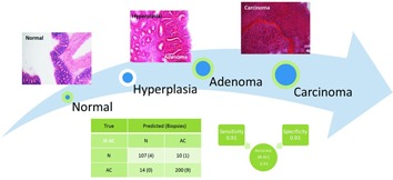
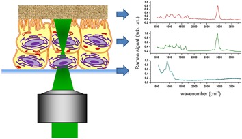
Raman microspectroscopy is combined with a specifically designed statistical analysis workflow to establish a reliable diagnosis for colon cancer. A systematic evaluation of different statistical analysis approaches while using a colon cancer mouse model with genotypic identical individuals yields a measure for the biological variance and a clinically relevant discrimination of healthy versus tumorous tissue with an accuracy of 95%.
Quantitative multi-image analysis for biomedical Raman spectroscopic imaging
- Pages: 542-550
- First Published: 02 February 2016
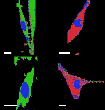
A broadly applicable methodology is described that enables the combination of a series of Raman images into a single image for analysis. This approach takes into account the entire Raman spectral profile of multiple images in order to identify, quantify and cluster the biochemical distribution across a series of Raman images.
Assessment of conjunctival microvilli abnormality by micro-Raman analysis – by G. Rusciano et al
- Pages: 551-559
- First Published: 15 February 2016
Conjunctival microvilli are microscopic cellular membrane protrusions on apical epithelial cells, which increase the surface area available for tear adherence. Pathological alterations of microvilli structure affect the tear film stability and, conversely, dysfunctions of tear film composition can lead to a suffering epithelium (dry-eye syndrome). In this work we demonstrated the effectiveness of micro-Raman analysis to reveal conjunctival microvilli abnormalities in donors affected by dry-eye syndrome.
Sign up for email alerts
Tools
More from this journal
Related Titles
 Volume 19, Issue 14July 22, 2025
 Volume 5, Issue 3-4August-November 2023
 Volume 13, Issue 21July 25, 2025