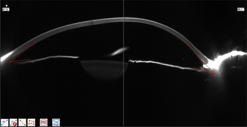
Application of corneal tomography before keratorefractive procedure for laser vision correction
Corresponding Author
Allan Luz
- [email protected]
- Phone: 55-79-3212-0800 | Fax: Fax: 55-79-3212-0844
Department of Ophthalmology of the Federal University of Sao Paulo, Sao Paulo, Brazil
Cornea and Refractive Surgery Department, Hospital de Olhos de Sergipe, Aracaju, Brazil
Rio de Janeiro Corneal Tomography and Biomechanics Study Group, Rio de Janeiro, Brazil
Corresponding author: e-mail: [email protected], Phone: 55-79-3212-0800, Fax: 55-79-3212-0844
Search for more papers by this authorBernardo Lopes
Department of Ophthalmology of the Federal University of Sao Paulo, Sao Paulo, Brazil
Rio de Janeiro Corneal Tomography and Biomechanics Study Group, Rio de Janeiro, Brazil
Search for more papers by this authorMarcela Salomão
Rio de Janeiro Corneal Tomography and Biomechanics Study Group, Rio de Janeiro, Brazil
Search for more papers by this authorRenato Ambrósio
Department of Ophthalmology of the Federal University of Sao Paulo, Sao Paulo, Brazil
Rio de Janeiro Corneal Tomography and Biomechanics Study Group, Rio de Janeiro, Brazil
Instituto de Olhos Renato Ambrósio and Visare Personal Laser Rio de Janeiro, Rio de Janeiro, Brazil
Search for more papers by this authorCorresponding Author
Allan Luz
- [email protected]
- Phone: 55-79-3212-0800 | Fax: Fax: 55-79-3212-0844
Department of Ophthalmology of the Federal University of Sao Paulo, Sao Paulo, Brazil
Cornea and Refractive Surgery Department, Hospital de Olhos de Sergipe, Aracaju, Brazil
Rio de Janeiro Corneal Tomography and Biomechanics Study Group, Rio de Janeiro, Brazil
Corresponding author: e-mail: [email protected], Phone: 55-79-3212-0800, Fax: 55-79-3212-0844
Search for more papers by this authorBernardo Lopes
Department of Ophthalmology of the Federal University of Sao Paulo, Sao Paulo, Brazil
Rio de Janeiro Corneal Tomography and Biomechanics Study Group, Rio de Janeiro, Brazil
Search for more papers by this authorMarcela Salomão
Rio de Janeiro Corneal Tomography and Biomechanics Study Group, Rio de Janeiro, Brazil
Search for more papers by this authorRenato Ambrósio
Department of Ophthalmology of the Federal University of Sao Paulo, Sao Paulo, Brazil
Rio de Janeiro Corneal Tomography and Biomechanics Study Group, Rio de Janeiro, Brazil
Instituto de Olhos Renato Ambrósio and Visare Personal Laser Rio de Janeiro, Rio de Janeiro, Brazil
Search for more papers by this authorAbstract
Ectasia after refractive surgery represents a major concern among refractive surgeons. Corneal abnormalities and preexisting corneal ectasia are the most important risk factors. Corneal topography and central corneal thickness are the factors traditionally screening for in refractive surgery candidates. Study of the anterior surface by Placido topography allows for identification of keratoconus before biomicroscopy. However, this is insufficient for the evaluation of pre-operative refractive surgery. There are cases of ectasia after laser in situ keratomilusis (LASIK) without identifiable risk factors such that there is a need to go beyond the corneal surface. A key requirement is quantifying susceptibility to corneal biomechanical instability and progression to ectasia. Tomographic indices derived from elevation maps and pachymetry spatial variation produce a Belin Ambrosio display final D index (BAD-D index), which has shown better results compared to surface curvature indices for detecting very mild forms of ectasia. A logistic regression formula, integrating age, residual stromal bed, and BAD-D (Ectasia Susceptibility Score, ESS) resulted in a significant improvement in accuracy, leading to 100% sensitivity and 94% specificity for detecting susceptible cases. A comprehensive corneal structural analysis based on corneal segmental tomography can detect susceptible corneas, which increases safety for refractive surgery patients.
Supporting Information
| Filename | Description |
|---|---|
| jbio201500236-sup-0001-author-biographies.pdfPDF document, 208.9 KB | Author Biographies |
Please note: The publisher is not responsible for the content or functionality of any supporting information supplied by the authors. Any queries (other than missing content) should be directed to the corresponding author for the article.
References
- 1R. Ambrósio Jr., L. P. Nogueira, D. L. Caldas, B. M. Fontes, A. Luz, J. O. Cazal, M. R. Alves, and M. W. Belin, Evaluation of corneal shape and biomechanics before LASIK. International ophthalmology clinics. 51 (2), 11–38 (2011).
- 2R. Ambrosio Jr., B. F. Valbon, F. Faria-Correia, I. Ramos, and A. Luz, Scheimpflug imaging for laser refractive surgery. Curr Opin Ophthalmol. 24 (4), 310–320 (2013).
- 3R. Ambrosio Jr. and J. B. Randleman, Screening for ectasia risk: what are we screening for and how should we screen for it? J Refract Surg. 29 (4), 230–232 (2013).
- 4R. Ambrosio Jr., S. D. Klyce, and S. E. Wilson, Corneal topographic and pachymetric screening of keratorefractive patients. J Refract Surg. 19 (1), 24–29 (2003).
- 5S. E. Wilson and R. Ambrosio Jr., Computerized corneal topography and its importance to wavefront technology. Cornea. 20 (5), 441–454 (2001).
- 6S. E. Wilson, D. T. Lin, S. D. Klyce, J. J. Reidy, and M. S. Insler, Topographic changes in contact lens-induced corneal warpage. Ophthalmology. 97 (6), 734–744 (1990).
- 7Y. S. Rabinowitz, H. Yang, Y. Brickman, J. Akkina, C. Riley, J. I. Rotter, and J. Elashoff, Videokeratography database of normal human corneas. Br J Ophthalmol. 80 (7), 610–616 (1996).
- 8R. Ambrosio Jr., D. G. Dawson, M. Salomao, F. P. Guerra, A. L. Caiado, and M. W. Belin, Corneal ectasia after LASIK despite low preoperative risk: tomographic and biomechanical findings in the unoperated, stable, fellow eye. J Refract Surg. 26 (11), 906–911 (2010).
- 9J. B. Randleman, W. B. Trattler, and R. D. Stulting, Validation of the Ectasia Risk Score System for preoperative laser in situ keratomileusis screening. Am J Ophthalmol. 145 (5), 813–818 (2008).
- 10P. S. Binder, R. L. Lindstrom, R. D. Stulting, E. Donnenfeld, H. Wu, P. McDonnell, Y. Rabinowitz. Keratoconus and corneal ectasia after LASIK. J Cataract Refract Surg. 31 (11), 2035–2038 (2005).
- 11P. Padmanabhan, R. Aiswaryah, V. Abinaya Priya, Post-LASIK keratectasia triggered by eye rubbing and treated with topography-guided ablation and collagen cross-linking – a case report. Cornea. 31 (5), 575–580 (2012).
- 12R. Ambrosio Jr., A. Luz, B. Lopes, I. Ramos, and M. W. Belin, Enhanced ectasia screening: the need for advanced and objective data. J Refract Surg. 30 (3), 151–152 (2014).
- 13R. Ambrosio Jr.,
I. Ramos,
B. Lopes,
A. L. C. Canedo,
R. Correa,
F. Guerra,
A. Luz,
F. Price,
M. Price,
S. Schallhor, and
M. W. Belin,
Assessing ectasia susceptibility prior to LASIK: the role of age and residual stromal bed (RSB) in conjunction to Belin-Ambrósio deviation index (BAD-D).
Revista Brasileira de Oftalmologia.
73
(2),
75–80
(2014).
10.5935/0034-7280.20140018 Google Scholar
- 14B. Lopes, I. Ramos, and R. Ambrosio Jr., Corneal densitometry in keratoconus. Cornea. 33 (12), 1282–1286 (2014).
- 15J. Buhren, T. Schaffeler, and T. Kohnen, Preoperative topographic characteristics of eyes that developed postoperative LASIK keratectasia. J Refract Surg. 29 (8), 540–549 (2013).
- 16R. Ambrosio Jr. and M. W. Belin, Imaging of the cornea: topography vs tomography. J Refract Surg. 26 (11), 847–849 (2010).
- 17S. N. Rao, T. Raviv, P. A. Majmudar, R. J. Epstein, Role of Orbscan II in screening keratoconus suspects before refractive corneal surgery. Ophthalmology. 109 (9), 1642–1646 (2002).
- 18R. Ambrosio Jr.,
F. Faria-Correia,
I. Ramos,
B. F. Valbon,
B. Lopes,
D. Jardim, and
A. Luz,
Enhanced Screening for Ectasia Susceptibility Among Refractive Candidates: The Role of Corneal Tomography and Biomechanics.
Current Ophthalmology Reports.
1
(1),
28–38
(2013).
10.1007/s40135-012-0003-z Google Scholar
- 19F. F. Correi, I. Ramos, B. Lopes, M. Q. Salomão, A. Luz, R. O. Correa, M. W. Belin, and R. Ambrosio Jr., Topometric and tomographic indices for the diagnosis of keratoconus. Int J Kerat Ectatic Dis. 2012, 92–99 (2012).
- 20A. J. Kanellopoulos and G. Asimellis. Anterior segment optical coherence tomography: assisted topographic corneal epithelial thickness distribution imaging of a keratoconus patient. Case Rep Ophthalmol. 4 (1), 74–78 (2013).
- 21L. J. Maguire and W. M. Bourne, Corneal topography of early keratoconus. Am J Ophthalmol. 108(2), 107–112 (1989).
- 22N. Maeda, S. D. Klyce, and Y. Tano, Detection and classification of mild irregular astigmatism in patients with good visual acuity. Surv Ophthalmol. 43 (1), 53–58 (1998).
- 23A. Saad and D. Gatinel, Topographic and tomographic properties of forme fruste keratoconus corneas. Invest Ophthalmol Vis Sci. 51 (11), 5546–5555 (2010).
- 24M. W. Belin and S. S. Khachikian, An introduction to understanding elevation-based topography: how elevation data are displayed – a review. Clin Experiment Ophthalmol. 37 (1), 14–29 (2009).
- 25U. de Sanctis, C. Loiacono, L. Richiardi, D. Turco, B. Mutani, and F. M. Grignolo, Sensitivity and specificity of posterior corneal elevation measured by Pentacam in discriminating keratoconus/subclinical keratoconus. Ophthalmology. 115 (9), 1534–1539 (2008).
- 26M. W. Belin and S. S. Khachikia. New devices and clinical implications for measuring corneal thickness. Clin Experiment Ophthalmol. 4 (8), 729–731 (2006).
- 27I. Kremer, Y. Shochot, A. Kaplan, and M. Blumenthal, Three year results of photoastigmatic refractive keratectomy for mild and atypical keratoconus. J Cataract Refract Surg. 24 (12), 1581–1588 (1998).
- 28M. W. Belin and S. S. Khachikian, Keratoconus/ectasia detection with the Oculus Pentacam: Belin/Ambrósio enhanced ectasia display. Highlights of Ophthalmology. 35 (6) (2007).
- 29R. Ambrosio Jr., R. S. Alonso, A. Luz, and L. G. Coca Velarde, Corneal-thickness spatial profile and corneal-volume distribution: tomographic indices to detect keratoconus. J Cataract Refract Surg. 32 (11), 1851–1859 (2006).
- 30A. Luz, M. Ursulio, D. Castaneda, R. Ambrosio Jr. [Corneal thickness progression from the thinnest point to the limbus: study based on a normal and a keratoconus population to create reference values]. Arq Bras Oftalmol. 69 (4), 579–583 (2006).
- 31R. Ambrosio Jr., Percentage thickness increase and absolute difference from thinnest to describe thickness profile. J Refract Surg. 26 (2), 84–86 (2010) author reply 6–7.
- 32R. Ambrosio Jr., A. L. Caiado, F. P. Guerra, R. Louzada, A. S. Roy, A. Luz, W. Dupps, and M. W. Belin, Novel pachymetric parameters based on corneal tomography for diagnosing keratoconus. J Refract Surg. 27 (10), 753–758 (2011).
- 33O. Muftuoglu, O. Ayar, V. Hurmeric, F. Orucoglu, and I. Kilic, Comparison of multimetric D index with keratometric, pachymetric, and posterior elevation parameters in diagnosing subclinical keratoconus in fellow eyes of asymmetric keratoconus patients. J Cataract Refract Surg. 41 (3), 557–565 (2015).
- 34P. R. Ruisenor Vazquez, J. D. Galletti, N. Minguez, M. Delrivo, F. Fuentes Bonthoux, T. Pfortner, and J. G. Galletti, Pentacam Scheimpflug tomography findings in topographically normal patients and subclinical keratoconus cases. Am J Ophthalmol. 158 (1), 32–40 (2014).
- 35D. Z. Reinstein, Therapeutic refractive surgery. J Refract Surg. 31 (1), 6–8 (2015).
- 36D. Z. Reinstein, T. J. Archer, Z. I. Dickeson, and M. Gobbe, Transepithelial phototherapeutic keratectomy protocol for treating irregular astigmatism based on population epithelial thickness measurements by artemis very high-frequency digital ultrasound. J Refract Surg. 30 (6), 380–387 (2014).
- 37R. H. Silverman, R. Urs, A. Roychoudhury, T. J. Archer, M. Gobbe, and D. Z. Reinstein, Epithelial remodeling as basis for machine-based identification of keratoconus. Invest Ophthalmol Vis Sci. 55 (3), 1580–1587 (2014).
- 38D. Z. Reinstein, T. J. Archer, and M. Gobbe. Corneal epithelial thickness profile in the diagnosis of keratoconus. J Refract Surg. 25 (7) 604–610 (2009).
- 39D. Z. Reinstein, R. H. Silverman, S. L. Trokel, and D. J. Coleman, Corneal pachymetric topogr aphy. Ophthalmology. 101 (3), 432–438 (1994).
- 40K. Azartash, J. Kwan, J. R. Paugh, A. L. Nguyen, J. V. Jester, and E. Gratton, Pre-corneal tear film thickness in humans measured with a novel technique. Mol Vis. 17, 756–767 (2011).
- 41D. Z. Reinstein, T. E. Yap, T. J. Archer, M. Gobbe, and R. H. Silverman, Comparison of Corneal Epithelial Thickness Measurement Between Fourier-Domain OCT and Very High-Frequency Digital Ultrasound. J Refract Surg. 31 (7), 438-445 (2005).
- 42J. Fujimoto, W. Drexler, Introduction to optical coherence tomography. Optical coherence tomography (Springer, Berlin, Heidelberg, 2008) p. 1–45.
- 43Y. Li, O. Tan, R. Brass, J. L. Weiss, D. Huang, Corneal epithelial thickness mapping by Fourier-domain optical coherence tomography in normal and keratoconic eyes. Ophthalmology. 119 (12), 2425–24 33 (2012).
- 44C. Temstet, O. Sandali, N. Bouheraoua, T. Hamiche, A. Galan, M. El Sanharawi, E. Basli, L. Larache, V. Borderie, Corneal epithelial thickness mapping using Fourier-domain optical coherence tomography for detection of form fruste keratoconus. Journal of Cataract & Refractive Surgery. 41 (4), 812–8 20 (2015).
- 45D. Z. Reinstein, T. J. Archer, M. Gobbe, R. H. Silverman, D. J. Coleman, Epithelial thickness in the normal cornea: three-dimensional display with very high frequency ultrasound. Journal of refractive surgery 24 (6), 571 (2008).


