Determining in vivo elasticity and viscosity with dynamic Scheimpflug imaging analysis in keratoconic and healthy eyes
Abstract
The paper presents a novel analysis method of corneal elasticity and viscosity based on corneal visualization Scheimpflug technology (CorVis ST) for keratoconus diagnosis. Methods for air puff force measurement and corneal imaging boundary extraction were proposed. Corneal biomechanical properties, described as tangent stiffness coefficient (STSC) and energy absorbed area (Aabsorbed), were assessed using the curves of the applied air puff force with corneal displacement to form a loading-unloading cycle. Twenty-five patients with keratoconus and 34 healthy control subjects, matched for intraocular pressure (IOP), were enrolled in this prospective study. The results showed that the intraclass correlation coefficients (ICC) of the STSC and Aabsorbed were 0.941 and 0.878 in Healthy group; and were 0.891 and 0.809 in Keratoconus group, respectively. Both STSC and Aabsorbed of keratoconus patients were significantly different from that of controls (both probability value P < 0.001). ROC curve analysis showed that the area under curve for STSC was 0.918 and for Aabsorbed was 0.894, which reached a good level of predictive accuracy for detecting keratoconus. Our results demonstrated that this new analysis method could be used to characterize the biomechanical properties of corneas.
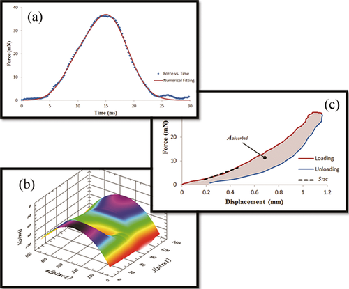
(a) The air puff force of CorVis ST was measured by a custom-designed force detection system. (b) Corneal displacement was extracted from CorVis ST using a proposed imaging analysis. (c) With the utilization of the air puff force and corneal dynamic displacement, an analysis method was developed to introduce new corneal biomechanical parameters – STSC and Aabsorbed.


