

Journal list menu

 Issue
IssueHuman Brain Mapping: Volume 42, Issue 4
837-1222March 2021
Export Citations
Download PDFs
COVER IMAGE
Cover Image
- First Published: 03 February 2021

COVER ILLUSTRATION Migraine is a primary headache disorder characterized by attacks of moderate to severe intensity. To represent gray matter structure, the cortical curvature, cortical thickness (top left), surface area (top center) and gray matter volume were used as morphometry parameters. Structural connectivity was obtained with tractography (top right) and represented by the number of streamlines. Simultaneous changes of gray matter morphometry and structural connectivity were analyzed using multimodal Canonical Correlation Analysis followed by joint Independent Component Analysis. Higher curvature values in migraine patients in the frontal pole and the cingulate gyrus (bottom center) were associated with connectivity alterations. Lower number of streamlines, possibly representing debilitated connectivity (blue, bottom left), and also higher streamline count, possibly representing strengthened connectivity (red, bottom right), were identified in migraine.
ISSUE INFORMATION
RESEARCH ARTICLES
Subject-specific segregation of functional territories based on deep phenotyping
- Pages: 841-870
- First Published: 24 December 2020
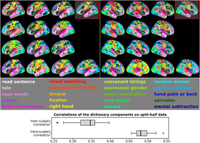
Although functional magnetic resonance imaging provides means to accurately characterize brain activation in response to behavior and ensuing mental functions, most studies are still bound to one single task and only report group-average brain maps that hinder effect specificity and a fine demarcation of brain regions. Here, we develop an individual-functional-atlasing approach to link functional segregation of specialized brain regions to elementary mental functions, by leveraging the Individual Brain Charting dataset. Results show that individual cognitive topographies, common to all tasks, are consistently mapped within as well as across participants and they reveal the contribution of each task to these common representations through the contrast-maps prediction scores of every task obtained from contrast maps of the remaining tasks.
Cardiorespiratory fitness, hippocampal subfield volumes, and mnemonic discrimination task performance in aging
- Pages: 871-892
- First Published: 16 December 2020
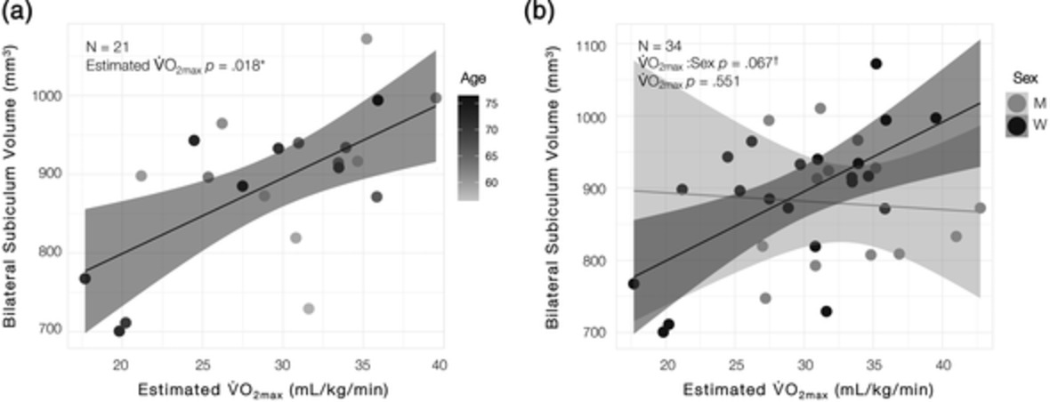
Sixty-five older adults (aged 55–85 years) completed a submaximal treadmill test to estimate cardiorespiratory fitness, a mnemonic discrimination task, and a high-resolution MRI scan to determine hippocampal subfield volumes. Left dentate gyrus/CA3 body volume predicted accuracy in the task condition with greatest stimuli similarity, and cardiorespiratory fitness predicted bilateral subiculum volume in older adult women, not men. Altogether, these findings provide further support for a role of the dentate gyrus in behavioral pattern separation in humans and suggest that cardiorespiratory fitness may have differential effects on hippocampal subfield integrity in older adult men and women.
Language distance in orthographic transparency affects cross-language pattern similarity between native and non-native languages
- Pages: 893-907
- First Published: 28 October 2020
Greater cross-language pattern similarity was associated with a smaller language distance in orthographic transparency in brain areas for phonological processing, especially in the left hemisphere. Further analysis confirmed that those brain regions represented phonological information. This study provides direct neuroimaging evidence for the modulatory effect of language distance in orthographic transparency on cross-language pattern similarity.
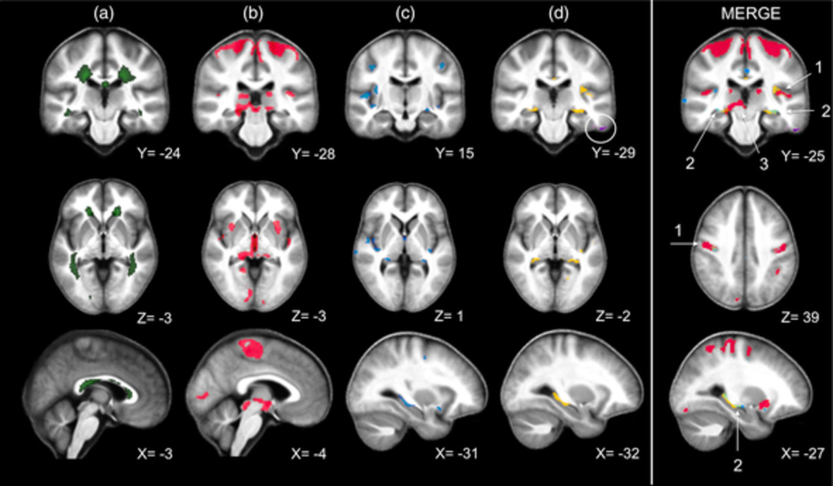
Multimodal fusion analysis of structural connectivity and gray matter morphology in migraine
- Pages: 908-921
- First Published: 08 December 2020
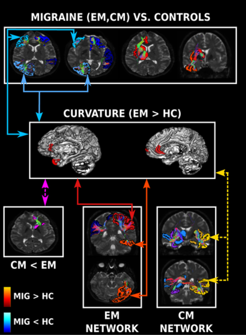
Planchuelo-Gómez et al. associate structural connectivity with gray matter morphometry. In chronic and episodic migraine, strengthened connectivity with pain processing regions, and higher cortical curvature related to weakened connectivity within each lobe were found. These results give a new insight about migraine neuroimaging biomarkers.
Prevent breaking bad: A proof of concept study of rebalancing the brain's rumination circuit with real-time fMRI functional connectivity neurofeedback
- Pages: 922-940
- First Published: 10 November 2020
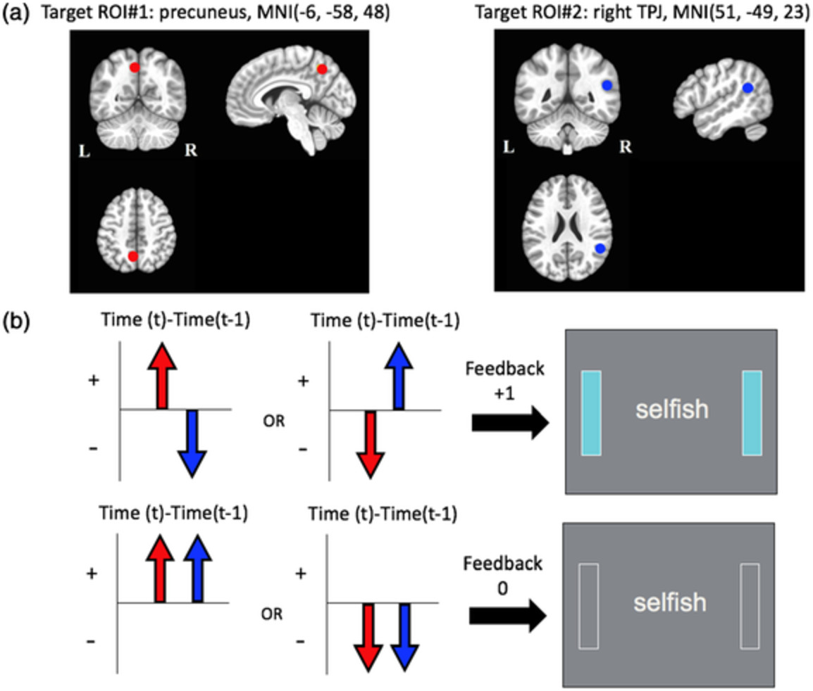
Regions of interest (ROI) for the connectivity-based rtfMRI-nf and the neurofeedback algorithm. (a) ROIs for the connectivity-based rtfMRI-nf. (b) Neurofeedback algorithm and display. Red circles and arrows indicate the precuneus ROI and BOLD activities, and blue circles and arrows indicate the right temporoparietal junction (rTPJ) ROI and BOLD activities. The sidebars on the screen were updated every 2-s with positive feedback (+1: light blue color) or no feedback (0: blank color).
Perceptual learning of tone patterns changes the effective connectivity between Heschl's gyrus and planum temporale
- Pages: 941-952
- First Published: 04 November 2020
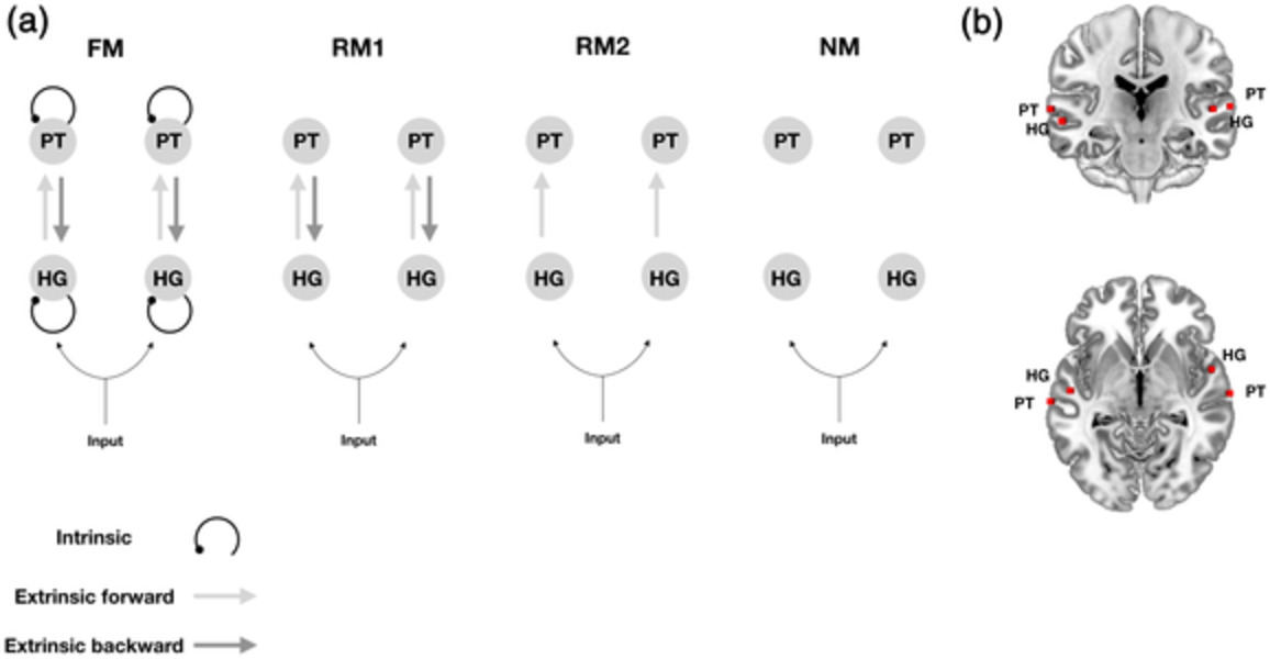
Perception of complex auditory stimuli requires the brain to update a model of its environment. Using dynamic causal modeling for functional magnetic resonance imaging during a melodic oddball paradigm, we show increased intrinsic connectivity within Heschl's gyrus (HG) and a concomitant increase in forward connectivity from HG to planum temporale in the left cerebral hemisphere. We did not find evidence of frontal activity. Our results are consistent with a predictive coding account of perceptual learning within the superior temporal gyrus.
Examining volumetric gradients based on the frustum surface ratio in the brain in autism spectrum disorder
- Pages: 953-966
- First Published: 09 December 2020
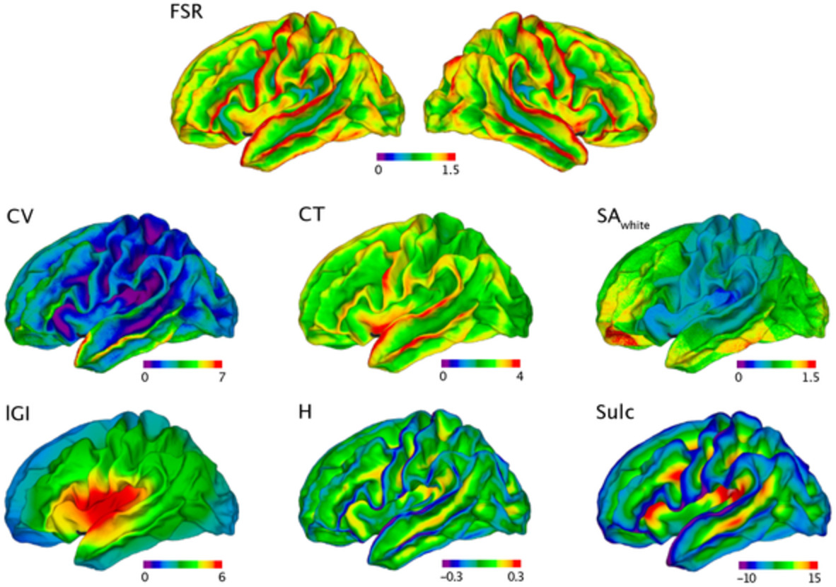
During human brain development, the grey matter volume (CV) expands extensively, leading to formation of the gyral-sulcal pattern of the cortical surface. Given the folded architecture of the cortex, the distribution of CV at a given location on the surface of the brain is likely to differ, depending on whether the cortex is outward-folded (as in gyri) or inward-folded (as in sulci). We have designed a novel imaging feature (FSR) to capture volumetric gradients from the outer to the inner surface of the brain, and to quantify the distortion of CV across cortical layers. This may be of relevance to neurodevelopmental disorders associated with atypical cortical expansion and lamination, as has been suggested for Autism Spectrum Disorder.
Detection of Alzheimer's Disease using cortical diffusion tensor imaging
- Pages: 967-977
- First Published: 11 November 2020
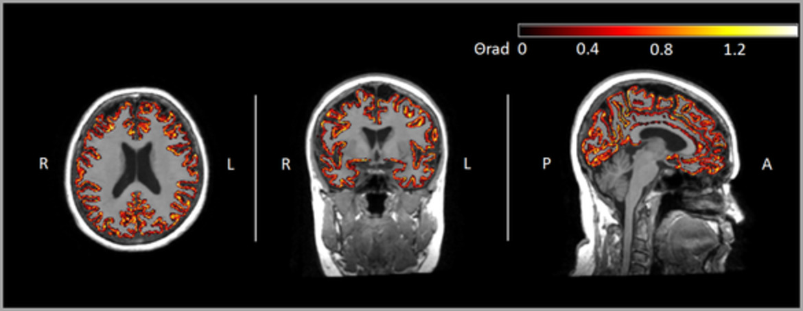
The aim of this research was to test a novel in-vivo brain MRI analysis method that could be used in clinical cohorts, to investigate cortical architecture changes in patients with Alzheimer's Disease (AD).The new measures could be used within the “ATN” framework as an index of cortical microstructure quality and a marker of Neurodegeneration. Further development may aid diagnosis, patient selection, and quantification of the “Neurodegeneration” component in response to therapies in clinical trials.
A comprehensive study on electroencephalography and magnetoencephalography sensitivity to cortical and subcortical sources
- Pages: 978-992
- First Published: 06 November 2020

Signal-to-noise ratio (SNR) maps are a good way to visualize electroencephalography (EEG) and magnetoencephalography (MEG) sensitivity. In our work, we computed EEG and MEG sensitivity maps in a six-compartment anisotropic head model. We show that ignoring the cerebrospinal fluid leads to an overestimation of EEG SNR values. For cortical sources, we found that radial and deep are more visible to EEG, tangential (the majority) more to MEG. Finally, our results show that MEG is not fully insensitive to subcortical sources.
White matter hyperintensities at critical crossroads for executive function and verbal abilities in small vessel disease
- Pages: 993-1002
- First Published: 24 November 2020
The manuscript describes a study of the relation between white matter hyperintensity (WMH) locations and executive and language abilities in small vessel disease patients without dementia with varying burden of WMH.
Voxel-Based quantitative MRI reveals spatial patterns of grey matter alteration in multiple sclerosis
- Pages: 1003-1012
- First Published: 06 November 2020
This cross-sectional study investigates the spatial distribution of grey matter microstructural damage in multiple sclerosis, using voxel-based quantification of three MRI parameters, sensitive to myelin and iron contents: magnetization transfer saturation (MT sat), effective transverse (R2*) and longitudinal (R1) relaxation rates. Potential gray matter atrophy was also investigated by a concurrent voxel-based morphometry analysis. We identify three different spatially segregated combinations of GM alterations, common to this MS patient cohort and potentially associated with different neuropathological characteristics: primary cortices, deep grey matter nuclei and hippocampus. The evidence of substantial demyelination of hippocampi beyond atrophic areas constitutes a key contribution of this study and usefully complements previous characterization of hippocampal damage in MS.
Cortical reorganization after motor stroke: A pilot study on differences between the upper and lower limbs
- Pages: 1013-1033
- First Published: 09 November 2020
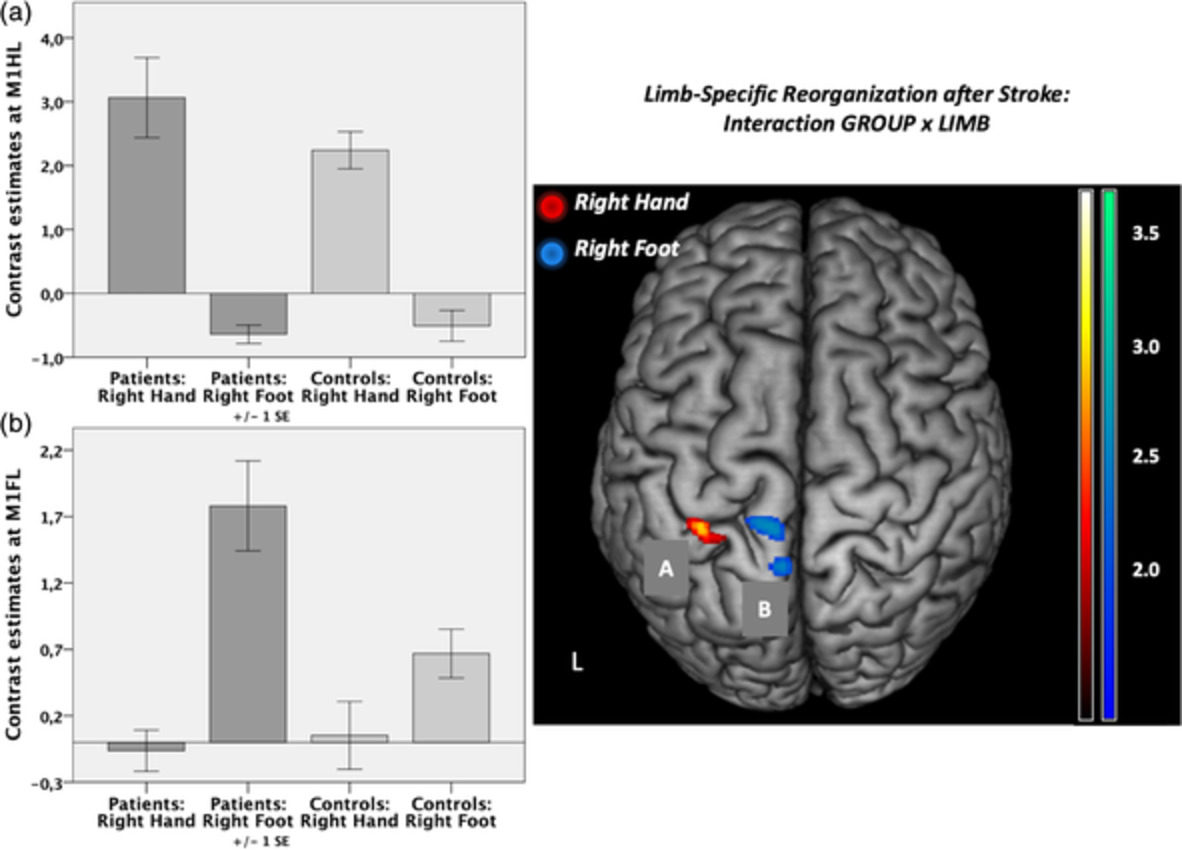
The present pilot study aimed to gain insights into the neural reorganization patterns of lower limb motor function in comparison to upper limb function in well-recovered chronic stroke patients compared to healthy participants using functional magnetic resonance imaging and dynamic causal modeling. For the first time, we thus investigated unilateral movements of both the hands and feet in one common paradigm. This enabled direct comparisons of the respective reorganization patterns underlying hand and foot movements as well as differential conclusions regarding their functional relevance.
Multimodal MRI assessment for first episode psychosis: A major change in the thalamus and an efficient stratification of a subgroup
- Pages: 1034-1053
- First Published: 30 December 2020
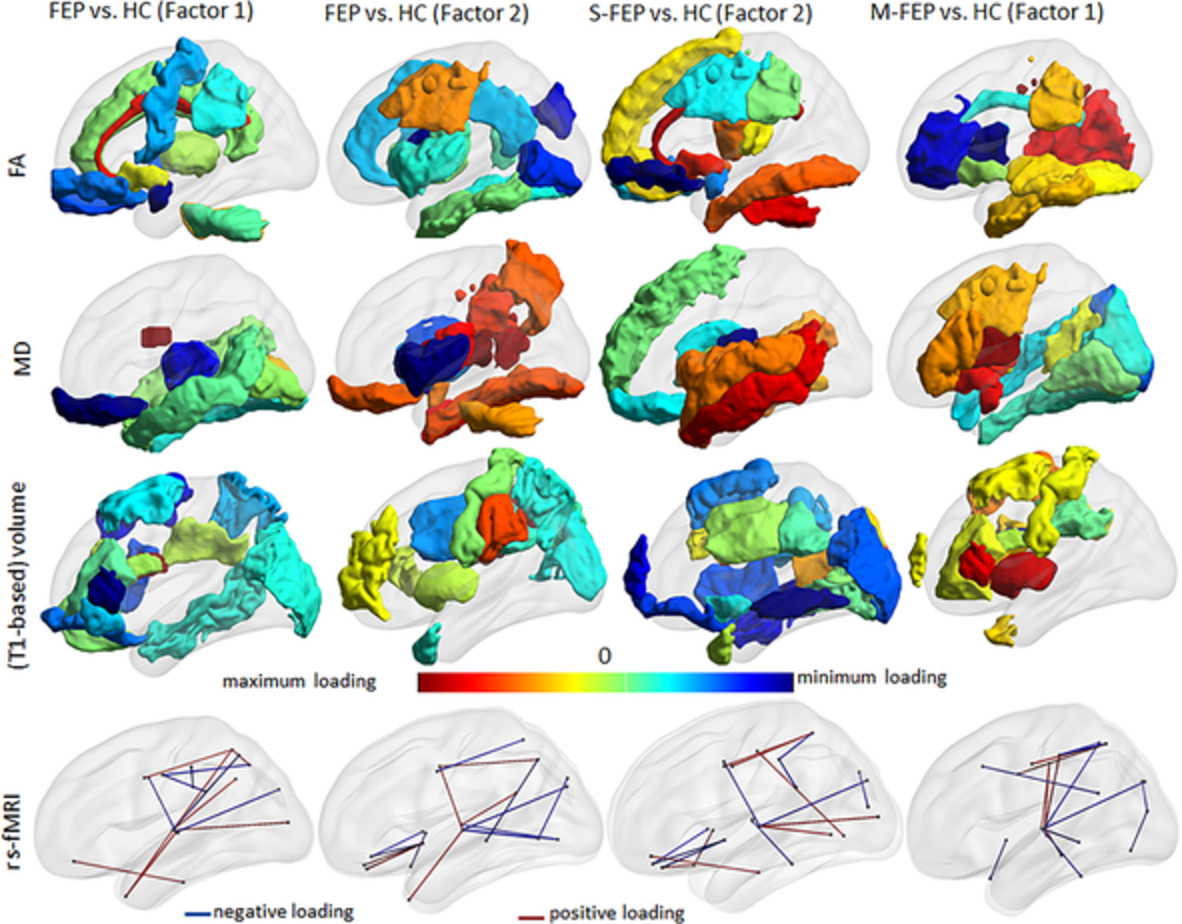
We combined automated structure-based analysis with supervised integrated factor analysis to examine multiple MRI features (volume, DTI, resting state fMRI) in the whole brain of individuals with first-episode psychosis. We successfully characterized this group, and subgroups with schizophrenia- and mood-associated psychosis. We found robust abnormalities centered in the thalamus.
Brain functional connectivity dynamics at rest in the aftermath of affective and cognitive challenges
- Pages: 1054-1069
- First Published: 24 November 2020
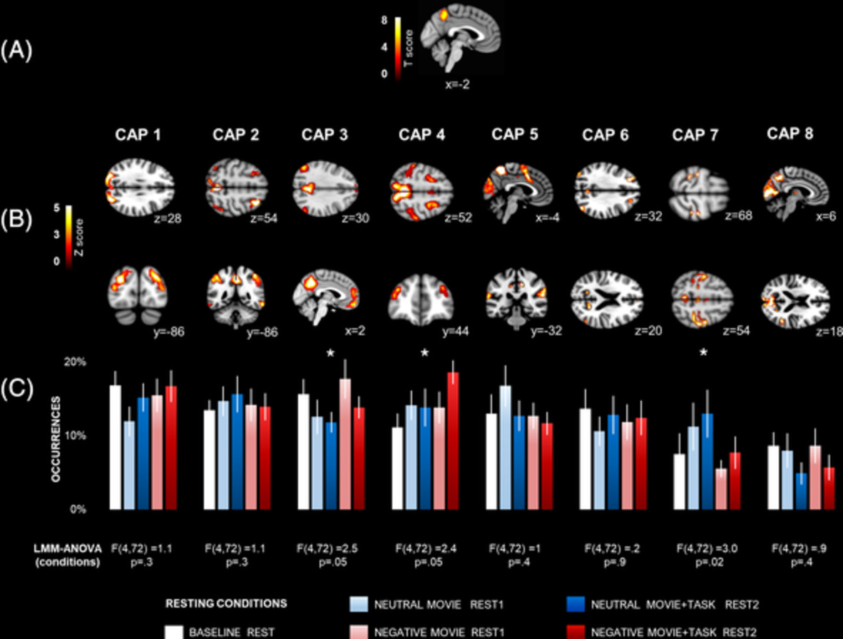
Dynamic intrinsic functional connectivity brain networks were altered after exposure to negative emotional content and after emotionally influenced cognitive control. Both brain networks fluctuations were associated with changes in self-reported affect state and cognitive control performance.
An empirical comparison of univariate versus multivariate methods for the analysis of brain–behavior mapping
- Pages: 1070-1101
- First Published: 20 November 2020
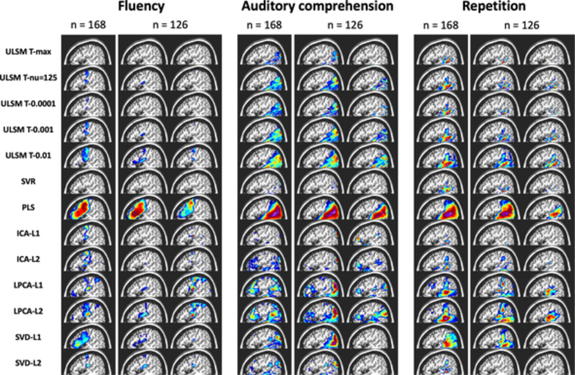
In the current study, we conducted the first comprehensive, empirical comparison of several univariate and multivariate lesion symptom mapping (LSM) methods using both synthetic and real behavioral data to identify the potential strengths and weaknesses of both approaches. Cumulatively, our analyses indicated that both univariate and multivariate methods can be equally robust in locating brain–behavior relationships, depending on the design of the study, the research question being asked, and with proper spatial metrics. The results provide crucial insights into the accuracy of different LSM methods and their susceptibility to artifact, providing a first of its kind data-driven navigational guide for users of LSM analyses.
Cortico-striato-thalamo-cerebellar networks of structural covariance underlying different epilepsy syndromes associated with generalized tonic–clonic seizures
- Pages: 1102-1115
- First Published: 29 December 2020
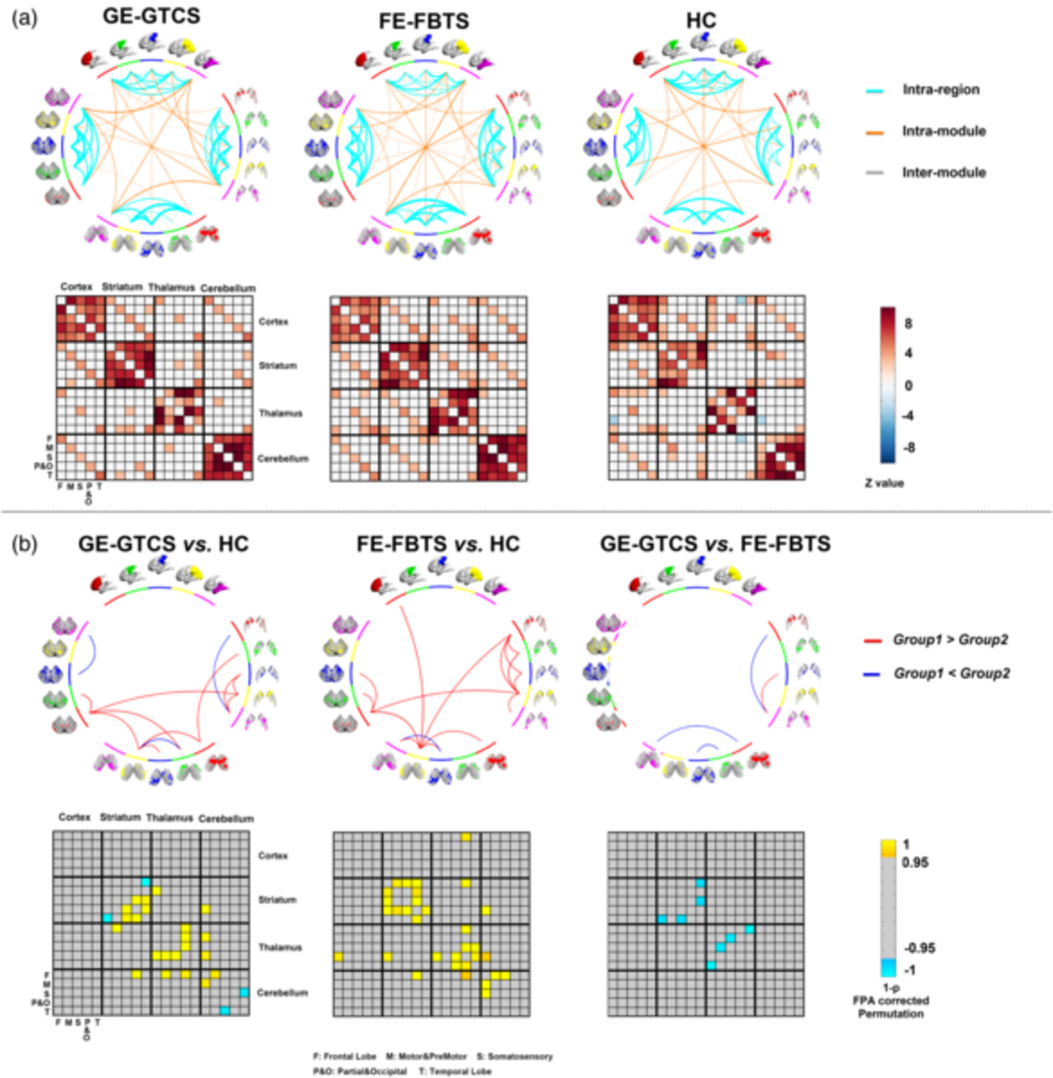
In this work, we used a winner-take-all strategy-based structural covariance connectivity analysis, to delineate the cortico-striato-thalamo-cerebellar networks in two syndromes of generalized epilepsy. Our findings implicated cortico-striato-thalamo-cerebellar network changes at a large temporal scale in GTCS, with FE-FBTS showing more severe network disruption. The study contributes novel imaging evidence for understanding the different epilepsy syndromes associated with generalized seizures.
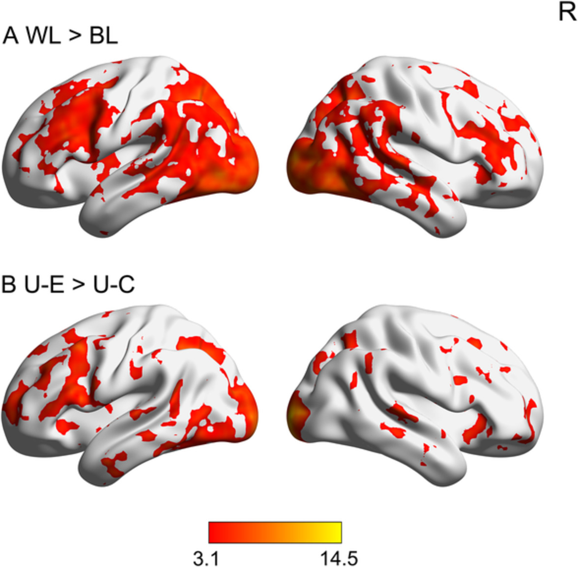
A method to mitigate spatio-temporally varying task-correlated motion artifacts from overt-speech fMRI paradigms in aphasia
- Pages: 1116-1129
- First Published: 19 November 2020
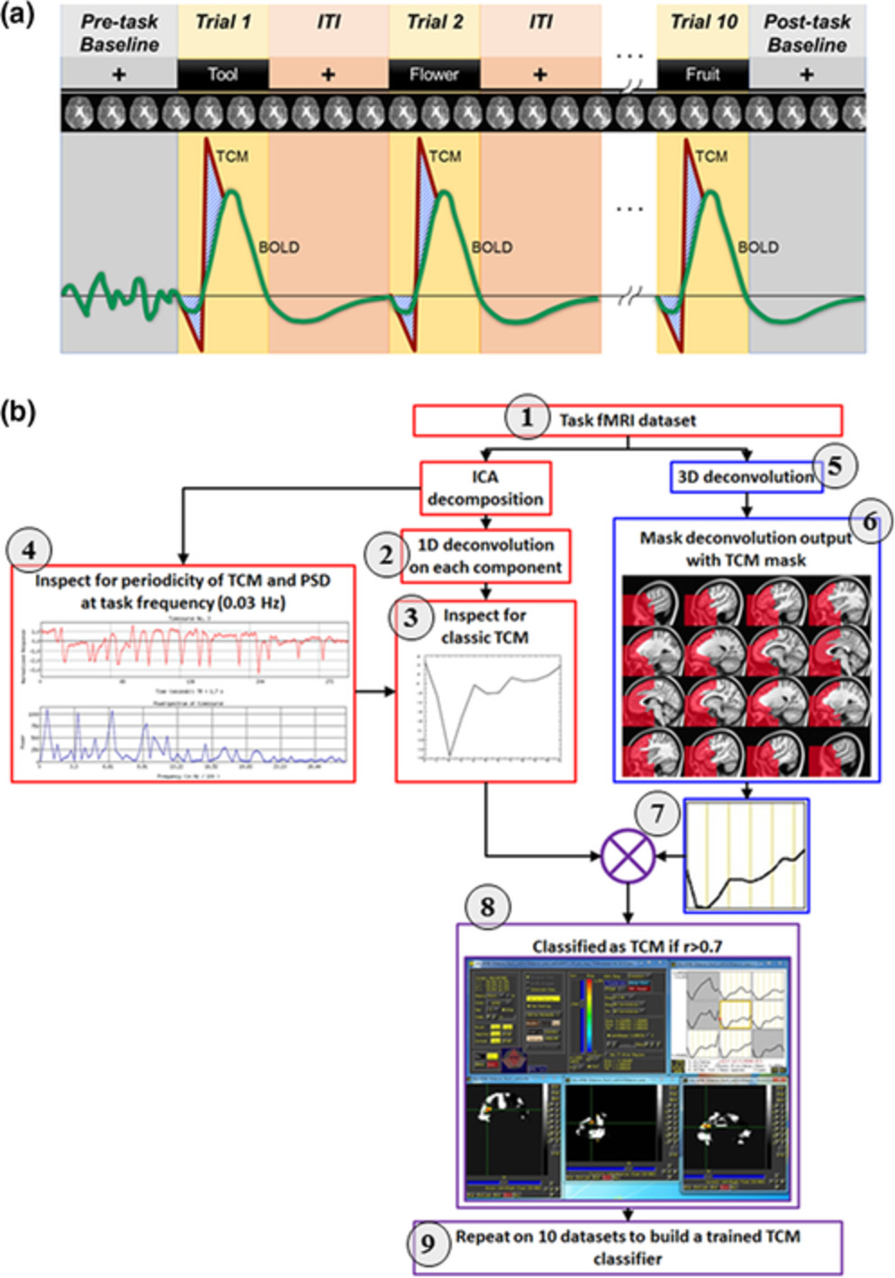
Quantifying accurate functional magnetic resonance imaging (fMRI) activation maps can be dampened by spatio-temporally varying task-correlated motion (TCM) artifacts in certain task paradigms (e.g., overt speech). In this study, we developed a novel independent component analysis (ICA)-based approach to denoise spatio-temporally varying TCM artifacts in fourteen persons with aphasia who participated in an overt language fMRI paradigm. Results show that the proposed methodology outperforms other approaches in removing TCM-related false positive activity (i.e., improved detectability power) with high spatial specificity. The proposed method was also effective in maintaining a balance between removal of TCM-related trial-by-trial variability and signal retention. Finally, we show that the TCM artifact is related to clinical metrics, such as speech fluency and aphasia severity, and the implication of TCM denoising on such relationship is also discussed.
Microstructural alterations in medial forebrain bundle are associated with interindividual pain sensitivity
- Pages: 1130-1137
- First Published: 10 November 2020
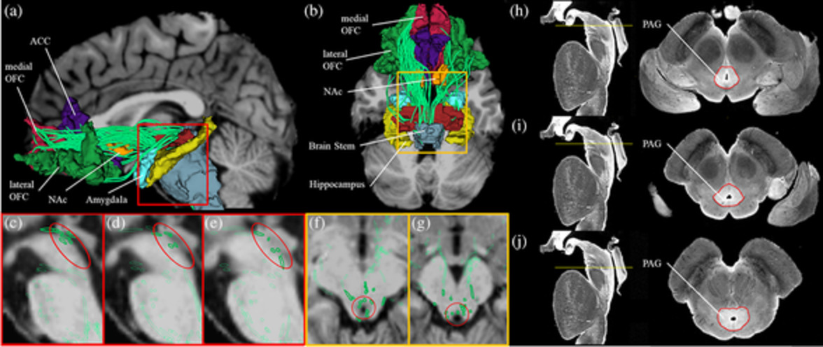
We aimed to investigate whether variation of microstructure of the medial forebrain bundle (MFB) is associated with interindividual pain sensitivity, by assessing interindividual pain sensitivity as a rating of pain intensity to heat stimuli and reconstructing the MFB using multitensor tractography from diffusion magnetic resonance imaging (dMRI) in 38 healthy men. Lower ratings of interindividual pain intensity correlated with higher fractional anisotropy (FAt) and lower radial diffusivity (RDt) of the MFB. As changes in FAt and RDt may reflect abnormalities in myelination, the results might be interpreted as that a lower pain rating is associated with higher degree of myelination of the MFB and could represent an inhibitory pathway of pain.
Rapid invisible frequency tagging reveals nonlinear integration of auditory and visual information
- Pages: 1138-1152
- First Published: 18 November 2020
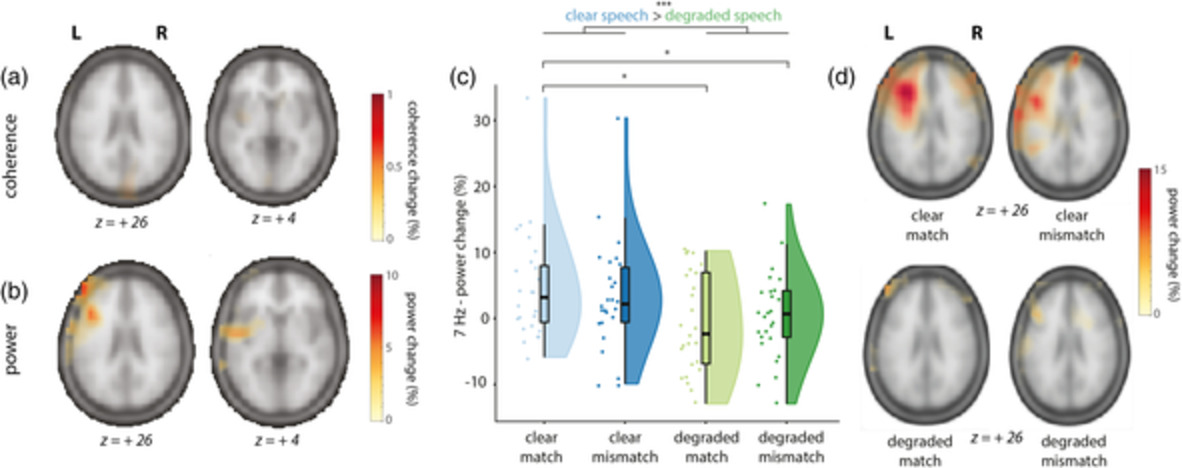
First, we provided a proof of principle that rapid invisible frequency tagging (RIFT) can be used to tag visual and auditory inputs at high frequencies, resulting in clear spectral peaks in the MEG signal, localized to early sensory cortices. Second, we demonstrated that RIFT can be used to identify intermodulation frequencies in a multimodal, semantic context. The observed intermodulation frequency was the result of the nonlinear interaction between visual and auditory tagged stimuli, and was localized to LIFG/pSTS/MTG, areas known to be involved in the multimodal integration of speech and gestures.
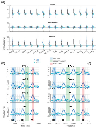
Modulation of neural oscillations during working memory update, maintenance, and readout: An hdEEG study
- Pages: 1153-1166
- First Published: 17 November 2020
Specific spectral signatures are associated with updating of memory information, working memory maintenance, and readout in a large fronto-parietal network, including also the insula and the cerebellum.
Reliability, sensitivity, and predictive value of fMRI during multiple object tracking as a marker of cognitive training gain in combination with tDCS in stroke survivors
- Pages: 1167-1181
- First Published: 20 November 2020

In this randomized double-blind study, we have investigated the feasibility and clinical utility of fMRI as a predictor and marker of response to cognitive training and transcranial direct current stimulation (tDCS) in chronic stroke patients. Linear mixed effects models revealed beneficial training effects on the trained tasks, with no additional effect of tDCS. There was no significant association between fMRI activation patterns and training gains. Intraclass correlation coefficients suggested satisfactory reliability across assessments and experimental contrasts, supporting the feasibility of fMRI as a marker for longitudinal alterations in a clinical context.
Transdiagnostic time-varying dysconnectivity across major psychiatric disorders
- Pages: 1182-1196
- First Published: 19 November 2020
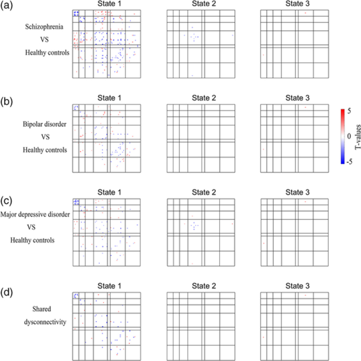
We identified three functional connectivity states across multiple psychiatric disorders and healthy controls: one was a more frequent state with weak connectivity, and the other two were less frequent states with strong connectivity. Dysconnectivity in the psychiatric patients was found nearly only in the weak connectivity state. Psychiatric patients spent less time in the weak connectivity state than HC patients.
Comparing machine and deep learning-based algorithms for prediction of clinical improvement in psychosis with functional magnetic resonance imaging
- Pages: 1197-1205
- First Published: 13 November 2020
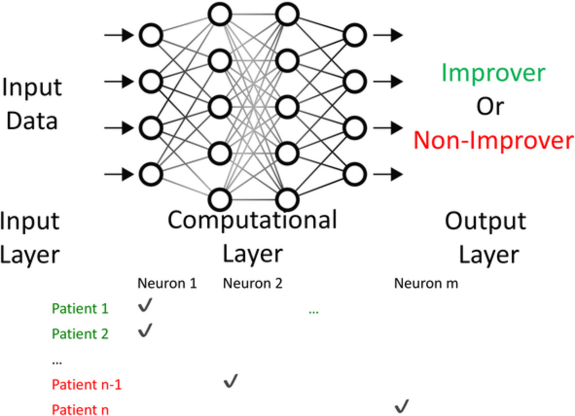
The performance of shallow machine learning algorithms was compared to deep learning when predicting symptomatic improvement in early psychosis using frontoparietal activation data from a cognitive control task. Deep learning outperformed all shallow algorithms for both binary and continuous classifiers. Explainable artificial intelligence (XAI) methods suggested the deep learner primarily used left dorsolateral prefrontal cortex activation to predict clinical improvement, providing an example of how XAI can be used to "demystify" the "black box" problem inherent in all machine learning algorithms.
Reorganization of functional brain network architecture in chronic osteoarthritis pain
- Pages: 1206-1222
- First Published: 19 November 2020
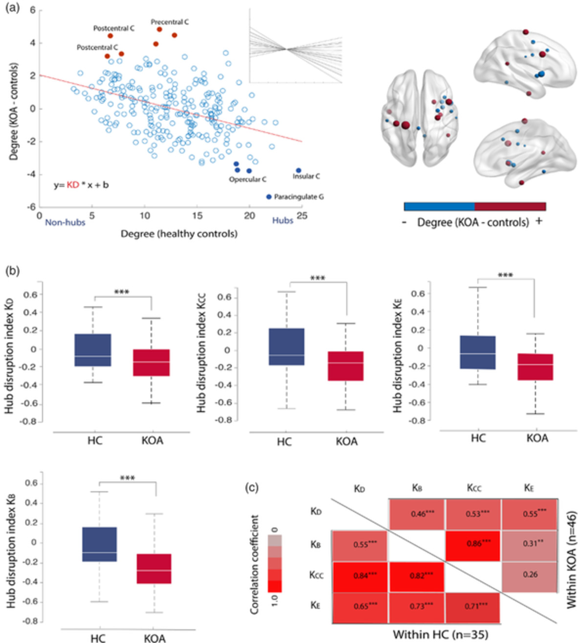
Osteoarthritis (OA) manifests with chronic pain, motor impairment and proprioceptive changes. However, the role of the brain in the disease is largely unknown. Here, we studied brain networks using the mathematical properties of graphs in knee and hip OA. We show that while conserving global topological properties, brain network architecture reorganizes in OA, at both global and local scale. Network connectivity related to OA pain intensity is dissociated from the major hub disruptions found:insula/M1/S1/parahippocampus; challenging the extent of dependence of OA pain on nociceptive signaling.