

Journal list menu

 Issue
IssueSmall: Volume 6, Issue 3
331-478February 5, 2010
Export Citations
Download PDFs
Cover Picture
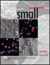
Microparticles: Small 3/2010
- First Published: 27 January 2010
The cover image depicts a collage of confocal laser scanning micrographs and scanning electron micrographs of biodegradable bicompartmental microparticles of different shapes and sizes made from poly(lactide-co-glycolide) produced via electrohydrodynamic co-jetting. In this process, two polymer solutions labeled with different fluorophores are introduced via two parallel capillaries to generate a composite “bicompartmental” droplet. Application of an electric field results in an electrified jet, wherein solvent evaporation causes the formation of particles, which are confined in bicompartmental architecture. Control over the solution and process parameters creates an array of particle sizes and controls the shapes of the bicompartmental particles. Such particles may find applications in drug delivery, diagnostics, and biosensing. For more information, please read the Full Paper “Towards Designer Microparticles: Simultaneous Control of Anisotropy, Shape, and Size” by J. Lahann et al., beginning on page 404.
Inside Cover
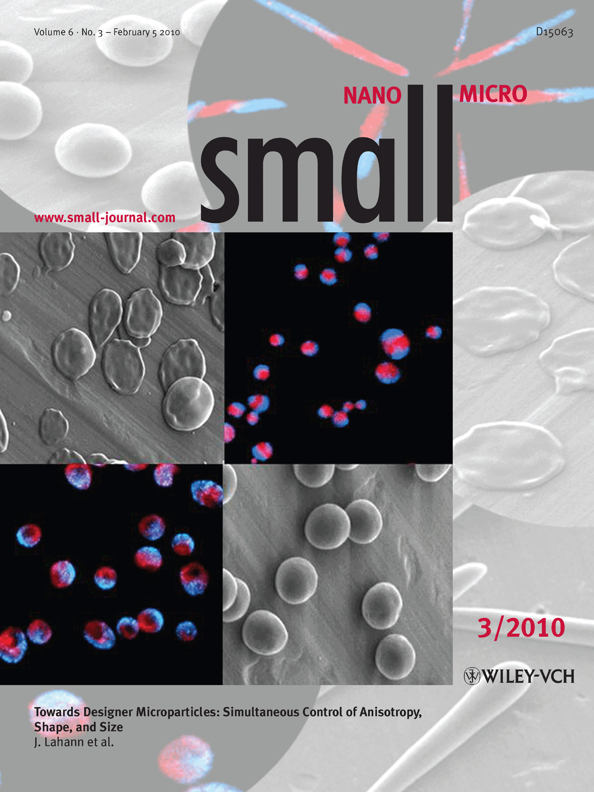
Stem cells: Small 3/2010
- First Published: 27 January 2010
The cover picture shows several human mesenchymal stem cells (green with blue nuclei) clustered around a stiff polymeric microstructure (blue) in a three-dimensional matrix. The microstructures are fabricated using photolithographic techniques and then suspended with stem cells in the matrix. The stem cells migrate and attach to the microstructures, and although the volume percentage of the microstructures within the matrix is quite low (0.07%), stem-cell morphology, clustering, and gene expression are significantly different compared to without the microstructures. The knowledge of how physical and mechanical cues in three dimensions can influence cellular function is necessary for the development of novel platforms for regenerative therapy. For more information, please read the Communication “Three-Dimensional Culture with Stiff Microstructures Increases Proliferation and Slows Osteogenic Differentiation of Human Mesenchymal Stem Cells by B. Russell et al., beginning on page 355.
Contents
Review
Nanomotors
Motion Control at the Nanoscale†
- Pages: 338-345
- First Published: 27 January 2010

This Review highlights recent progress towards controlling the movement of fuel-driven nanomotors and discusses the challenges and opportunities associated with the achievement of such nanoscale motion control. Regulating the movement of artificial nanomotors often follows nature's elegant and remarkable approach for motion control. Such control of the movement of synthetic nanomotors is essential for performing various tasks and diverse applications.
Communications
Biosensors
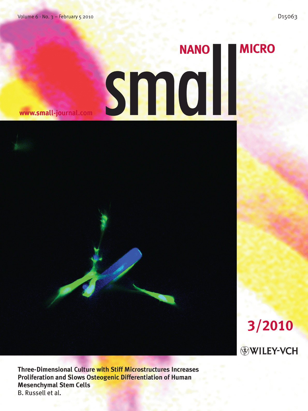
Single-Molecule FRET Imaging for Enzymatic Reactions at High Ligand Concentrations†
- Pages: 346-350
- First Published: 27 January 2010
Single-molecule Förster resonance energy transfer (FRET) imaging at micromolar concentrations of an analyte is demonstrated. A quantum dot (QD)–dye donor–acceptor pair permits the approach of physiological levels of fluorescently labeled analytes when imaging molecular dynamics. Single-molecule FRET imaging of myosin ATP hydrolysis is performed using QDs attached to myosin as a donor enzyme and Cy3-labeled ATP as an acceptor ligand at micromolar levels (see image).
Viral nanoparticles
Functional Single-Virus–Polyelectrolyte Hybrids Make Large-Scale Applications of Viral Nanoparticles More Efficient†
- Pages: 351-354
- First Published: 27 January 2010
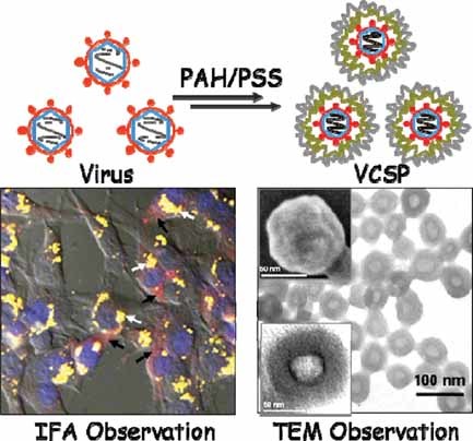
Single enveloped viruses can be prepared on a large scale with high efficiency by a layer-by-layer method. The virus/polyelectrolyte core/shell nanoparticles (VCSPs) exhibit unique characteristics, such as direct observation by electron microscopy without staining, easy separation and concentration, rapid perinuclear delivery, and improved biological safety, which resolve the conventional shortcomings of viruses in nanoscale applications.
Stem cells
Three-Dimensional Culture with Stiff Microstructures Increases Proliferation and Slows Osteogenic Differentiation of Human Mesenchymal Stem Cells†
- Pages: 355-360
- First Published: 27 January 2010
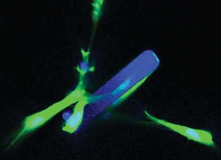
A small quantity of stiff microstructures in a 3D matrix regulates human mesenchymal stem cells to increase proliferation and slow differentiation after 10 days in culture. Cell morphology, clustering, and gene expression (see image) are significantly different although the stiff microstructures are only 0.07% of the total gel volume.
Nanochannels
Fabrication of Stable Single Nanochannels with Controllable Ionic Rectification†
- Pages: 361-365
- First Published: 27 January 2010
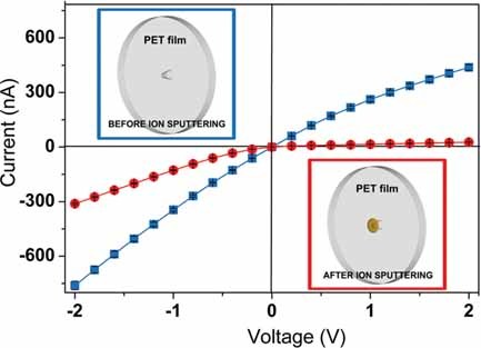
Artificial single nanochannels with chemical/structural asymmetry and controllable continuous change of ionic-current rectification are prepared by ion-sputtering technology. The nanochannel is embedded in a track-etched poly(ethylene terephthalate) (PET) membrane and sputtered with Pt on the tip side of the nanochannel.
Nanomedicine
Bovine Serum Albumin-Based Magnetic Nanocarrier for MRI Diagnosis and Hyperthermic Therapy: A Potential Theranostic Approach Against Cancer†
- Pages: 366-370
- First Published: 27 January 2010
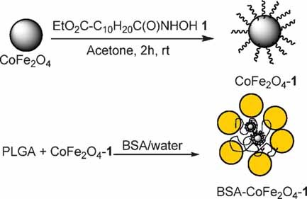
Chemical synthesis, stability, and characterization of a new albumin-based magnetic nanocarrier containing cobalt ferrite nanoparticles is reported. The BSA–cobalt-based nanocarrier is tested as a theranostic nanomedicine: both diagnostic abilities in vivo and therapeutic hyperthermic effects on standard human tumor cell line (HeLa cells, see image) are investigated.
Inkless contact printing
High-Aspect-Ratio Imageable Top-Surface Lithography Using UV-Assisted Inkless Contact Printing
- Pages: 371-375
- First Published: 27 January 2010
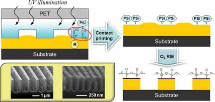
The simple production of micro/nanoscale patterns with very high aspect ratios exceeding that of the mold used is demonstrated using unconventional top-surface lithography. The method combines inkless contact printing and subsequent dry pattern development by oxygen reactive ion etching (see image), making it possible to form high-resolution, high-aspect-ratio patterns without the aid of expensive optical protocols.
Nanowire heterostructures
One-Dimensional Heterostructures of Single-Walled Carbon Nanotubes and CdSe Nanowires
- Pages: 376-380
- First Published: 27 January 2010
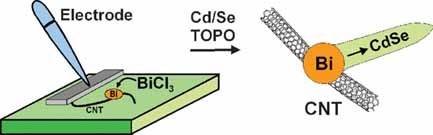
A one-dimensional heterostructure comprising single-walled carbon nanotubes (CNTs) and CdSe nanowires is prepared via electrochemical deposition of Bi nanoparticles onto the nanotubes (see image), followed by solution–liquid–solid synthesis of the semiconductor nanowires.
Nanotubes
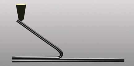
Synthesis and Thermal Expansion of Copper Nanotubes and Nanowires with Y- and Step-Shaped Topologies†
- Pages: 381-385
- First Published: 27 January 2010
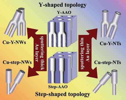
Copper nanotubes (NTs) and nanowires (NWs) with Y- and step-shaped topologies (see image) are built via electrodeposition inside anodic aluminum oxide templates. Thermal-expansion measurements demonstrate that their thermal-expansion coefficients (TECs) are temperature dependent and that the TECs of the stem/large-diameter segments are more sensitive than those of the branch/small-diameter segments.
Nanohybrids
Hierarchically Constrained Dynamics and Emergence of Complex Behavior in Nanohybrids†
- Pages: 386-390
- First Published: 27 January 2010
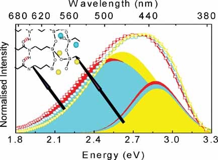
The emitted relaxation energy of an organic–inorganic nanohybrid upon repeated heating/cooling cycles exhibits a logarithmic time dependence (associated with hierarchically constrained dynamics), thereby providing a conspicuous fingerprint of emergent complex behavior. It is proposed that the emergence of complexity may be the rule, rather than the exception, concerning the interplay of individual and collective behavior.
Self-assembled nanostructures
A Supramolecular Network as Sacrificial Mask for the Generation of a Nanopatterned Binary Self-Assembled Monolayer†
- Pages: 391-394
- First Published: 27 January 2010
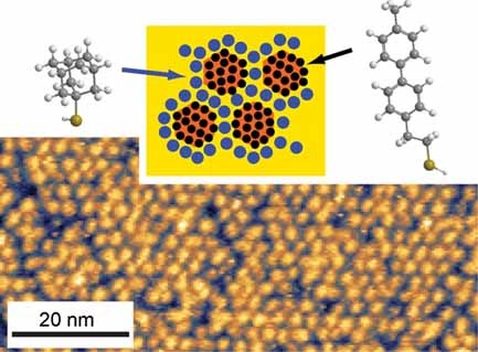
A supramolecular hydrogen-bonded network adsorbed on Au(111) can corral thiol self-assembled monolayers (SAMs) inside its pores. Enhancing the thermodynamic stability of these SAM nanoislands by Cu underpotential deposition allows selective replacement of the network by a second thiol species. The resulting binary SAM (see image) retains the periodicity and symmetry of the original supramolecular mask.
Frontispiece
Nanocrystal formation: Small 3/2010
- First Published: 27 January 2010
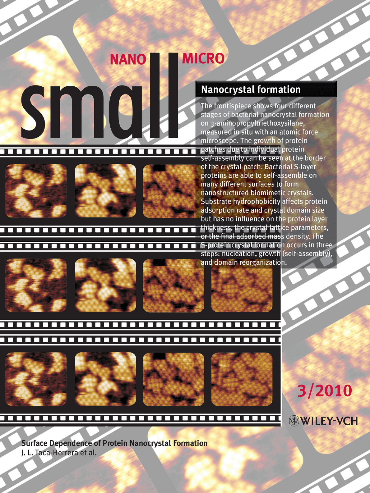
The frontispiece shows four different stages of bacterial nanocrystal formation on 3-aminopropyltriethoxysilane, measured in situ with an atomic force microscope. The growth of protein patches due to individual protein self-assembly can be seen at the border of the crystal patch. Bacterial S-layer proteins are able to self-assemble on many different surfaces to form nanostructured biomimetic crystals. Substrate hydrophobicity affects protein adsorption rate and crystal domain size but has no influence on the protein layer thickness, the crystal-lattice parameters, or the final adsorbed mass density. The S-protein crystal formation occurs in three steps: nucleation, growth (self-assembly), and domain reorganization. For more information, please read the Full Paper ‘Surface Dependence of Protein Nanocrystal Formation” by J. L. Toca-Herrera et al., beginning on page 396.
Full Papers
Nanocrystal formation
Surface Dependence of Protein Nanocrystal Formation†
- Pages: 396-403
- First Published: 27 January 2010
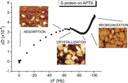
Nanocrystal formation of the bacterial protein SbpA on silicon dioxide and silane coupling agents is studied with high-resolution atomic force microscopy and quartz crystal microbalance with dissipation. S-layer formation occurs in three steps: protein adsorption, protein self-assembly, and crystalline-domain reorganization (see image). Substrate chemistry influences crystalline-domain size and layer compliance but not the crystal lattice parameters.
Microparticles
Towards Designer Microparticles: Simultaneous Control of Anisotropy, Shape, and Size
- Pages: 404-411
- First Published: 27 January 2010
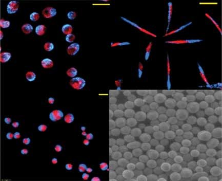
Simultaneous control over the anisotropy, shape, and size of poly(lactide-co-glycolide) (PLGA) microparticles via electrohydrodynamic co-jetting is demonstrated by the fabrication of bicompartmental discoid and rod-shaped particles in addition to spheres (see image). This is achieved by controlling different solution and process parameters during co-jetting. Such designer particles can be employed as novel therapeutic agents for drug delivery and diagnostics.
Multifunctional nanotubes
Water-Dispersible, Multifunctional, Magnetic, Luminescent Silica-Encapsulated Composite Nanotubes
- Pages: 412-420
- First Published: 27 January 2010
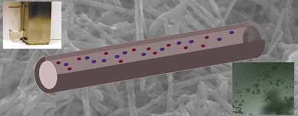
A multifunctional SiO2-based nanostructure is synthesized by a novel method. Preformed CdSe quantum dots and magnetite nanoparticles are incorporated within the channels of SiO2 nanotubes (see image). The resulting nanostructure possesses intriguing photoluminescent and superparamagnetic properties characteristic of its constituent components.
Biomaterials
Conducting-Polymer Nanotubes Improve Electrical Properties, Mechanical Adhesion, Neural Attachment, and Neurite Outgrowth of Neural Electrodes
- Pages: 421-429
- First Published: 27 January 2010
The superiority of deposited conducting-polymer nanotubes of poly(3,4-ethylenedioxythiophene) (PEDOT) and poly(pyrrole) (PPy) on neural microelectrodes (see image) compared to their film counterparts is reported in terms of resistance to delamination, reduced impedance, increased charge-capacity density, neural attachment, and neurite outgrowth.
Nanocarriers
HDL-Mimicking Peptide–Lipid Nanoparticles with Improved Tumor Targeting
- Pages: 430-437
- First Published: 27 January 2010
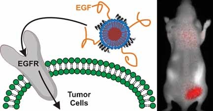
Fluorescent nanocarriers that mimic high-density lipoprotein can target epidermal growth factor receptor (EGFR)-expressing cancer cells (see picture). The nanocarriers have controllable size, cargo loading, and shielding properties. The small size and surface properties result in excellent tumor accumulation, the modular targeting of the nanoparticle being essential for tumor cell uptake.
Nanomechanical peeling
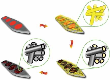
Mechanical Peeling of Free-Standing Single-Walled Carbon-Nanotube Bundles
- Pages: 438-445
- First Published: 27 January 2010
Nanomechanical peeling of thin free-standing bundles of single-walled carbon nanotubes is performed by in situ nanomanipulation techniques (see image) inside a high-resolution scanning electron microscope. The adhesion strength between bundled nanotubes at various peel-front positions is determined from the deformation curvatures of the delaminated nanotube bundle based on a nonlinear-elastica model.
Plasmon coupling
Visualizing Plasmon Coupling in Closely Spaced Chains of Ag Nanoparticles by Electron Energy-Loss Spectroscopy
- Pages: 446-451
- First Published: 27 January 2010
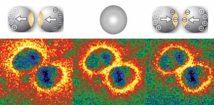
Plasmon coupling through chains of Ag nanoparticles is investigated using electron energy-loss spectroscopy. In-phase and anti-phase coupling modes are identified in dimers by anisotropic plasmon mapping. The in-phase mode is found to be optically active and the path of light transportation is visualized.
Nanotransport
Utilization of Myosin and Actin Bundles for the Transport of Molecular Cargo
- Pages: 452-457
- First Published: 27 January 2010
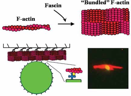
The utility of actin bundles for nanotransport applications is examined. Actin bundling affects the translational behavior of the actin filaments and may be useful for the transport of molecular cargo (streptavidin-functionalized beads) while the myosin motor may be regulated photonically. These data suggest that actin bundling may significantly improve the applicability of the myosin motor for future nanotechnological applications.
Graphene
Flexible, Transparent, Conducting Films of Randomly Stacked Graphene from Surfactant-Stabilized, Oxide-Free Graphene Dispersions
- Pages: 458-464
- First Published: 27 January 2010
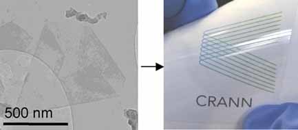
Graphene thin films are prepared using surfactant-stabilized dispersions of graphene in water. Electromechanical stability coupled with high transparency make these films attractive as transparent flexible conductors (see image).
Microcontact printing
The Janus-SAM Approach for the Flexible Functionalization of Gold and Titanium Oxide Surfaces
- Pages: 465-470
- First Published: 27 January 2010
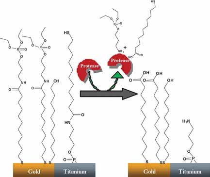
Microcontact printing is used to create a Janus self-assembled monolayer on both gold and titanium oxide surfaces (see image). The newly formed surfaces are subjected to surface reactions and, as an example application, are used for the chemisorption of bovine serum albumin. At each stage, the JSAMs are characterized by X-ray photoelectron spectroscopy and dynamic water-contact-angle measurements.
Drug delivery
Rattle-Type Fe3O4@SiO2 Hollow Mesoporous Spheres as Carriers for Drug Delivery
- Pages: 471-478
- First Published: 27 January 2010
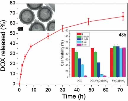
Hollow mesoporous spheres of Fe3O4@SiO2 exhibit relatively fast cell uptake and no cytotoxicity up to a concentration of 150 µg mL−1 after 48 h of incubation. Doxorubicin hydrochloride (DOX)-loaded spheres exhibit a somewhat higher cytotoxicity than free DOX (see picture). The Fe3O4SiO2 spheres have potential for drug loading and delivery into cancer cells to induce cell death.
Sign up for email alerts
Tools

Related Titles
- Advanced Materials
- Advanced Functional Materials
- Advanced Energy Materials
- Advanced Electronic Materials
- Advanced Engineering Materials
- Advanced Healthcare Materials
- Advanced Materials Interfaces
- Advanced Materials Technologies
- Advanced Optical Materials
- Advanced Physics Research
- Advanced Science
- Advanced Sensor Research
- Nano Select
- Particle & Particle Systems Characterization
- Small Methods
- Small Science
- Small Structures