

Journal list menu

 Issue
IssueSmall: Volume 5, Issue 18
2023-2119September 18, 2009
Export Citations
Download PDFs
Cover Picture
Multiple emulsions: Small 18/2009
- First Published: 14 September 2009
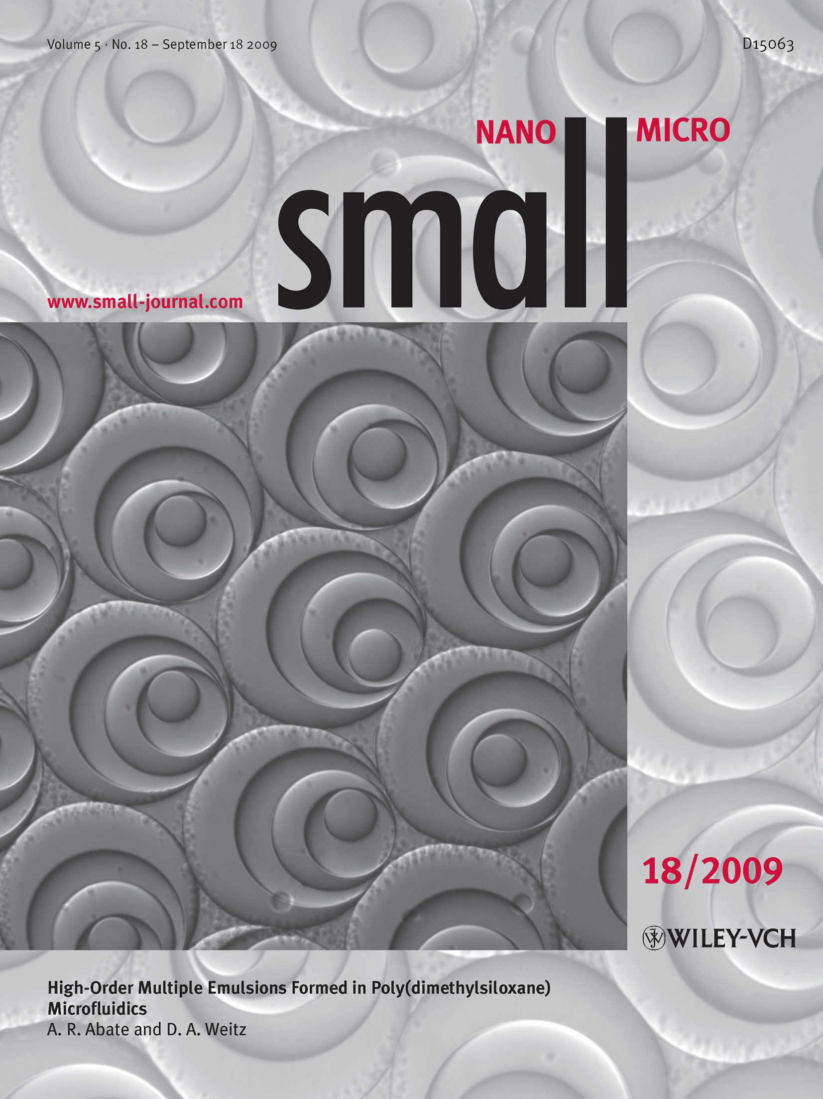
The cover picture shows an optical microscopy image of monodisperse W/O/W/O/W/O quintuple emulsions formed using a glass-coated poly(dimethylsiloxane) device. The device consists of five sequential drop makers functionalized to have alternating hydrophilic/hydrophobic wettability. The outermost drop diameter is about 120 micrometers. For more information, please read the Communication “High-Order Multiple Emulsions Formed in Poly(dimethylsiloxane) Microfluidics” by A. R. Abate and D. A. Weitz, beginning on page 2030.
Inside Cover
Protein nanotubes: Small 18/2009
- First Published: 14 September 2009

The cover picture shows a surface representation of a self-assembled nanotube made from a modified form of the ring-shaped bacterial protein, TRAP. The crystal structure of TRAP is shown as a backbone trace with each ring a different color to its neighbors. The tube-forming TRAP rings contain a small number of point mutations, which drive the self-assembly process. The resulting tubes are ≈8 nm in diameter and up to 1 µm or more in length, with a central cavity ≈2 nm in diameter. Since it is trivial to add further mutations to the protein surface, it is hoped that this tube may become a useful nanocomponent with a wide range of applications. For more information, please read the Full Paper “A Self-Assembled Protein Nanotube with High Aspect Ratio” by J. R. H. Tame, J. G. Heddle, et al., beginning on page 2077.
Contents
Communications
Multiple emulsions
High-Order Multiple Emulsions Formed in Poly(dimethylsiloxane) Microfluidics†
- Pages: 2030-2032
- First Published: 14 September 2009
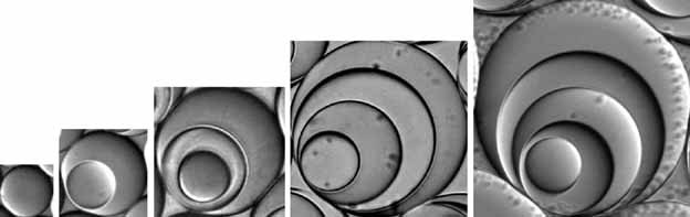
Multiple emulsions are formed using poly(dimethylsiloxane) microfluidic devices. The single emulsions (see image, left) are formed using a single drop maker with uniform wettability. The double, triple, quadruple, and quintuple emulsions (right) are formed using linear sequences of drop makers with alternating wettability.
Biomimetic nanoparticle synthesis
Nanoparticle Formation in Giant Vesicles: Synthesis in Biomimetic Compartments†
- Pages: 2033-2037
- First Published: 14 September 2009
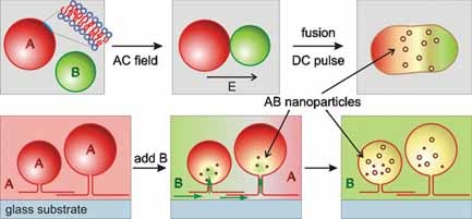
Nanoparticles of CdS with radii of 4 or 50 nm are formed in a controlled fashion inside lipid giant vesicles. For this purpose, two protocols are developed: electrofusion of differently loaded vesicles and slow vesicle content exchange via lipid nanotubes (see image). The process of particle formation can be directly monitored with fluorescence microscopy. The approach can be used to form any kind of nanoparticle.
Protein filaments
Biotemplated Metal Nanowires Using Hyperthermophilic Protein Filaments†
- Pages: 2038-2042
- First Published: 14 September 2009
Self-assembled hyperthermophilic γ-prefoldin protein filaments are used to template different metal nanoparticle wires. Filaments are uniformly decorated with small nanoparticles along the length of the protein and exhibit good conductivity behavior when aligned across an electrode gap.
Holey nanowires
Holey Gold Nanowires Formed by Photoconversion of Dissipative Nanostructures Emerged at the Aqueous–Organic Interface†
- Pages: 2043-2047
- First Published: 14 September 2009
Gold nanowires are obtained by photoreduction of linear self-assemblies formed from Au(OH)4− and tetra-alkyl ammonium ions at the water–chloroform interface. They are dissipative nanostructures formed only under non-equilibrium conditions, which require continuous vectorial transport of ammonium ions across the interface. Nanocavities are observed in nanowires at almost regular intervals, which is a salient feature of dissipative structures.
Photonic crystals
Nanoparticle One-Dimensional Photonic-Crystal Dye Laser†
- Pages: 2048-2052
- First Published: 14 September 2009
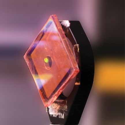
Nanoparticle one-dimensional photonic crystals (see image) possess high reflectivity arising from Bragg diffraction of light incident on a photonic lattice comprising nanoparticle layers of alternating refractive index. The nanoparticle layers provide mesoporosity, allowing for the introduction of a variety of functional molecules and materials into the intrananoparticle voids, creating myriad opportunities for the development of new kinds of optical and optoelectronic devices.
Combinatorial libraries
Synthesis and Application of FRET Nanoparticles in the Profiling of a Protease†
- Pages: 2053-2056
- First Published: 14 September 2009
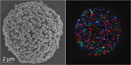
Fluorescent silica nanoparticles incorporating unique ratios of energy-transfer dyes are synthesized and applied as colloidal barcodes to encode a microsphere-bound combinatorial peptide library. The affinity of the West Nile virus protease is profiled using this library with cleavage of the peptide detected by flow cytometry. The cleaved peptide substrates are sorted and then identified through decoding by confocal microscopy combined with spectral unmixing (see image).
Hollow nanospheres
Solid-to-Hollow Single-Particle Manipulation of a Self-Assembled Luminescent NaYF4:Yb,Er Nanocrystal Monolayer by Electron-Beam Lithography†
- Pages: 2057-2060
- First Published: 14 September 2009
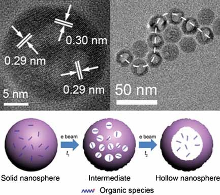
A hollow victory: Hollow-sphere nanocrystals are fabricated by electron-beam lithography in a self-assembled luminescent NaYF4:Yb,Er nanocrystal monolayer (see picture). The formation mechanism is a heat-induced inner acid-etching process. Pattern formation can be achieved with single-nanoparticle resolution. The 20-nm β-NaYF4:Yb,Er nanocrystal monolayer acts as a data-storage medium.
Nanotube arrays
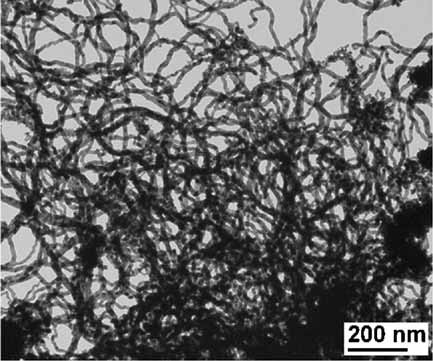
Facile “Needle-Scratching” Method for Fast Catalyst Patterns Used for Large-Scale Growth of Densely Aligned Single-Walled Carbon-Nanotube Arrays†
- Pages: 2061-2065
- First Published: 14 September 2009
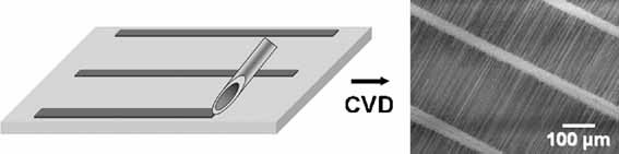
Scratching the surface: A simple needle-scratching method (NSM) generates large-area catalyst patterns on solid substrates for the growth of densely aligned single-walled carbon-nanotube (SWCNT) arrays by chemical vapor deposition (CVD, see picture). A high density of well-aligned, ultralong SWCNTs is obtained on single-crystal quartz. This NSM could allow the fast, cheap, and large-area fabrication of CNT-based nanodevices.
Frontispiece
Cytotoxicity: Small 18/2009
- First Published: 14 September 2009
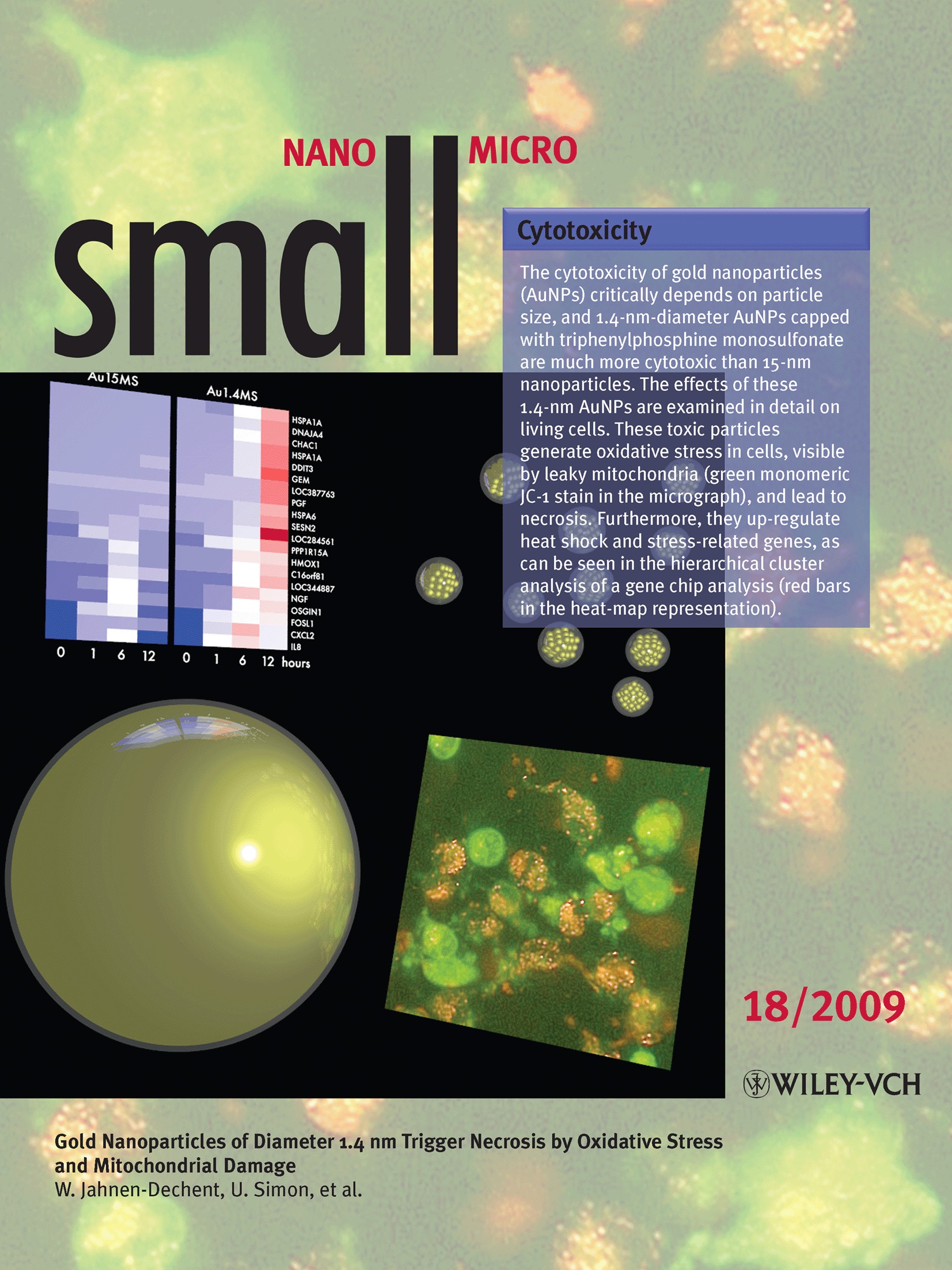
The frontispiece shows gold nanoparticles (AuNPs), the cytotoxicity of which critically depends on particle size, and 1.4-nm-diameter AuNPs capped with triphenylphosphine monosulfonate are much more cytotoxic than 15-nm nanoparticles. The effects of these 1.4-nm AuNPs are examined in detail on living cells. These toxic particles generate oxidative stress in cells, visible by leaky mitochondria (green monomeric JC-1 stain in the micrograph), and lead to necrosis. Furthermore, they up-regulate heat shock and stress-related genes, as can be seen in the hierarchical cluster analysis of a gene chip analysis (red bars in the heat-map representation). For more information, please read the Full Paper “Gold Nanoparticles of Diameter 1.4 nm Trigger Necrosis by Oxidative Stress and Mitochondrial Damage” by W. Jahnen-Dechent, U. Simon, et al., beginning on page 2067.
Full Papers
Cytotoxicity
Gold Nanoparticles of Diameter 1.4 nm Trigger Necrosis by Oxidative Stress and Mitochondrial Damage
- Pages: 2067-2076
- First Published: 14 September 2009
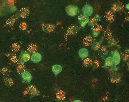
Cells set alight: Ultrasmall triphenylphosphine-capped gold nanoparticles continuously generate reactive oxygen species, which trigger leakiness of mitochondria and cell necrosis. This is visualized by discharge into the cytoplasm of green monomeric JC-1 stain from mitochondria (see picture), where it exists as a red dimer in undamaged cells.
Protein nanotubes
A Self-Assembled Protein Nanotube with High Aspect Ratio
- Pages: 2077-2084
- First Published: 14 September 2009
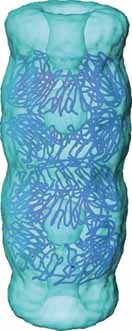
Using the X-ray crystal structure as a guide, the nanometric, ring-shaped protein TRAP can be modified so that, upon addition of reducing agent, it can spontaneously form long, regular, nanotubes with a 2.5-nm cavity (see image), which may have a wide variety of nanotechnological applications.
Gene expression
In situ Visualization of Gene Expression Using Polymer-Coated Quantum–Dot–DNA Conjugates
- Pages: 2085-2091
- First Published: 14 September 2009
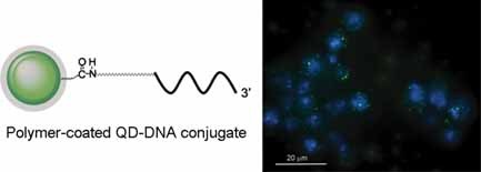
The selective binding of quantum-dot (QD)–DNA conjugates with their specific target sequence of various genes is demonstrated. Fluorescence in situ hybridization using QD-based probes shows the potential of detecting low-expressing genes often involved in various disease-related biological processes (see image). Quantitative image analysis of inducible genes shows good correlation with the quantification results obtained by real-time RT-PCR.
Helical nanostructures
Switchable Helical Structures Formed by the Hierarchical Self-Assembly of Laterally Tethered Nanorods
- Pages: 2092-2098
- First Published: 14 September 2009
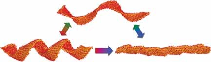
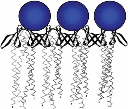
Flat bilayer sheets formed by the self-assembly of laterally tethered nanorod amphiphiles and their molecular analogs are predicted to fold into distinct helical structures depending on solvent conditions. Transformation between helical morphologies can be induced by switching solvent selectivity, as indicated by the color gradient in the arrows (see image).
Laser patterning
Direct Laser Patterning of Soft Matter: Photothermal Processing of Supported Phospholipid Multilayers with Nanoscale Precision
- Pages: 2099-2104
- First Published: 14 September 2009
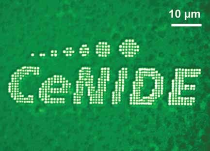
Photothermal laser processing is a facile, powerful tool for rapid large-area micro- and nanopatterning of supported phospholipid membranes (see image). Despite a laser-spot diameter of a few micrometers, voids with diameters of 300 nm and below are fabricated. This opens up an avenue towards optical engineering of biointerfaces with subwavelength resolution.
Nanocrescent arrays
Parallel Preparation of Densely Packed Arrays of 150-nm Gold-Nanocrescent Resonators in Three Dimensions
- Pages: 2105-2110
- First Published: 14 September 2009
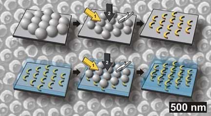
Fabrication of ordered three-dimensional arrangements of gold-nanocrescent resonators by repeated nanosphere lithography is demonstrated (see picture). The mutual orientation between the single layers can be adjusted to give chiral metamaterials.
Nanocube clusters
Plasmon Coupling in Clusters Composed of Two-Dimensionally Ordered Gold Nanocubes
- Pages: 2111-2119
- First Published: 14 September 2009
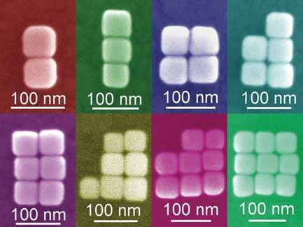
Gold nanocubes are assembled on substrates into clusters of varying numbers and ordering (see picture). Plasmon coupling among the gold nanocubes is investigated by dark-field scattering and electrostatic calculations. Gold-nanocube clusters exhibit various plasmon resonance modes, the energy and intensity of which depend on the number and ordering of nanocubes in the cluster.
Sign up for email alerts
Tools

Related Titles
- Advanced Materials
- Advanced Functional Materials
- Advanced Energy Materials
- Advanced Electronic Materials
- Advanced Engineering Materials
- Advanced Healthcare Materials
- Advanced Materials Interfaces
- Advanced Materials Technologies
- Advanced Optical Materials
- Advanced Physics Research
- Advanced Science
- Advanced Sensor Research
- Nano Select
- Particle & Particle Systems Characterization
- Small Methods
- Small Science
- Small Structures