

Journal list menu

 Issue
IssueJournal of Comparative Neurology: Volume 527, Issue 1
Retinal Special Issue I: Mammals
C1, 1-344January 1, 2019
Export Citations
Download PDFs
COVER IMAGE
Cover Image, Volume 527, Issue 1
- Page: C1
- First Published: 30 December 2018
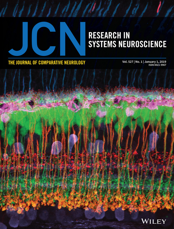
Cover image by Luca Della Santina, PhD. UCSF Ophthalmology. Vertical section of the adult mouse retina, labeled for the major neuronal cell types: Photoreceptors (cones in blue), Horizontal Cells (magenta), Cone bipolar cells (ON bipolars in green, OFF bipolars in red), Rod bipolar cells (orange), Amacrine and Ganglion cells (purple). Copyright 2019, Wiley Periodicals, Inc.
ISSUE INFORMATION - TOC
The Journal of Comparative Neurology, Table of Content, Vol. 527, No. 1, January 1, 2019
- Pages: 1-2
- First Published: 30 December 2018
EDITORIALS
Introduction to retinal special issue I
- Pages: 7-8
- First Published: 30 December 2018
Dedication of Retinal Special Issue to: Harvey J. Karten, M.D.
- Pages: 9-10
- First Published: 30 December 2018
OBITUARY
RESEARCH ARTICLES
Photoreceptor
Diversity of photoreceptor arrangements in nocturnal, cathemeral and diurnal Malagasy lemurs
- Pages: 13-37
- First Published: 05 January 2017
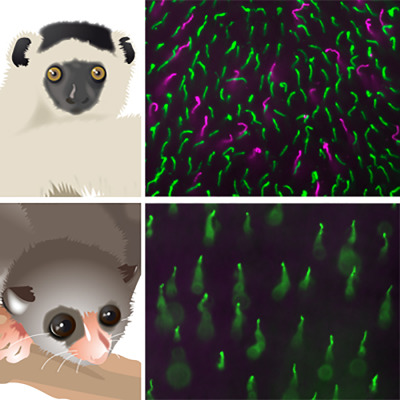
Retinal cone photoreceptor arrangements were studied in 11 species of Malagasy lemurs. The diversity of cone patterns ranges from relatively high cone densities and the presence of medium-to-longwave-sensitive (green) as well as shortwave-sensitive cones (magenta) in the diurnal sifaka (top) to low cone densities and an absence of shortwave-sensitive cones in the nocturnal dwarf lemurs (bottom).
Development of cone photoreceptors and their synapses in the human and monkey fovea
- Pages: 38-51
- First Published: 11 January 2017
Using immunocytochemistry and electron microscopy at various gestational time points in human and monkey retinas, this study suggests an intrinsically programmed, nonsynchronous expression of molecules in cone synaptic development. Ribbon synapses at foveal cone pedicles are present at midgestation in monkeys, and perhaps also in humans.
Bipolar
Synaptogenesis and synaptic protein localization in the postnatal development of rod bipolar cell dendrites in mouse retina
- Pages: 52-66
- First Published: 25 May 2017
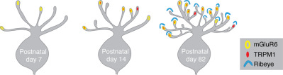
We examined the postnatal development, dendritic protein distribution and synaptogenesis of mouse rod bipolar cells by using 3D reconstructions of both conventional and high-resolution confocal images. We demonstrate that rod bipolar cells: (a) target the appropriate number of rods during development, (b) independently traffic mGluR6 and TRPM1 to dendrites, and (c) may connect to a larger number of rods than previously reported.
CNS synapses are stabilized trans-synaptically by laminins and laminin-interacting proteins
- Pages: 67-86
- First Published: 12 October 2017
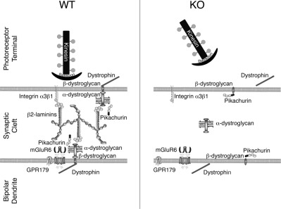
This study explores the roles of the extracellular matrix in the organization of the photoreceptor synapse. In wild-type (WT) retina, proteins associated with the release mechanism in the photoreceptor terminal (kinesin and bassoon) are apposed to components of the post-synaptic signaling cascade (mGluR6, GPR179) in the bipolar dendrite. The extracellular complex (including laminins and pikachurin) interacts with putative receptors (integrins, α- and β- dystroglycan). In the absence of laminin β2 (KO), this trans-synaptic organization is disrupted and the scaffolding of the post-synaptic cascade is disorganized, resulting in decreased signaling between photoreceptors and depolarizing bipolar cells.
Amacrine
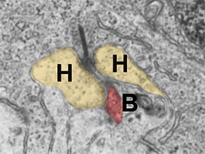
Rod-cone crossover connectome of mammalian bipolar cells
- Pages: 87-116
- First Published: 22 July 2016
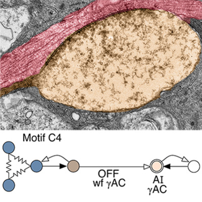
Ultrastructural and network graph visualization allows mapping of amacrine cell motifs whereby cone bipolar cells inhibit rod bipolar cells (C motifs) and vice versa (R motifs). Many crossover synapses are exceptionally large, which may be a signature for synaptic winner-take-all switches.
Selective synaptic connections in the retinal pathway for night vision
- Pages: 117-132
- First Published: 30 August 2017
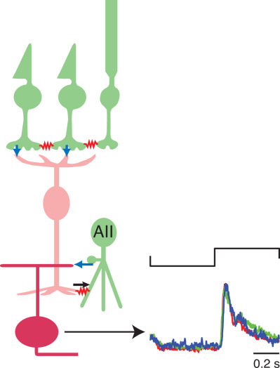
This study examined synaptic connections between retinal ganglion cells and the AII amacrine cell, which is a component of the night vision circuit in mammalian retina (rods → rod bipolar cells → AII amacrine cell). Using a physiological assay, combined with morphological analysis, we show that a direct inhibitory synapse from AII amacrine cells is limited to a privileged minority of ganglion cell types. More generally, co-stratification did not reliably predict synaptic connection in the inner plexiform layer.
Multiple cell types form the VIP amacrine cell population
- Pages: 133-158
- First Published: 04 May 2017

VIP-Confetti fluorescent amacrine cell types in the VIP-Confetti (Brainbow2.1) retina and their stratification. (a, c, d) VIP-1 cells. (b, e, f) VIP-2 cells. VIP cells are in magenta and calretinin-immunoreactive cells are in green. Scale bar: 25 μm.
Amacrine cells coupled to ganglion cells via gap junctions are highly vulnerable in glaucomatous mouse retinas
- Pages: 159-173
- First Published: 13 July 2016
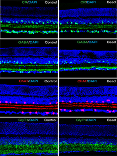
In this study we show that the structural and functional integrity of retinal amacrine cells are compromised in a mouse model of glaucoma. Specific immunocytochemical markers were used to identify amacrine cell subpopulations in both the inner nuclear and ganglion cell layers. Calretinin- and GABA- immunoreactive cells were highly vulnerable to glaucomatous damage, whereas ChAT-positive and glycinergic amacrine cell subtypes were largely unaffected. This difference between amacrine cell subtypes appears to be related to their gap junctional coupling patterns. Amacrine cells that are coupled to ganglion cells were lost in glaucoma, whereas uncoupled amacrine cells were not. Our results suggest that amacrine cell loss in glaucoma occurs secondary to ganglion cell death through the gap junction–mediated bystander effect.
Development of ON and OFF cholinergic amacrine cells in the human fetal retina
- Pages: 174-186
- First Published: 06 February 2018
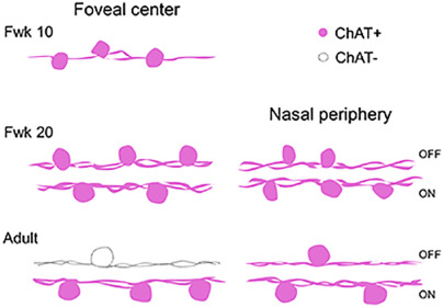
Human cholinergic (ChAT) amacrine cells are first observed in the fovea at early-gestation around fetal week (Fwk) 10, and become evident across the retina with age. Characteristic ON and OFF populations of ChAT cells with processes stratifying in separate bands in the synaptic neuropil emerge in the fovea by Fwk 11, and are present as late as mid-gestation (Fwk 20). Previous work, however, demonstrate very few, if any, OFF ChAT-immunoreactive cells in the adult human fovea (Rodieck & Marshak, ). Thus, mechanisms must act to regulate ChAT expression differentially in the ON and OFF populations as the retina matures beyond mid-gestation.
RGCs
Development
Brn3a and Brn3b knockout mice display unvaried retinal fine structure despite major morphological and numerical alterations of ganglion cells
- Pages: 187-211
- First Published: 08 July 2016
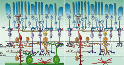
The retina constitutes a highly organized, multilayered outpost of the CNS (left). Individual cell types commit to their fate and migrate to their final locations at different stages during development and within precise time frames. In this study we show that, even though ganglion cells are the first neurons to form and to assume their final location in the retina, a severe decrease in their number and major abnormalities in their dendritic arbors affect minimally the remaining retinal cells types, including their major synaptic partners (i.e., bipolar and amacrine cells) (right). Failure of proper ganglion cell development appears to have negligible consequences on overall retinal layering and synaptic architecture, suggesting that these retinal features follow intrinsic, hard-wired rules.
Diversity
Distinct timing of neurogenesis of ipsilateral and contralateral retinal ganglion cells
- Pages: 212-224
- First Published: 15 May 2018
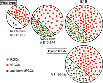
With specific markers and birthdating methods (EdU), we charted the neurogenesis of ipsi- and contralateral RGCs. Ipsi RGC neurogenesis lags behind that of contra RGCs elsewhere in the retina but is similar to contra RGCs in ventrotemporal retina. Cyclin D2 is important for the production of RGCs from VT retina.
Expression of transcription factors divides retinal ganglion cells into distinct classes
- Pages: 225-235
- First Published: 12 January 2017
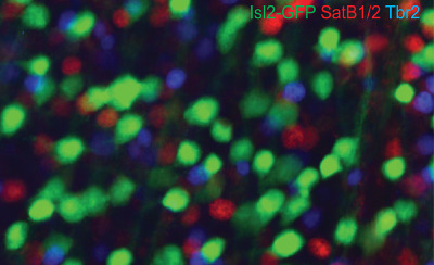
The authors show that in the mouse, retinal ganglion cells can be divided into three classes, each expressing just one of the transcription factors: Isl2, Tbr2 and SatB1 or SatB2. These classes emerge post-mitotically and cells from each class are born over highly overlapped periods of retinal development.
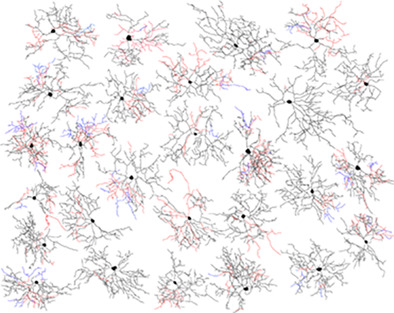
Survey of retinal ganglion cell morphology in marmoset
- Pages: 236-258
- First Published: 20 December 2016
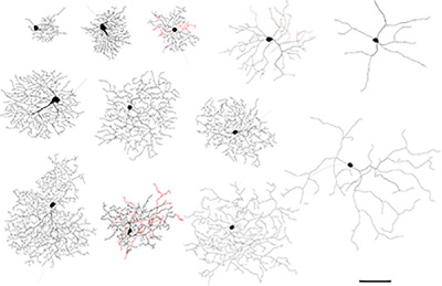
Over 17 types of ganglion cell were distinguished in marmoset retina. The study shows that the fovea is dominated by midget and parasol cells, but outside the fovea wide-field ganglion cell types make up a significant proportion of the total ganglion cell population. Scale bar = 100 μm.
Sub-topographic maps for regionally enhanced analysis of visual space in the mouse retina
- Pages: 259-269
- First Published: 20 April 2018
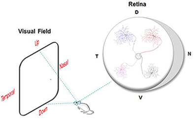
All visual information is processed through the activity of various subtypes of retinal ganglion cells (RGCs). As a prominent model for studying the visual system, the mouse retina was thought to encode visual space in a uniform manner. Here, we discovered that three morphologically and functionally distinct subtypes of RGCs show distinct retinotopic organizations. Therefore, signals located in different portions of the visual space are conveyed and analyzed differently in the brain.
Direction selectivity
Directional excitatory input to direction-selective ganglion cells in the rabbit retina
- Pages: 270-281
- First Published: 14 March 2017
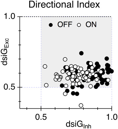
Directional tuning of excitatory currents recorded in direction-selective ganglion cells (DSGCs) is thought to be an artifact produced by the strong directional tuning of inhibitory inputs leading to directionally-asymmetric voltage-clamp errors. If this hypothesis is correct, then the strength of excitatory and inhibitory directional tuning should be positively correlated. The graph shows the results of experiments in a large sample of DSGCs that failed to detect the expected positive correlation.
Genetic access to neurons in the accessory optic system reveals a role for Sema6A in midbrain circuitry mediating motion perception
- Pages: 282-296
- First Published: 03 August 2018
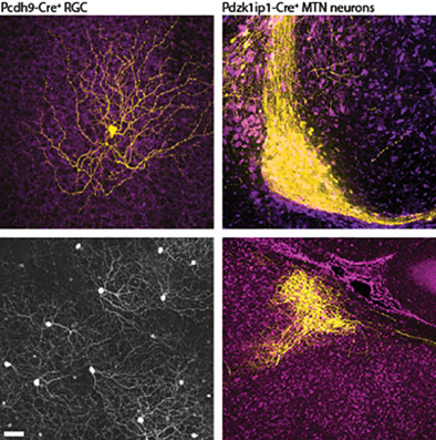
The authors characterize mouse lines labeling retinal ganglion cells and brain neurons in the Accessory Optic System. Pcdh9-Cre+ RGCs prefer ventral motion, target the medial terminal nucleus (MTN) and survive after optic nerve crush. Postsynaptic MTN targets of these RGCs require Sema6A to form connections with other AOS nuclei.
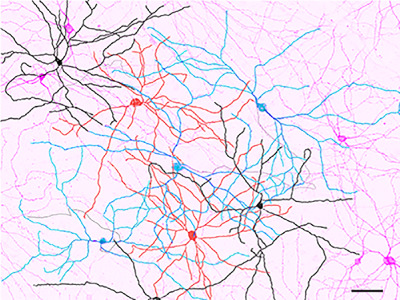
ipRGCs
The M6 cell: A small-field bistratified photosensitive retinal ganglion cell
- Pages: 297-311
- First Published: 12 October 2018
Please welcome the sixth member of the family of photosensitive retinal ganglion cells (ipRGCs) of the mouse retina, named the M6 cell. This cell is morphologically unique among ipRGCs with a small diameter, highly branched, spiny, bistratified dendritic arbor (ON-black; OFF-red), with frequent appearance of recursive “diving” dendrites (blue).
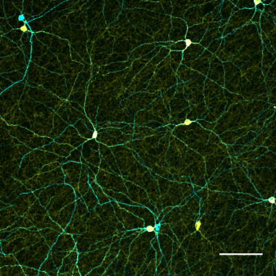
Melanopsin-expressing ganglion cells in human retina: Morphology, distribution, and synaptic connections
- Pages: 312-327
- First Published: 18 January 2017
Using immunohistochemistry and confocal microscopy the authors describe two morphological types of melanopsin-expressing cells with overlapping dendritic fields across human retina. Scale bar = 100 ?m.
Distribution and diversity of intrinsically photosensitive retinal ganglion cells in tree shrew
- Pages: 328-344
- First Published: 14 December 2017
We identify and distinguish multiple types of intrinsically photosensitive retinal ganglion cells in tree shrew. The tree shrew is a highly visual diurnal mammal with a cone-dominated retina, and is an evolutionary intermediate between rodents and primates. Thus, these animals provide insight in the evolution of mammalian and primate vision. One population of melanopsin expressing cells (cyan in accompanied image) also expresses tyrosine hydroxylase (yellow). This expression pattern suggests an intrinsically photosensitive ganglion cell that is dopaminergic.