Journal list menu

 Issue
IssueJournal of Biophotonics: Volume 10, Issue 1
3-165January 2017
Export Citations
Download PDFs
Cover Pictures
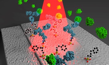
Front Cover: Three-dimensional mapping of the orientation of collagen corneal lamellae in healthy and keratoconic human corneas using SHG microscopy (J. Biophotonics 1/2017)
- First Published: 18 January 2017
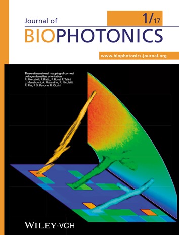
Graphic composition representing a three-dimensional view of corneal structural lamellae with different axial orientation, realized by properly re-slicing the portion of corneal tissue scanned by second-harmonic generation microscopy. The sutural lamellae have been placed onto an image representing their axial orientation in a colorcoded scale. The same color-coded scale has been used for coloring also the background rainbow and the depicted lamellae, which are represented with their real measured orientation.
Further details can be found in the article by Raffaella Mercatelli et al. on pp. 75–83.
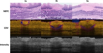
Back Cover: Laser thermal therapy monitoring using complex differential variance in optical coherence tomography (J. Biophotonics 1/2017)
- First Published: 18 January 2017

The cover image demonstrates the direct, label-free visualization of laser thermal coagulation in the esophagus using dynamic OCT measurements based on complex differential variance (CDV), potentially enabling microscopic image-guided procedures in numerous epithelial applications. The temporal evolution of the expanding thermal lesion, confirmed by NBTC histology (top panel), is clearly delineated using instantaneous CDV and the history of CDV values (middle panels), which is difficult to visualize using conventional intensity OCT (bottom panel).
Further details can be found in the article by William C. Y. Lo et al. on pp. 84–91.
Issue Information
Contents
Editorial
2017 – One Decade of Journal of Biophotonics
- Pages: 9-10
- First Published: 18 January 2017
Review Articles
Fluorescent probes for nanoscopy: four categories and multiple possibilities
- Pages: 11-23
- First Published: 25 May 2016
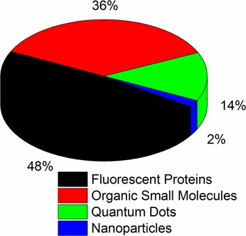
Nanoscopy enables breaking down the light diffraction limit and reveals the nanostructures of objects being studied using light. In 2014, three scientists pioneered the development of nanoscopy and won the Nobel Prize in Chemistry. This recognized the achievement of the past twenty years in the field of nanoscopy. However, fluorescent probes used in the field of nanoscopy are still numbered. Here, we review the currently available four categories of probes and existing methods to improve the performance of probes.
Three-dimensional imaging technologies: a priority for the advancement of tissue engineering and a challenge for the imaging community
- Pages: 24-45
- First Published: 25 April 2016
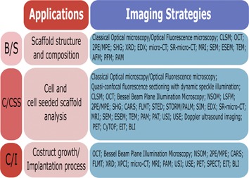
Tissue engineering/regenerative medicine is an interdisciplinary field that applies engineering and life sciences principles to restore/replace damaged tissues/organs with in vitro artificially-created ones. Imaging techniques are fundamental tools for tissue engineering products to monitor morphological, functional and molecular features, to analyze their pre-implantation quality assessment and their fate after implantation. This review focuses on imaging technologies developments and applications describing strengths and weaknesses of the current imaging approaches.
Visualizing liver anatomy, physiology and pharmacology using multiphoton microscopy
- Pages: 46-60
- First Published: 17 June 2016
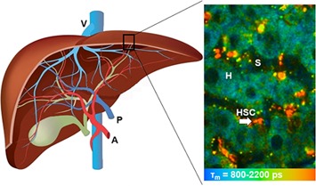
Multiphoton microscopy is a new tool which provides insights into deep live tissues to understand the cellular morphology, microenvironment, immune responses and spatiotemporal dynamics of drugs and therapeutic cells in the liver. This review summarizes its principles, opportunities, applications and limitations in hepatology. A key emphasis is on the use of fluorescence lifetime imaging to add additional quantification and specificity to the detection of fluorescent species in the liver.
Letters
Direct and label-free detection of the human growth hormone in urine by an ultrasensitive bimodal waveguide biosensor
- Pages: 61-67
- First Published: 27 September 2016
Graphical abstract of the hGH direct detection using a bimodal nanometric waveguide in which anti-hGH are immobilizing on by a silane coupling agent.
Bessel-beam illumination in dual-axis confocal microscopy mitigates resolution degradation caused by refractive heterogeneities
- Pages: 68-74
- First Published: 26 September 2016
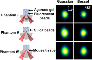
The propagation-invariant and self-reconstructing features of Bessel beams have been shown to benefit volumetric microscopy of tissues with refractive heterogeneities. This study examined the resolution degradation that occurs when performing dual-axis confocal (DAC) microscopy within tissues using Gaussian or Bessel illumination. Results indicate Bessel beams are useful for preserving resolution in DAC microscopy, in which tissue-imaging performance is sensitive to positional changes and distortions of off-axis illumination and collection beams.
Full Articles
Three-dimensional mapping of the orientation of collagen corneal lamellae in healthy and keratoconic human corneas using SHG microscopy
- Pages: 75-83
- First Published: 29 July 2016
Keratoconus is an ophthalmic disease consisting in an abnormal conical shape of cornea that causes visual impairment. A cross-correlation analysis performed on SHG images allowed diagnosing keratoconus against healthy cornea on the basis of the inclination of corneal lamellae. For the first time, the analysis was carried out in conjunction with epi-detection, paving the way for the ophthalmic clinical use of SHG microscopy.
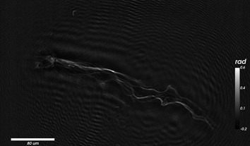
Laser thermal therapy monitoring using complex differential variance in optical coherence tomography
- Pages: 84-91
- First Published: 14 September 2016
Dynamic OCT measurements based on complex differential variance (CDV) are demonstrated to directly and accurately visualize the thermal coagulation zone during laser thermal therapy in 3 different tissue types: retina, skin, and esophagus. The ability to guide thermal therapy at high resolution opens up interesting opportunities for precisely targeting epithelial lesions in many clinical applications including ophthalmology, dermatology, gastroenterology and beyond.
Cancer cell identification by bi-color ZnO and TiO2 nanowires
- Pages: 92-97
- First Published: 08 January 2016
The microscopic image of the co-cultured SCC and HS68 cells which are applied with ZnO-EGFR and TiO2-Vimentin nanoprobes under the illumination of a laser. Among them, purple emission from ZnO, green emission from TiO2 can be seen. The position of SCC and Hs68 cells can be easily verified with the help of the two probes.
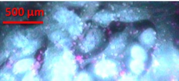
Real-time endoscopic Raman spectroscopy for in vivo early lung cancer detection
- Pages: 98-110
- First Published: 08 January 2016
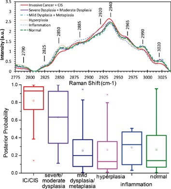
A custom-made endoscopic Raman spectroscopy system has been used to study the Raman spectroscopy properties of lung tissues in vivo in central airways of 80 patients. Multivariate statistical analysis of the acquired spectral data covering various pathologies including normal, inflammation, hyperplasia, metaplasia, mild/moderate/severe dysplasia, carcinoma-in-situ, and invasive cancer suggests that lung cancers and precancerous lesions can be differentiated from benign lesions and normal tissues with high sensitivity (90%) and good specificity (65%).
Quantitative phase measurements of tendon collagen fibres
- Pages: 111-117
- First Published: 29 January 2016
Quantitative phase imaging using digital inline holography (DIHM) allows the direct determination of the optical index of micron scale objects. Using this technique we measure the optical index of collagen fibres extracted from tendons in water. Heat exposure and tensile overload, both decrease the optical index of the fibres by 0.008 or less, demonstrating that DIHM can be used to quantify changes in collagen fibres a few microns in diameter.
High-content cell death imaging using quantum dot-based TIRF microscopy for the determination of anticancer activity against breast cancer stem cell
- Pages: 118-127
- First Published: 15 January 2016
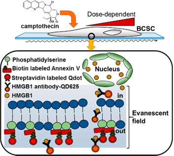
This study attempted a two color monitoring of drug-induced cell deaths using quantum dot probe-based total internal reflection fluorescence (TIRF) as a novel optical detection method to determine anticancer activity against breast cancer stem cell (BCSC). The apoptotic and necrotic BCSC deaths induced by camptothecin were concurrently monitored. Qdot-based TIRF as an improved alternative to MTT assay was successfully demonstrated for the quantitative determination of anticancer activity.
Identification of ex-vivo confocal laser scanning microscopic features of melanocytic lesions and their histological correlates
- Pages: 128-142
- First Published: 19 April 2016

The ex-vivo confocal laser scanning microscope allows rapid tissue examination. Along with its in-vivo counterpart it opens new horizons for diagnostic techniques in dermatology. In contrast to basal cell carcinoma diagnostics, the role of ex-vivo CLSM in melanocytic lesions remains unstudied. More experience is needed to characterize the main features of benign and malignant entities and their histopathological correlates. This study provides the first overview on this fast emerging topic.
Label-free in vivo cellular-level detection and imaging of apoptosis
- Pages: 143-150
- First Published: 19 April 2016
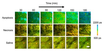
Apoptosis, or programmed cell death plays a critical role in health and homeostasis as well as in the treatment of many diseases. While this process has been characterized extensively in vitro, label-free detection of cells in vivo has yet to be shown. In this study, fluorescence lifetime imaging microscopy of reduced nicotinamide adenine dinucleotide is utilized to assess the metabolic response of in vivo mouse keratinocytes following induction of apoptosis.
Differentiating responses of lung cancer cell lines to Doxorubicin exposure: in vitro Raman micro spectroscopy, oxidative stress and bcl-2 protein expression
- Pages: 151-165
- First Published: 18 April 2016
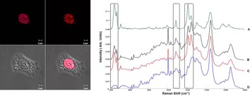
Raman microspectroscopy is employed to investigate and compare the in vitro mechanisms of action of Doxorubicin and cellular resistances of cancer cell lines A549 and Calu-1.
Results show the potential of Raman not only to distinguish the different mechanisms of action and subcellular response but also to elucidate drug resistance in the different cell lines.
Sign up for email alerts
Tools
More from this journal
Related Titles
 Volume 19, Issue 14July 22, 2025
 Volume 5, Issue 3-4August-November 2023
 Volume 13, Issue 21July 25, 2025