Cover Picture
Free Access
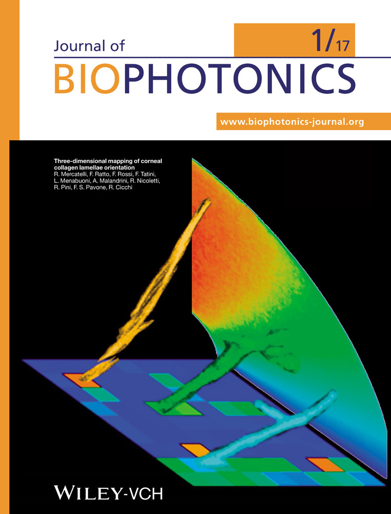
Front Cover: Three-dimensional mapping of the orientation of collagen corneal lamellae in healthy and keratoconic human corneas using SHG microscopy (J. Biophotonics 1/2017)
First published: 18 January 2017
Graphical Abstract
Graphic composition representing a three-dimensional view of corneal structural lamellae with different axial orientation, realized by properly re-slicing the portion of corneal tissue scanned by second-harmonic generation microscopy. The sutural lamellae have been placed onto an image representing their axial orientation in a colorcoded scale. The same color-coded scale has been used for coloring also the background rainbow and the depicted lamellae, which are represented with their real measured orientation.
Further details can be found in the article by Raffaella Mercatelli et al. on pp. 75–83.