High-content cell death imaging using quantum dot-based TIRF microscopy for the determination of anticancer activity against breast cancer stem cell
Abstract
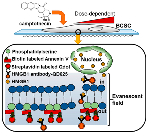
We report a two color monitoring of drug-induced cell deaths using total internal reflection fluorescence (TIRF) as a novel method to determine anticancer activity. Instead of cancer cells, breast cancer stem cells (CSCs) were directly tested in the present assay to determine the effective concentration (EC50) values of camptothecin and cisplatin. Phosphatidylserine and HMGB1 protein were concurrently detected to observe apoptotic and necrotic cell death induced by anticancer drugs using quantum dot (Qdot)-antibody conjugates. Only 50-to-100 breast CSCs were consumed at each cell chamber due to the high sensitivity of Qdot-based TIRF. The high sensitivity of Qdot-based TIRF, that enables the consumption of a small number of cells, is advantageous for cost-effective large-scale drug screening. In addition, unlike MTT assay, this approach can provide a more uniform range of EC50 values because the average values of single breast CSCs fluorescence intensities are observed to acquire EC50 values as a function of dose. This research successfully demonstrated the possibility that Qdot-based TIRF can be widely used as an improved alternative to MTT assay for the determination of anticancer drug efficacies.


