Journal list menu

 Issue
IssueJournal of Biophotonics: Volume 14, Issue 10
October 2021
Export Citations
Download PDFs
COVER PICTURE
Front Cover
- First Published: 04 October 2021
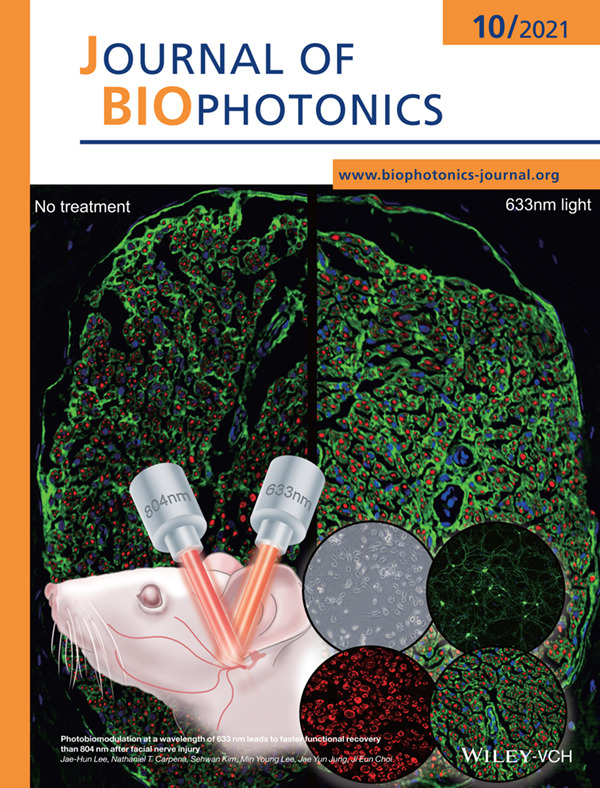
Photobiomodulation (PBM) therapy has been introduced as noninvasive treatment modality for peripheral nerve injuries. Despite numerous reports of the positive effect of PBM on peripheral nerve regeneration, there remains no agreement on the optimal parameters to support the best therapeutic activity in facial nerve regeneration. We assessed the effects of PBM of various wavelengths on cell viability under the oxidative stress and nerve regeneration after facial nerve injuries.
Further details can be found in the article by Jae-Hun Lee, Nathaniel T. Carpena, Sehwan Kim, Min Young Lee, Jae Yun Jung, and Ji Eun Choi (e202100159)
Inside Front Cover
- First Published: 04 October 2021
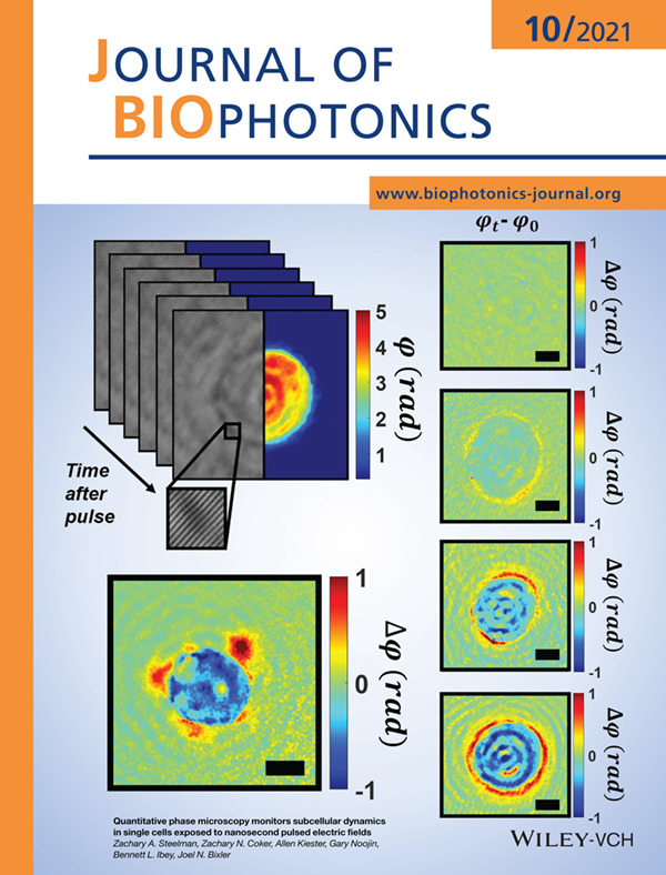
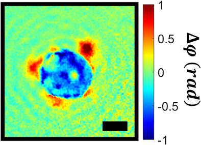
We introduce quantitative phase imaging (QPI) as a useful tool for the study of single cells undergoing cytoskeletal remodeling after nanosecond pulsed electric field (nsPEF) exposure. In particular, we use cell swelling, dry mass and disorder strength measurements derived from QPI phase images to monitor the cellular response to nsPEFs, augmenting more traditional fluorescence-based methods.
Further details can be found in the article by Zachary A. Steelman, Zachary N. Coker, Allen Kiester, Gary Noojin, Bennett L. Ibey, and Joel N. Bixler (e202100125)
ISSUE INFORMATION
LETTER
Intravascular molecular-structural imaging with a miniaturized integrated near-infrared fluorescence and ultrasound catheter
- First Published: 23 June 2021
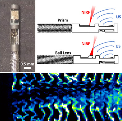
Coronary artery disease remains a leading cause of mortality and advanced in vivo imaging approaches are needed to enable better diagnosis for improved patient therapy and treatment. We demonstrate, characterize and compare optical prism and ball-lens sensor designs for a miniaturized hybrid intravascular ultrasound (IVUS) and near-infrared fluorescence (NIRF) catheter. In vivo experiments show co-registered molecular-structural information and suggest considerable potential for biological phenotyping of vascular disease.
RESEARCH ARTICLES
Photobiomodulation at a wavelength of 633 nm leads to faster functional recovery than 804 nm after facial nerve injury
- First Published: 12 July 2021
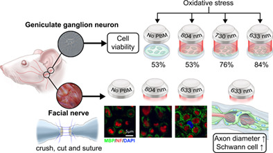
The 633-nm laser irradiation increases cell viability under oxidative stress, stimulates Schwann cells, induces axonal regeneration and consequently provides faster functional recovery of the facial nerve.
Quantitative phase microscopy monitors subcellular dynamics in single cells exposed to nanosecond pulsed electric fields
- First Published: 21 July 2021
In this work, we utilize quantitative phase imaging to study the bioeffects of nanosecond pulsed electric fields in single cells. In particular, the cell swelling, dry mass and disorder strength are monitored to observe changes in cell volume, membrane integrity and mechanobiology, respectively. We hope this pilot study will lead to further application of phase-based imaging tools for the study of electric pulse-induced biophysical change.
Sub millimetre flexible fibre probe for background and fluorescence free Raman spectroscopy
- First Published: 15 April 2021
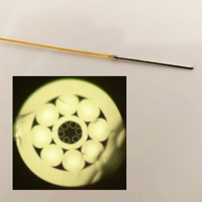
Using the shifted-excitation Raman difference spectroscopy technique and an optical fibre featuring a negative curvature excitation core and a coaxial ring of collection cores, we have developed a portable, background and fluorescence free, endoscopic Raman probe. The probe consists of a single fibre with a diameter of less than 0.25 mm packaged in a sub-millimetre tubing, making it compatible with standard bronchoscopes.
The measurement of ocular axial length in normal human eyes based on an improved Twyman-Green interferometer
- First Published: 22 June 2021
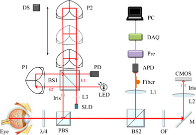
Schematic of the New AL based on improved Twyman-Green interferometer.
Deep-skin multiphoton microscopy in vivo excited at 1600 nm: A comparative investigation with silicone oil and deuterium dioxide immersion
- First Published: 23 June 2021
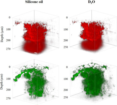
This study compares silicone oil and deuterium dioxide immersion in deep-skin MPM excited at the 1700-nm window. Silicone oil immersion yields higher signal levels, while maximum imaging depth and spatial resolution with both immersion media are similar. Besides, a feasible technique for characterizing spatial resolution in skin MPM in vivo is demonstrated, through local injection of fluorescent beads and subsequent MPM.
In vivo spectroscopic photoacoustic imaging and laser-induced nanoparticle vaporization for anti-HER2 breast cancer
- First Published: 09 July 2021
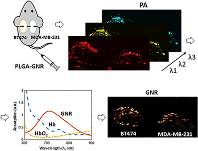
This study reports on the development and application of theragnostic agents targeting the HER2 receptors in breast tumors. Photoacoustic imaging and a linear spectral unmixing technique were performed to monitor the longitudinal nanoparticle uptake in tumors. The therapeutic efficacy of these nanoparticles was evaluated through optical droplet vaporization. Our results demonstrate the potential of these nanoparticles as photoacoustic imaging and therapeutic agents for anti-HER2 breast cancer in biomedical research.
Automatic lumen segmentation using uniqueness of vascular connected region for intravascular optical coherence tomography
- First Published: 29 June 2021
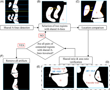
We present an automatic lumen segmentation method using uniqueness of connected region for intravascular optical coherence tomography (IVOCT), which can effectively remove the effect on lumen segmentation caused by blood artifacts. Utilizing the uniqueness of vascular wall on A-lines, we detect the A-lines shared by multiple connected regions, identify connected regions generated by blood artifacts using traversal comparison of connected regions' location, shared ratio and area ratio and then remove all artifacts.
Toward optical spectroscopy-guided lung biopsy: Demonstration of tissue-type classification
- First Published: 10 July 2021
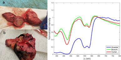
We investigated the feasibility of elastic scattering spectroscopy (ESS), coupled with machine learning, to distinguish lung lesions from the various nearby tissue types. Optical spectra were recorded with an ESS fiberoptic probe in different areas of freshly-excised lung tissues from surgical resections. A machine learning model was used to analyze, retrospectively, 2032 measurements from excised tissues of 35 patients. With high accuracy, ESS was able to distinguish alveolar tissue from bronchi, alveolar tissue from lesions, and bronchi from lesions.
Modeling the efficiency of UV at 254 nm for disinfecting the different layers within N95 respirators
- First Published: 29 June 2021
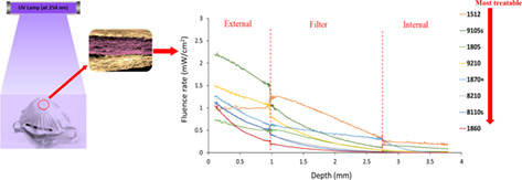
The study presented a Monte Carlo simulation of light transport in eight commonly used filtered facepiece respirators (FFRs) to assess the efficacy of UV at 254 nm for the inactivation of SARS-CoV-2. The results showed different fluence rates across the thickness of the eight different FFRs, implying that some FFR models may be more treatable than others.
High contrast imaging of dental fluorosis in the short wavelength infrared
- First Published: 24 July 2021
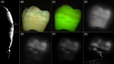
Dental fluorosis is an increasing problem due to over exposure to fluoride. The contrast of fluorosis of varying severity on extracted teeth was measured at SWIR wavelengths ranging from 400 to 2150-nm using an extended range of InGaAs camera and broadband light sources. The contrast of hypomineralization is significantly higher (P < 0.05) at 1460 and 1950 nm wavelengths than for the visible, fluorescence or other SWIR wavelengths. New clinical methods are needed for the measurement of fluorosis for surveillance at the community level.
In vitro evaluation of photodynamic activity of methylene blue against Trichophyton verrucosum azole-susceptible and -resistant strains
- First Published: 29 June 2021
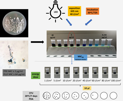
The search for the “Holy Grail” of antifungal therapy can be observed today. Interestingly, new therapeutic strategies can involve the use of chemosensitizers to achieve synergistic effect or physicochemical factors inducing stress conditions in fungal cells. In this study we examined in vitro antifungal effectiveness of photodynamic strategy with methylene blue using 635 nm light irradiation toward the T. verrucosum susceptible and itraconazole- and/or fluconazole-resistant strains.
Quantitative label-free optical technique to analyze the ultrastructure changes and spatiotemporal relationship of enamel induced by Msx2 deletion
- First Published: 09 July 2021
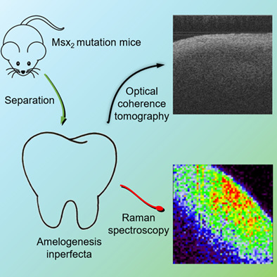
It is the first time that the structural difference of enamel mineralization imperfecta induced by gene defect was found by Raman spectroscopy and optical coherence tomography. And further improved the evaluation criteria of Raman spectroscopy and optical coherence tomography for biomineralization. Revealing the spatiotemporal difference of enamel mineralization caused by Msx2 gene knockout. This study proposes a set of optical analysis methods to solve the problem of enamel mineralization.
Sign up for email alerts
Tools
More from this journal
Related Titles
 Volume 19, Issue 14July 22, 2025
 Volume 5, Issue 3-4August-November 2023
 Volume 13, Issue 21July 25, 2025