

Journal list menu

 Issue
IssueEchocardiography: Volume 42, Issue 4
April 2025
Export Citations
Download PDFs
ISSUE INFORMATION
ORIGINAL ARTICLE
Meta-Analysis of the Association Between Left-Ventricular Late Gadolinium Enhancement on Cardiac MRI and Atrial Fibrillation in Patients With Hypertrophic Cardiomyopathy
- First Published: 28 March 2025
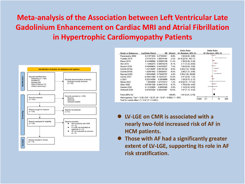
Meta-analysis of the association between left ventricular late gadolinium enhancement on cardiac MRI and atrial fibrillation in hypertrophic cardiomyopathy patients.
Impaired Aortic Biomechanical Properties in Patients With Severe Obstructive Sleep Apnea Syndrome
- First Published: 28 March 2025
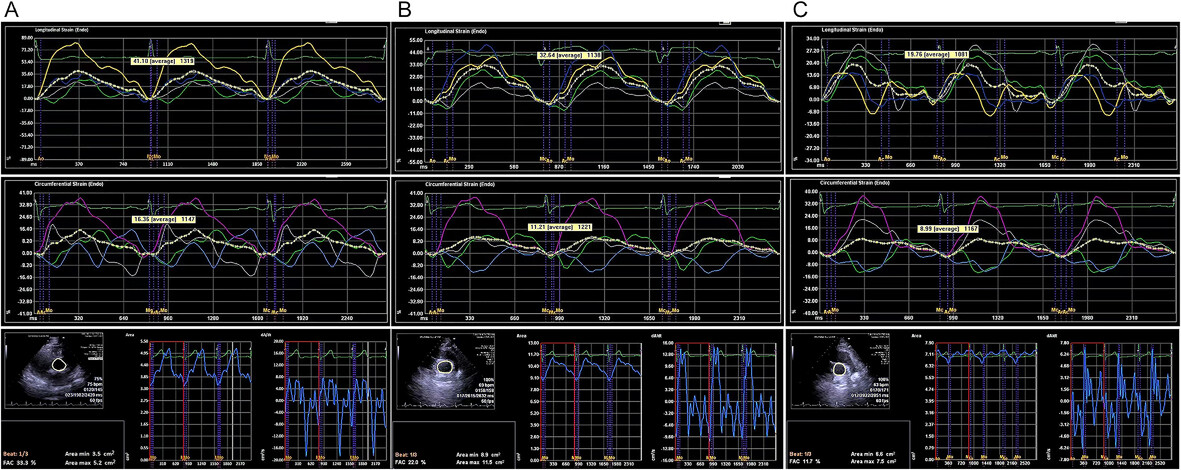
The longitudinal and circumferential strain curves are displayed in the segmental model with a specific color. The fractional area change was automatically calculated via VVI software. A, Control group; B, OSAS group; C, OSAS+HT group. ALS, ACS, and FAC were significantly lower in the patient groups (OSAS, OSAS + HT) than in the control group.
Impact of Various Doses of Dobutamine on Myocardial Viability and the Effects of β-Blockers in Patients With Ischemic Cardiomyopathy: An Assessment Using Stress Echocardiography
- First Published: 28 March 2025
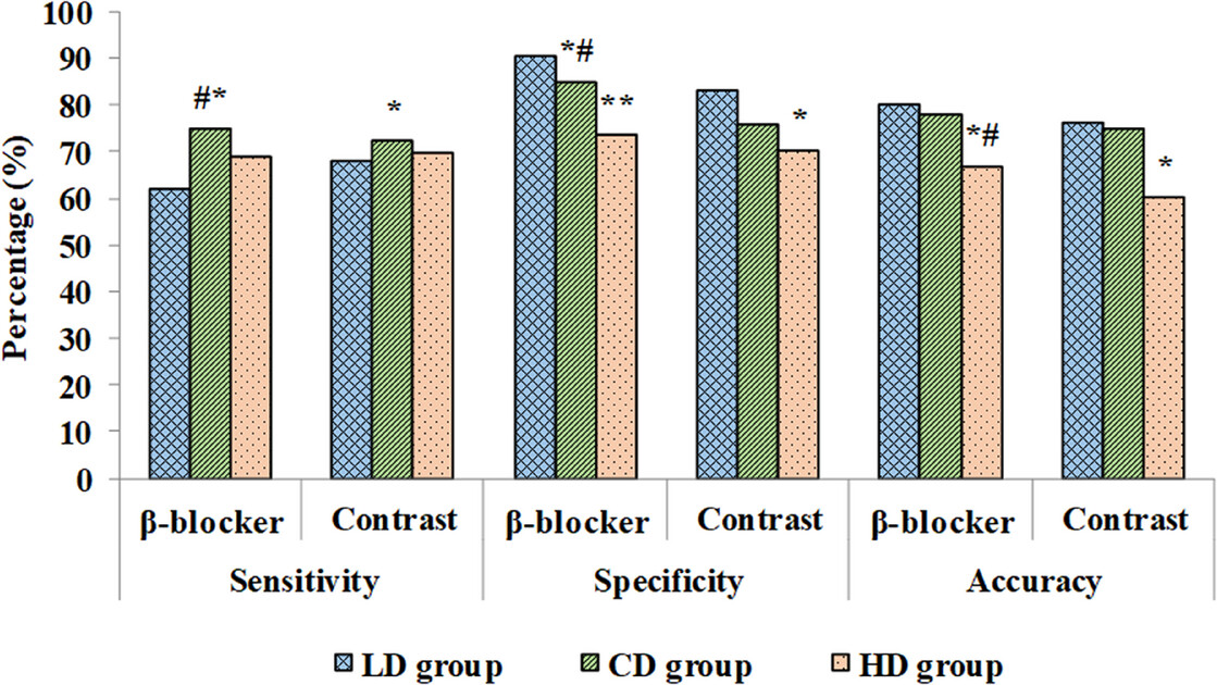
Conventional-dose dobutamine stress echocardiography (DSE) showed superior diagnostic accuracy for myocardial viability in ischemic cardiomyopathy compared to low-dose DSE, with β-blockers further enhancing diagnostic performance. Optimal DSE dosing improves patient assessment.
Clinical Study of Post-Chemotherapy Cardiotoxicity in Breast Cancer Patients Based on Ultrasound Radiomics
- First Published: 29 March 2025
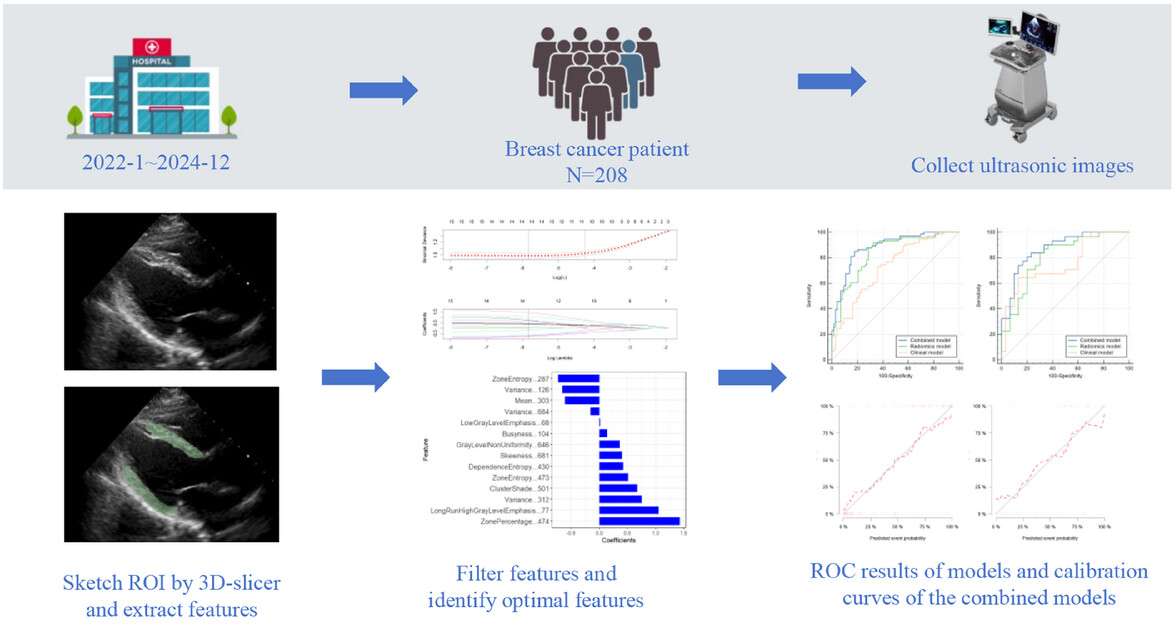
The aim of this study is to develop and validate a combined model based on ultrasound radiomics in order to detect early CTRCD. By analyzing detailed medical images, radiomics extracts quantitative features to identify subtle cardiac changes, improving early diagnosis.
COMMENTARY
Echocardiography and the Unmet Clinical Needs in Chronic Heart Failure
- First Published: 02 April 2025
CASE IMAGE
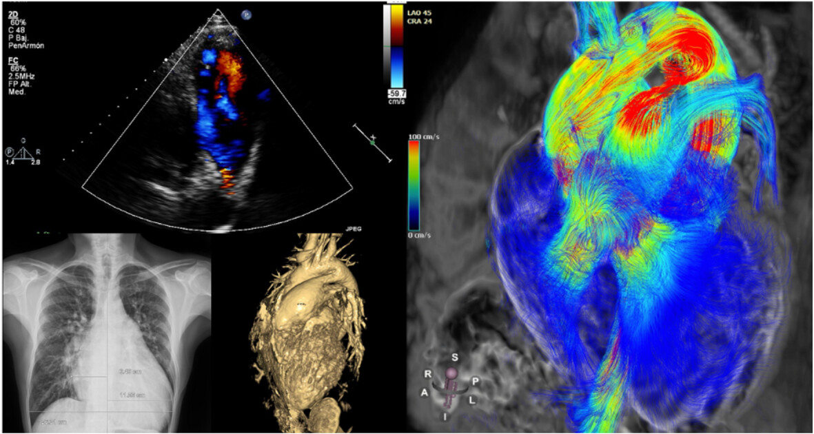
Congenital Atresia of the Left Main Coronary Artery: Multiple Imaging Diagnosis of a Rare Coronary Anomaly
- First Published: 29 March 2025
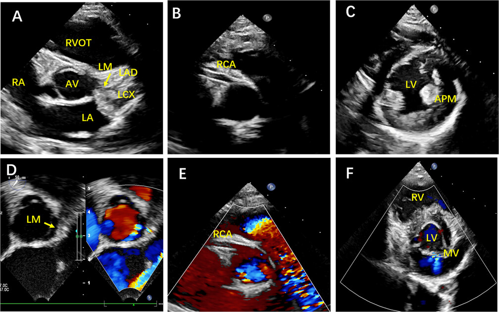
Left main coronary artery atresia (LMCAA) is a rare congenital coronary anomaly and sometimes presents with non-specific clinical symptoms that make the diagnosis challenging. We are presenting an interesting case that required multimodality imaging to establish the diagnosis.
Large Subcutaneous Mass Adjacent to the Left Ventricular Apex
- First Published: 02 April 2025
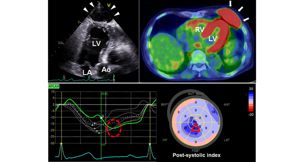
Two-dimensional speckle tracking helped determine whether the tumor adjacent to the left ventricular apex had infiltrated the pericardium. The peak strain value remains unaffected, although the peak timing in the area adjacent to the tumor is delayed. Consequently, the tumor was determined not to have infiltrated the pericardium.
Rheumatoid Inflammation Causing Aortic Valve Perforation
- First Published: 02 April 2025
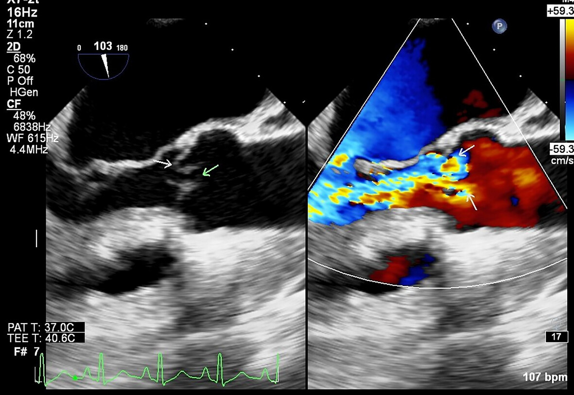
An elderly woman with rheumatoid arthritis presented with progressive dyspnea. Transoesophageal echocardiography revealed severe aortic regurgitation with perforation of the non-coronary and left coronary leaflet. After stabilization and a week of antibiotic therapy, aortic valve replacement was performed. The valve histology showed palisading granulomas typical of rheumatoid etiology.
COMMENTARY
How to Decide Right Heart Function After Left Ventricular Assist Device?
- First Published: 02 April 2025
REVIEW
Update on Imaging in Hypertrophic Cardiomyopathy
- First Published: 08 April 2025
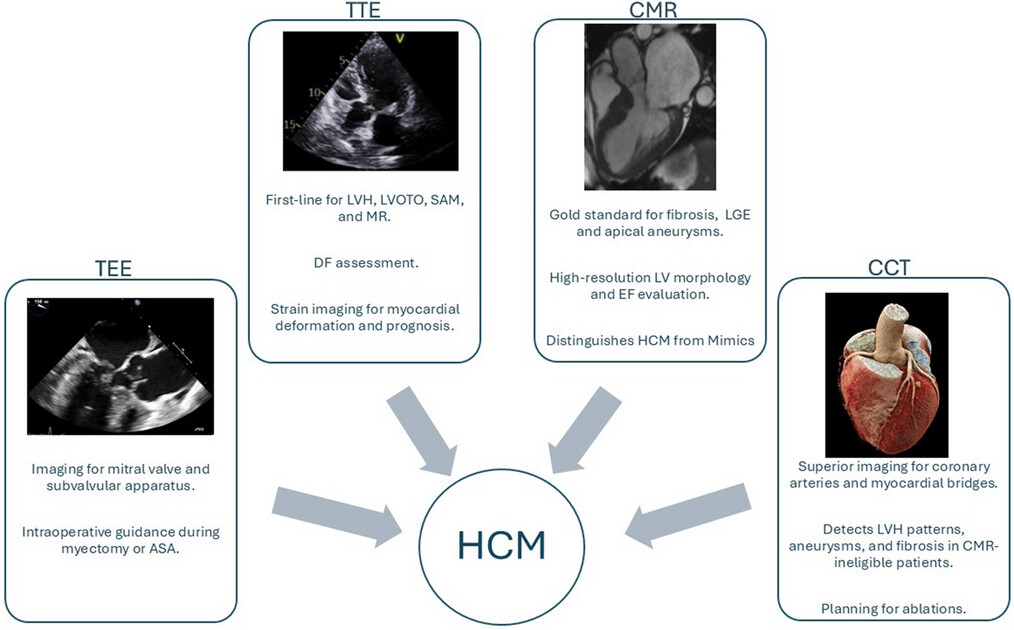
Comprehensive Imaging Modalities in HCM
Roles of different imaging modalities in HCM diagnosis and management. ASA, alcohol septal ablation; CCT, cardiac computed tomography; CMR, cardiac magnetic resonance imaging; DF, diastolic function; EF, ejection fraction; HCM, hypertrophic cardiomyopathy; LGE, late gadolinium enhancement; LVH, left ventricular hypertrophy; LVOTO, left ventricular outflow tract obstruction; MR, mitral regurgitation; SAM, systolic anterior motion; TEE, transesophageal echocardiography; TTE, transthoracic echocardiography .
Ablation of Ventricular and Atrial Arrhythmias in the Era of Cardiac Magnetic Resonance
- First Published: 07 April 2025
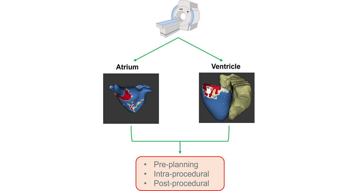
Cardiac magnetic resonance (CMR) has significantly advanced over recent decades, becoming indispensable in various settings. In electrophysiology (EP), CMR plays a pivotal role in procedural planning, real-time guidance, and post-procedural evaluation. Its precise definition of myocardial scarring tailor's ablation processes, while the possibility of crafting 3D models of the heart and merging them into the EP lab is emerging. Additionally, specialized sequences assess ablation outcomes and predict arrhythmia recurrence, highlighting its prognostic value.
ORIGINAL ARTICLE
Longitudinal Trajectory of Left Ventricular Systolic Function in Children During Anthracycline-Based Chemotherapy Assessed by Noninvasive Myocardial Work
- First Published: 07 April 2025
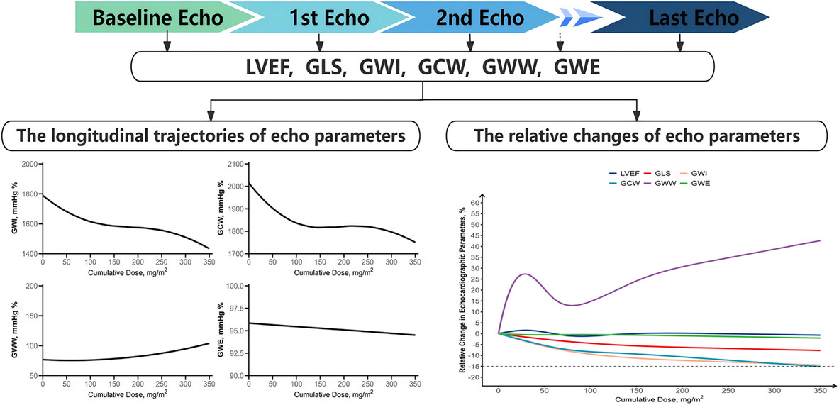
Myocardial work indices (GWI/GCW/GWE) detected cardiotoxicity earlier than LVEF in children receiving anthracyclines, with initial deterioration at 30–60 mg/m2. GWI and GCW showed the largest decline, plateauing at 100–250 mg/m2, while GLS and GWE declined consistently, highlighting myocardial work as a sensitive tool for assessing anthracycline-induced cardiotoxicity (AIC).
CASE IMAGE
Adult Patient With Systolic Murmur: An Unexpected Diagnosis of Congenital Heart Disease
- First Published: 07 April 2025
Unseen Window: Incidental Detection of Aortopulmonary Window in Tetralogy of Fallot: A Case Report
- First Published: 08 April 2025
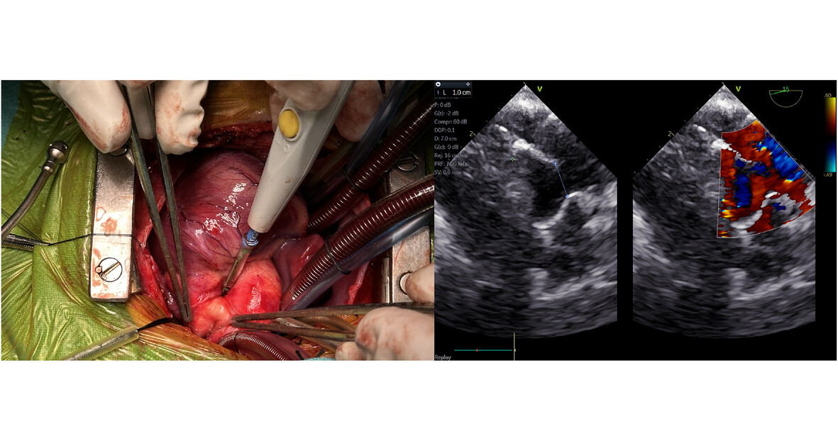
Image on the left shows the intraoperative image with the surgeon's electrosurgical unit pointing toward the aortopulmonary connection, and the corresponding image on the right is of the intraoperative transesophageal echocardiography showing the same communication, which has a width of 1 cm
REVIEW
Tricuspid Regurgitation: Knowledge of Tricuspid Valve Morphology, Etiology of Regurgitation, and Grading of Regurgitation Severity
- First Published: 08 April 2025
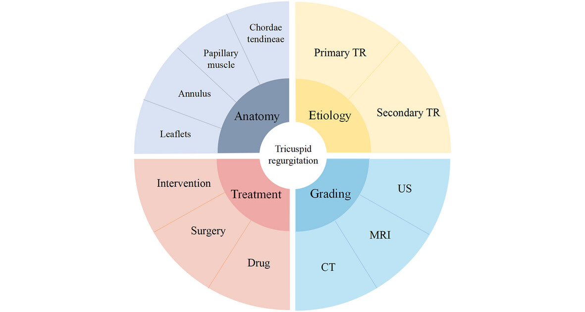
With the aging of the population, the number of patients with tricuspid regurgitation (TR) is increasing. Severe TR is associated with global morbidity and mortality of cardiovascular events. In recent years, the rapid development of transcatheter interventions for tricuspid valve disease has made TR a current research hotspot. More preoperative information about the patient's tricuspid valve anatomy, the etiology leading to TR, and the severity of TR will aid in intraoperative maneuvers and postoperative prognosis assessments.
ORIGINAL ARTICLE
Longitudinal Evaluation of Immune Checkpoint Inhibitor-Induced Fatigue Syndrome by Rest, Stress, and Speckle-Tracking Strain Echocardiography
- First Published: 08 April 2025
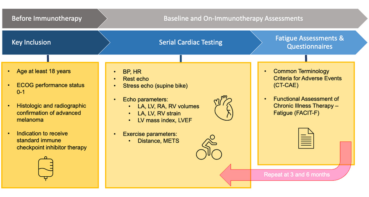
A prospective echocardiographic study monitored cardiac function following the initiation of immune checkpoint inhibitors in cancer patients. LVOT VTI and peak exercise stage achieved were reduced, but other biventricular structure/functional markers and LV/LA strain were unchanged .
COMMENTARY
What Is the Optimal Measurement Method for CT-Derived Fractional Flow Reserve?
- First Published: 08 April 2025
LETTER TO THE EDITOR
CASE IMAGE
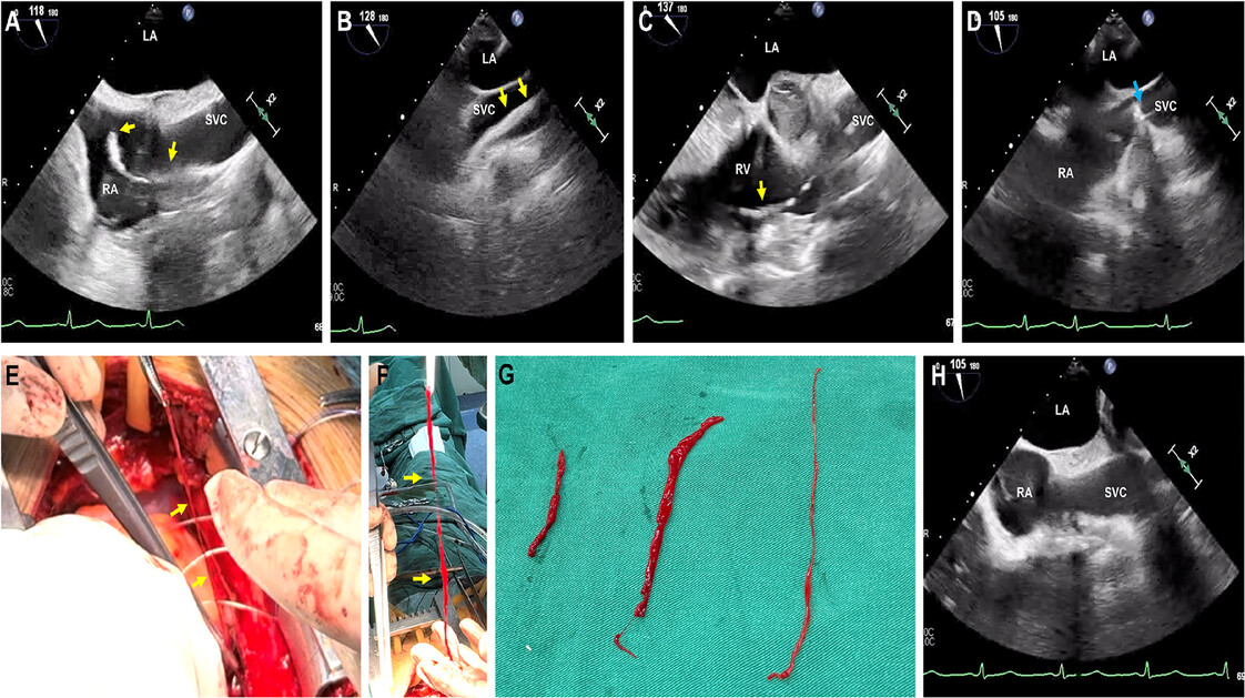
Cardiac Fresh Thrombus Following Cardiac Surgery: A Intraoperative Transesophageal Echocardiographic Diagnosis
- First Published: 13 April 2025
Pseudoaneurysms of the Ascending Aorta Following Coronary Artery Bypass Surgery, Presenting as Stroke
- First Published: 13 April 2025
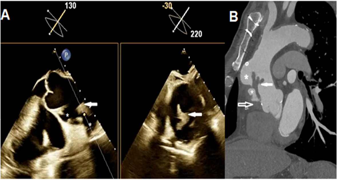
TEE simultaneous orthogonal long axis and short axis views and MDCT of ascending aorta sagittal view demonstrate two complicated pseudoaneurysm (*) in anterior wall of ascending aorta and intramural hematoma (open arrow) extending proximally to aortic root and a large thrombus (solid arrow) protruding into the lumen of ascending aorta
ORIGINAL ARTICLE
Comprehensive Quantification of Fetal Cardiac Function in Gestational Diabetes Mellitus Using Advanced Fetal HQ Imaging Techniques
- First Published: 11 April 2025
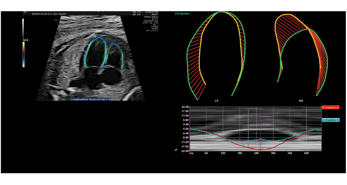
Gestational diabetes mellitus (GDM) may profoundly influence both the short-term and long-term health of mothers and their offspring. Maternal hyperglycemia in gestational diabetes impairs fetal left ventricular function, particularly myocardial deformation, as assessed by fetal HQ imaging.
COMMENTARY
Routine Echocardiographic Assessments of Single Ventricle Patients Should Include Atrial Strain
- First Published: 13 April 2025
ORIGINAL ARTICLE
Sex Differences in Diastolic Function Following Myocardial Infarction on Doppler Echocardiography
- First Published: 11 April 2025
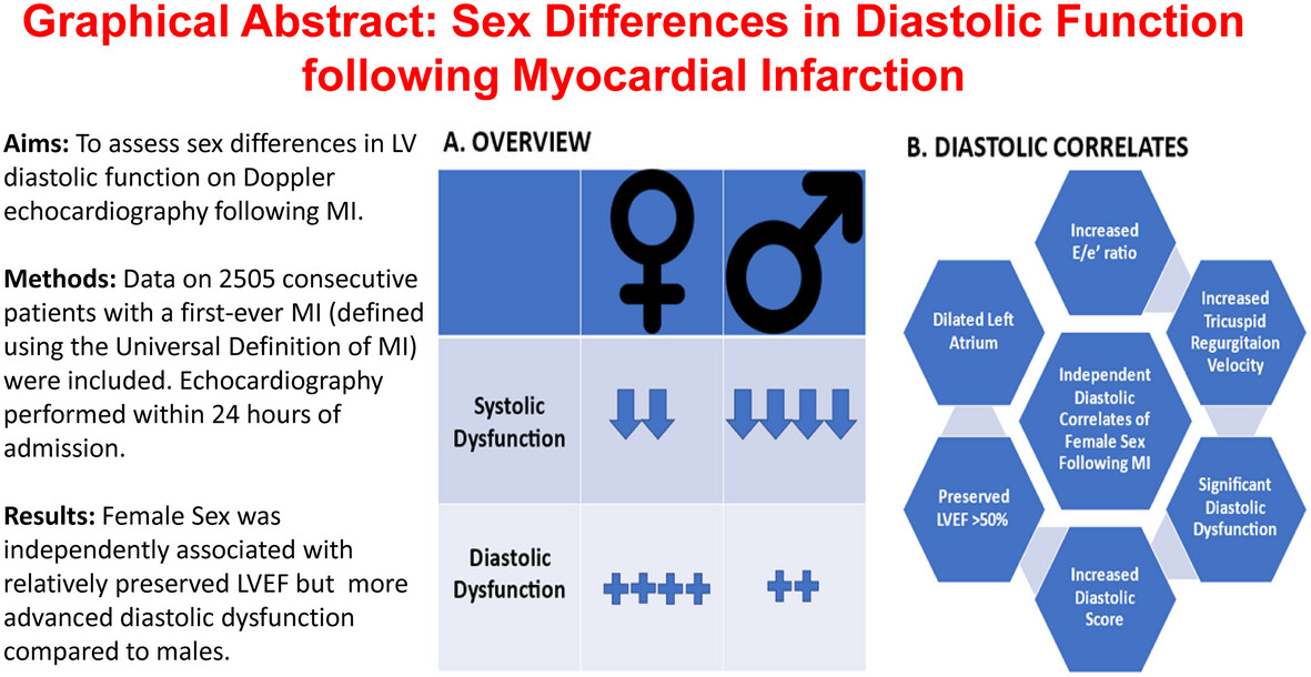
Study overview and main findings shown. The central figure illustrates the overall findings comparing systolic and diastolic function between sexes. The figure on the right shows a detailed breakdown of diastolic parameters that were independently associated with female sex.
Additional Predictive Importance of Middle Cerebral Artery, Umbilical Artery and Heart for Coarctation of the Aorta in Fetal Ultrasound Parameters
- First Published: 14 April 2025
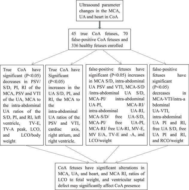
CoA fetuses have significant alterations in the ultrasound parameters of the MCA, UA, heart, and MCA RI, ratios of LCO to fetal weight, and ventricular septal defect may significantly affect CoA presence.
OPINION
CASE IMAGE
Incomplete Shone's Syndrome With Unicuspid Aortic Valve
- First Published: 22 April 2025
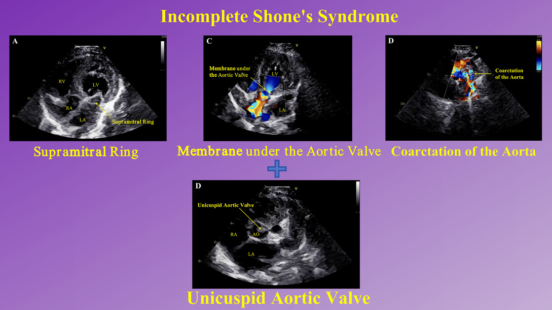
While this syndrome is associated with bicuspid aortic valve, here we report a case presenting with Shone's syndrome complicated by unicuspid aortic valve.
REVIEW
Before and Beyond Tissue Characterization: Cardiac Magnetic Resonance Imaging in the Morphological, Volumetric, and Functional Evaluation of the Right Ventricle in Arrhythmogenic Right Ventricular Cardiomyopathy, a Narrative Review
- First Published: 22 April 2025
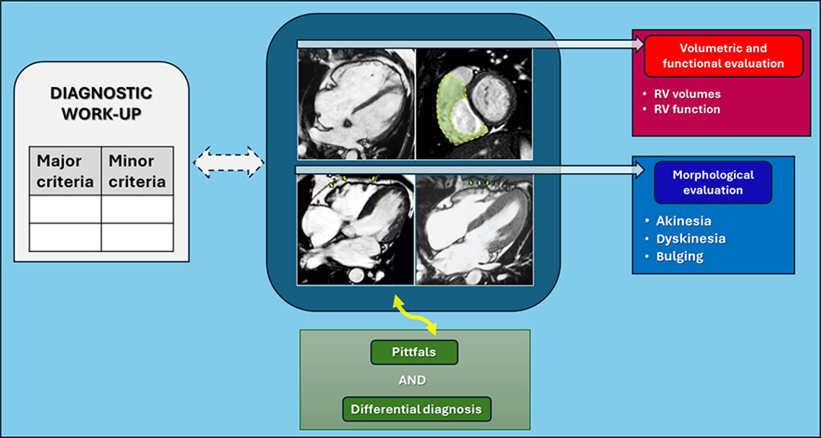
In patients with suspected arrhythmogenic right ventricular cardiomyopathy, evaluation of the right ventricle by cardiac magnetic resonance imaging includes volumetric, functional, and morphological studies. This is because much of the information obtained from magnetic resonance imaging constitutes some of the criteria required for the diagnostic work-up of the disease and must therefore be included and considered alongside all other criteria. It is essential to be mindful of technical and interpretative pitfalls and to always consider possible alternative diagnoses for an accurate differential diagnosis.
COMMENTARY
Is It Possible to Detect the Pathogenesis of Cardiac Amyloidosis by Echocardiography?
- First Published: 24 April 2025
REVIEW
Role of CT and CMR in the Management of Chronic Coronary Syndrome
- First Published: 24 April 2025
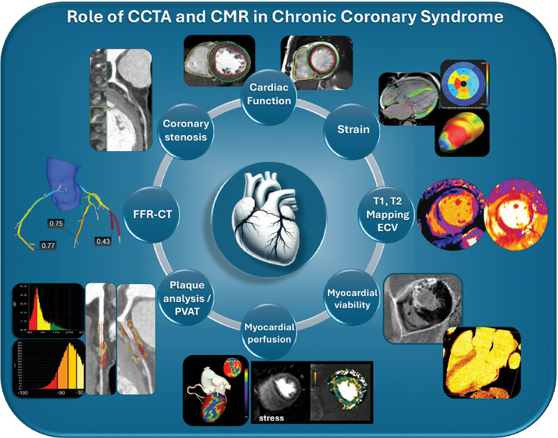
Advanced non-invasive imaging modalities, such as coronary computed tomography angiography (CCTA) and cardiac magnetic resonance (CMR), are pivotal in improving diagnostic precision and guiding personalized management strategies for patients with chronic coronary syndrome (CCS). These technologies enable comprehensive assessments, including coronary stenosis, plaque characteristics, perivascular adipose tissue (PVAT), CT-derived fractional flow reserve (FFR), cardiac function, strain imaging, parametric mapping (including extracellular volume [ECV]), tissue characterization, stress myocardial perfusion, and myocardial viability.
Cardiac Magnetic Resonance Imaging and Coronary Computed Tomography Angiography in Cardiomyopathy: Diagnostic and Prognostic Insights
- First Published: 22 April 2025
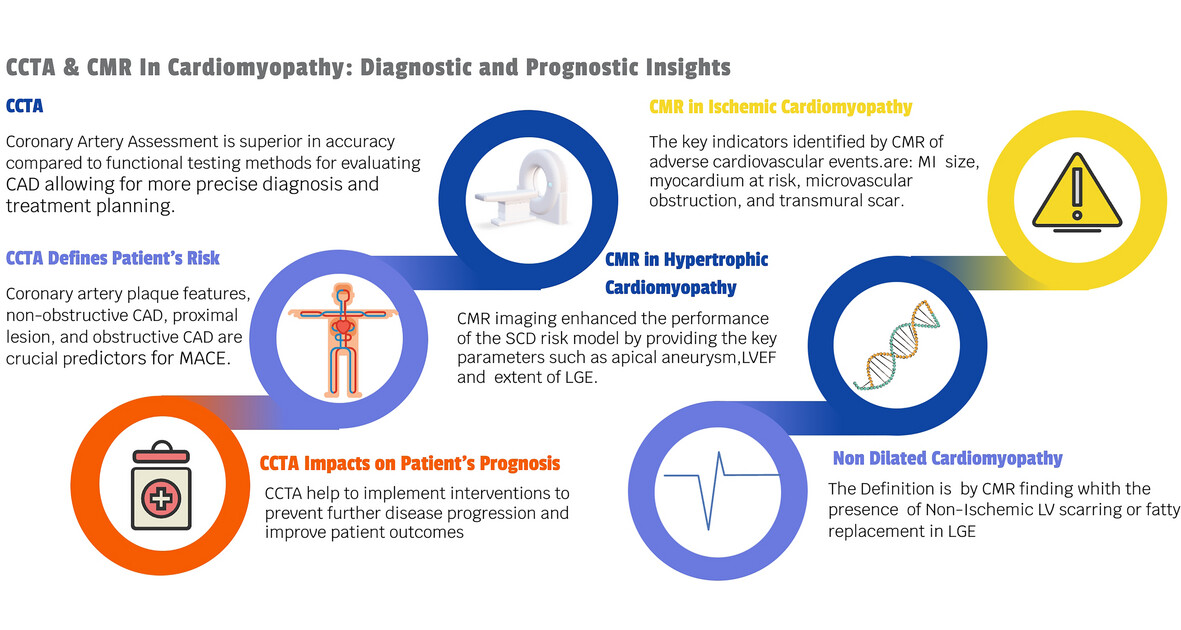
CCTA has demonstrated high sensitivity compared to functional testing modalities and a substantial negative predictive value. Identifying high-risk plaque features through CCTA guides the initiation of preventive therapy, which has proven effective for plaque regression, particularly with statin therapy. CCTA has shown that anatomical assessments, such as plaque location and coronary artery severity, correlate with MACE. Tissue characterization through CMR is crucial for identifying cardiomyopathy subtypes, as LGE may be the sole indicator of the condition and carries prognostic significance. CCTA, coronary computed tomography angiography; CMR, cardiac magnetic resonance; CAD, coronary artery disease; MACE, Major adverse cardiac events; LGE, Late Gadolinium enhancement; SCD, sudden cardiac death; LVEF, left ventricular ejection fraction.