Journal list menu

 Issue
IssueJournal of Magnetic Resonance Imaging: Volume 62, Issue 1
spcone, 1-306July 2025
Export Citations
Download PDFs
Cover Image
Presence of Fragmented Intratumoral Thrombosed Microvasculature in the Necrotic and Peri-Necrotic Regions on SWI Differentiates IDH Wild-Type Glioblastoma From IDH Mutant Grade 4 Astrocytoma
- Page: spcone
- First Published: 19 June 2025
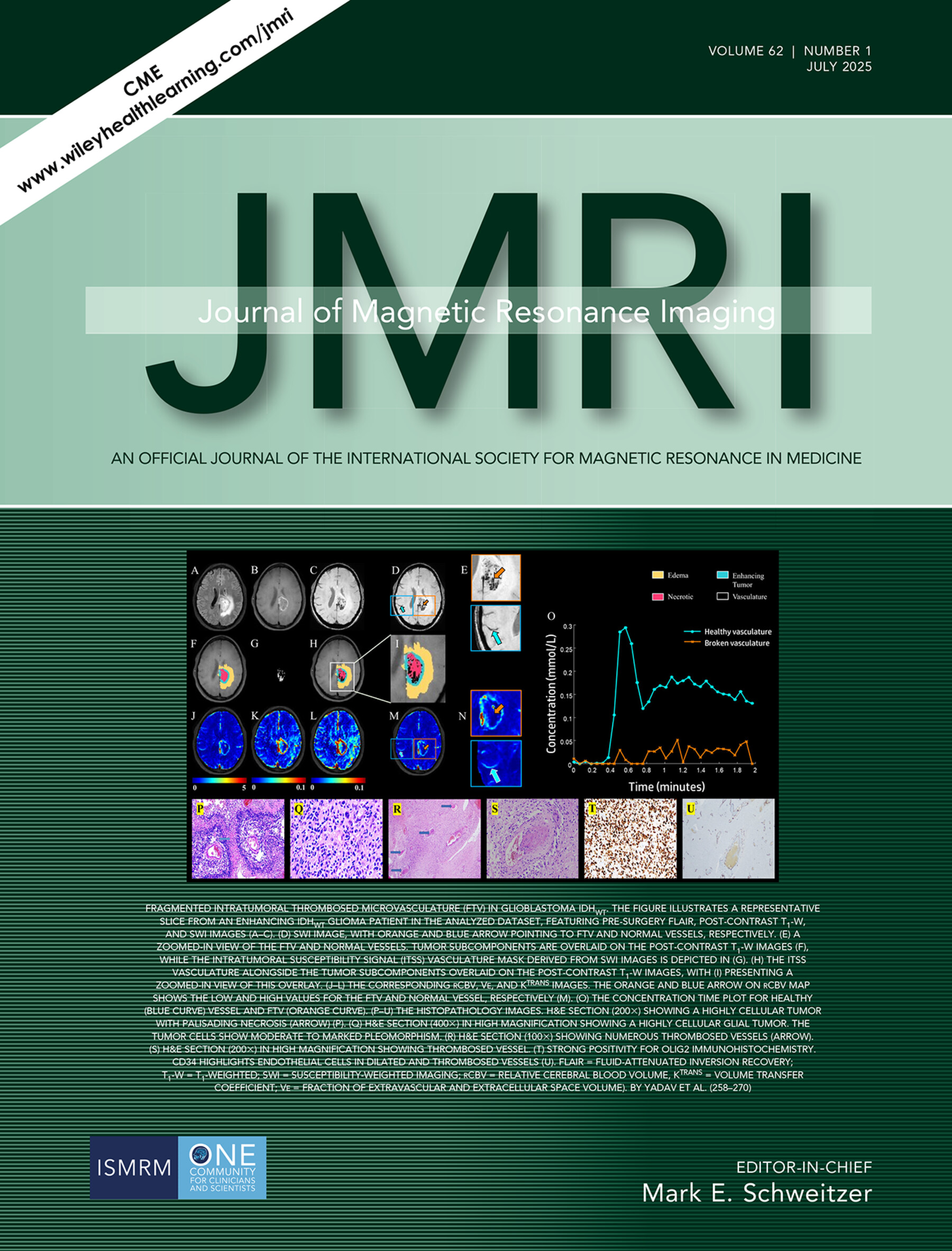
Fragmented Intratumoral Thrombosed Microvasculature (FTV) in Glioblastoma IDHWT. The Figure Illustrates a Representative Slice from an Enhancing IDHWT GLIOMA Patient in the Analyzed Dataset, Featuring Pre-Surgery Flair, Post-Contrast T1-W, and SWI Images (A-C). (D) SWI Image, with Orange and Blue Arrow Pointing to FTV and Normal Vessels, Respectively. (E) A Zoomed-In View of the FTV and Normal Vessels. Tumor Subcomponents are Overlaid on the Post-Contrast T1-W Images (F), While the Intratumoral Susceptibility Signal (ITSS) Vasculature Mask Derived from SWI Images is Depicted in (G). (H) The ITSS Vasculature Alongside the Tumor Subcomponents Overlaid on the Post-Contrast T1-W Images, with (I) Presenting a Zoomed-In View of this Overlay. (J-L) The Corresponding rCBV, Ve, and Ktrans Images. The Orange and Blue Arrow on rCBV Map Shows the Low and High Values for the FTV and Normal Vessel, Respectively (M). (O) The Concentration Time Plot for Healthy (Blue Curve) Vessel and FTV (Orange Curve). (P-U) The Histopathology Images. H&E Section (2003) Showing a Highly Cellular Tumor with Palisading Necrosis (Arrow) (P). (Q) H&E Section (4003) In High Magnification Showing a Highly Cellular Glial Tumor. The Tumor Cells Show Moderate to Marked Pleomorphism. (R) H&E Section (1003) Showing Numerous Thrombosed Vessels (Arrow). (S) H&E Section (2003) In High Magnification Showing Thrombosed Vessel. (T) Strong Positivity for OLIG2 Immunohistochemistry. CD34 Highlights Endothelial Cells in Dilated and Thrombosed Vessels (U). Flair = Fluid-Attenuated Inversion Recovery; T1-W = T1-Weighted; SWI = Susceptibility-Weighted Imaging; rCBV = Relative Cerebral Blood Volume, KTRANS = Volume Transfer Coefficient; Ve = Fraction of Extravascular and Extracellular Space Volume). By Yadav et al. (258-270)
Issue Information
Review
Multiparametric MRI for Assessment of the Biological Invasiveness and Prognosis of Pancreatic Ductal Adenocarcinoma in the Era of Artificial Intelligence
- Pages: 9-19
- First Published: 09 January 2025
Magnetic Resonance Acoustic Radiation Force Imaging (MR-ARFI)
- Pages: 20-39
- First Published: 22 January 2025
Visualizing Preosteoarthritis: Updates on UTE-Based Compositional MRI and Deep Learning Algorithms
- Pages: 40-57
- First Published: 10 January 2025
Breast
Breast MRI to Screen Women With Extremely Dense Breasts
- Pages: 58-72
- First Published: 24 January 2025
Research Article
Head and Neck
The Potential of Model-Free Parameters Derived From IVIM in Evaluating Pathological Indicators and Long-Term Survival in NPC
- Pages: 73-86
- First Published: 10 March 2025
Editorial
Editorial for “The Potential of Model-Free Parameters Derived From IVIM in Evaluating Pathological Indicators and Long-Term Survival in NPC”
- Pages: 87-88
- First Published: 10 March 2025
Research Article
Pediatrics
Prenatal MR Diagnosis of Total Anomalous Pulmonary Venous Connection and Related Brain Growth Changes
- Pages: 89-100
- First Published: 04 December 2024
Editorial
Editorial for “Prenatal MR Diagnosis of Total Anomalous Pulmonary Venous Connection and Related Brain Growth Changes”
- Pages: 101-102
- First Published: 06 December 2024
Research Article
Breast
Influence of Multiband Technique on Temporal Diffusion Spectroscopy and Its Diagnostic Value in Breast Tumors
- Pages: 103-113
- First Published: 31 January 2025
Editorial
Editorial for “Influence of Multiband Technique on Temporal Diffusion Spectroscopy and Its Diagnostic Value in Breast Tumors”
- Pages: 114-115
- First Published: 28 February 2025
Research Article
Cardiac
Left Ventricular Hemodynamic Forces Changes in Fabry Disease: A Cardiac Magnetic Resonance Study
- Pages: 116-127
- First Published: 22 January 2025
Editorial
Editorial for “Left Ventricular Hemodynamic Forces Changes in Fabry Disease: A Cardiac Magnetic Resonance Study”
- Pages: 128-129
- First Published: 28 January 2025
Research Article
Cardiac
Ventricular Discordance as an MRI Phenotype Provides Prognostic Value Among Arrhythmogenic Cardiomyopathy
- Pages: 130-141
- First Published: 30 December 2024
Editorial
Editorial for “Ventricular Discordance as an MRI Phenotype Provides Prognostic Value Among Arrhythmogenic Cardiomyopathy”
- Pages: 142-143
- First Published: 06 January 2025
Commentary
Cardiac
Beyond Coronary Artery Lesions, Unmasking Myocardial Abnormalities via Cardiac Magnetic Resonance Imaging in Kawasaki Disease
- Pages: 144-145
- First Published: 12 March 2025
Research Article
Musculoskeletal
Reference Values for Water-Specific T1, Intermuscular and Intramuscular Fat Content in Skeletal Muscle at 2.89 T
- Pages: 146-159
- First Published: 24 January 2025
Simultaneous Bilateral T1, T2, and T1ρ Relaxation Mapping of Hip Joint With 3D-MRI Fingerprinting
- Pages: 160-173
- First Published: 24 December 2024
Editorial
Editorial for “Simultaneous Bilateral T1, T2, and T1ρ Relaxation Mapping of Hip Joint With 3D-MRI Fingerprinting”
- Pages: 174-175
- First Published: 30 December 2024
Research Article
Musculoskeletal
Identifying Primary Sites of Spinal Metastases: Expert-Derived Features vs. ResNet50 Model Using Nonenhanced MRI
- Pages: 176-186
- First Published: 27 January 2025
Editorial
Editorial for “Identifying Primary Sites of Spinal Metastases: Expert-Derived Features vs. ResNet50 Model Using Nonenhanced MRI”
- Pages: 187-188
- First Published: 29 January 2025
Research Article
Vascular
Simultaneous Luminal and Hemodynamic Evaluation of the Cervical Arteries Using Nonenhanced 3D Quantitative Quiescent-Interval Slice-Selective Magnetic Resonance Angiography
- Pages: 189-200
- First Published: 09 January 2025
Morphological Study on Lenticulostriate Arteries in Patients With Middle Cerebral Artery Stenosis at 7 T MRI
- Pages: 201-212
- First Published: 09 January 2025
Editorial
Editorial for “Morphological Study on Lenticulostriate Arteries in Patients With Middle Cerebral Artery Stenosis at 7 T MRI”
- Pages: 213-214
- First Published: 10 January 2025
Research Article
Vascular
Associations of Central Arterial Stiffness With Brain White Matter Integrity and Gray Matter Volume in MRI Across the Adult Lifespan
- Pages: 215-229
- First Published: 10 January 2025
Editorial
Editorial for “Associations of Central Arterial Stiffness With Brain White Matter Integrity and Gray Matter Volume in MRI Across the Adult Lifespan”
- Pages: 230-231
- First Published: 04 February 2025
Research Article
Pelvis
MRI Signs Associated With Bladder Injury During Cesarean Delivery in Severe Placenta Accreta Spectrum Disorders
- Pages: 232-241
- First Published: 09 January 2025
Editorial
Editorial for “MRI Signs Associated With Bladder Injury During Cesarean Delivery in Severe Placenta Accreta Spectrum Disorders”
- Pages: 242-243
- First Published: 09 January 2025
Research Article
Safety
The Effects of Moderate to High Static Magnetic Fields on Pancreatic Damage
- Pages: 244-257
- First Published: 09 January 2025
Neuro
Presence of Fragmented Intratumoral Thrombosed Microvasculature in the Necrotic and Peri-Necrotic Regions on SWI Differentiates IDH Wild-Type Glioblastoma From IDH Mutant Grade 4 Astrocytoma
- Pages: 258-270
- First Published: 09 January 2025
Thalamic Magnetic Susceptibility (χ) Alterations in Neurodegenerative Diseases: A Systematic Review and Meta-Analysis of Quantitative Susceptibility Mapping Studies
- Pages: 271-294
- First Published: 20 January 2025
Effect of Body Position on Dynamic Apparent Diffusion Coefficient Changes During the Cardiac Cycle in the Human Brain
- Pages: 295-302
- First Published: 21 March 2025
Letter to the Editor
Correction
Correction to “Pediatric Z-Score Calculator of Cardiac MRI Volumetric Measurements”
- Page: 306
- First Published: 06 June 2025