Journal list menu

 Issue
IssueJournal of Morphology: Volume 279, Issue 6
707-860June 2018
Export Citations
Download PDFs
ISSUE INFORMATION
RESEARCH ARTICLES
Morphology and ultrastructure of the esophagus during the ontogeny of the spider crab Maja brachydactyla (Decapoda, Brachyura, Majidae)
- Pages: 710-723
- First Published: 01 March 2018
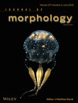
Maja brachydactyla. Esophagus. General diagram of the adult (left) and larvae (right). Abbreviations: BV, blood vessels; C, cuticle; CT, connective tissue; CM, circular muscles; DM, dilator muscles; E, epithelium; EN, endocuticle; EP, epicuticle; EX, exocuticle; G, rosette glands; LM, longitudinal muscles.
Development of the duct system during exocrine pancreas differentiation in the grass snake Natrix natrix (Lepidosauria, Serpentes)
- Pages: 724-746
- First Published: 21 February 2018

During the embryonic development, the pancreas of the grass snake embryos develops into the branched structure divided into the extralobular, intralobular, and intercalated ducts that pattern of branching is different than in other vertebrates. Within the pancreatic duct walls are located four cell types: principal, endocrine, goblet, and basal cells. The ductal lumen is formed by the process of cavitation during which the cells undergo cell death named anoikis.
Embryonic and early development of the Zagros tooth-carp, Aphanius vladykovi (Actinopterygii: Cyprinodontidae)
- Pages: 747-756
- First Published: 21 February 2018
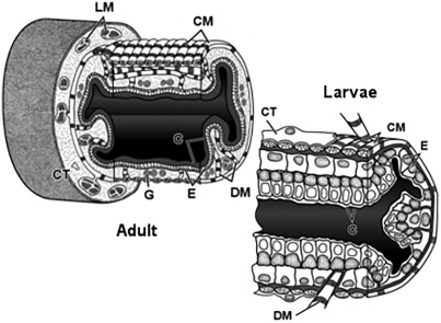
In this study, we have demonstrated that A. vladykovi embryos can be successfully reared in the laboratory and have provided a detailed description of embryonic development including zygote, blastula, gastrula, organogenesis, and early larvae. Understanding of its embryonic stages and development will help in use of techniques to increase its population. By providing such data, we can plan for its artificial propagation to restock it in its natural habitats and to establish gene banks.
Morphology of the core fibrous layer of the cetacean tail fluke
- Pages: 757-765
- First Published: 09 March 2018
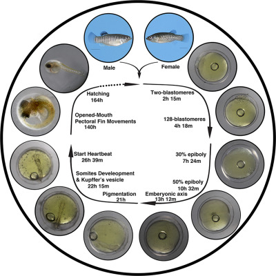
In place of skeletal elements, the arrangement of fibers within the core layer of the cetacean tail fluke blade allows the structure to maintain a specific flexibility profile. This arrangement is maintained across six species with only slight variations.
Evolution of hindlimb bone dimensions and muscle masses in house mice selectively bred for high voluntary wheel-running behavior
- Pages: 766-779
- First Published: 13 March 2018
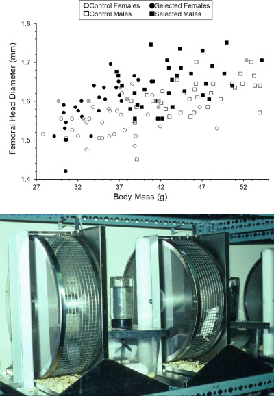
We used selective breeding of house mice to study coadaptation of morphology with the evolution of high voluntary exercise. We found that skeletal dimensions and muscle masses can evolve rapidly in response to directional selection on locomotor behavior.
Functional morphology of the cranio-mandibular complex of the Guira cuckoo (Aves)
- Pages: 780-791
- First Published: 13 March 2018
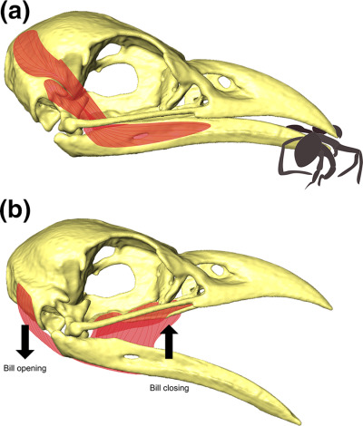
In Guira guira (a), the external adductor muscles work favoring the force (higher mechanical advantage values and perpendicular position when bill is closed), acting mainly during the food processing; while (b) depressor and pterygoid muscles prioritize velocity, being important in the prey capture moment.
Axial complex and associated structures of the sea urchin Strongylocentrotus pallidus (Sars, G.O. 1871) (Echinodermata: Echinoidea)
- Pages: 792-808
- First Published: 12 March 2018
The microscopic anatomy of the axial complex and associated structures of the sea urchin Strongylocentrotus pallidus is studied and compared with other eleutherozoans.
Prenatal cranial bone development of Thomas's horseshoe bat (Rhinolophus thomasi): with special reference to petrosal morphology
- Pages: 809-827
- First Published: 14 March 2018
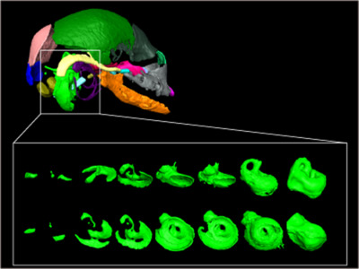
Relative cochlear size against skull size is maintained constantly throughout the postnatal ontogeny to adulthood in Rhinolophus, a pattern previously reported neither in any other bats nor mammals.
Scale diversity in bigeye tuna (Thunnus obesus): Fat-filled trabecular scales made of cellular bone
- Pages: 828-840
- First Published: 14 March 2018
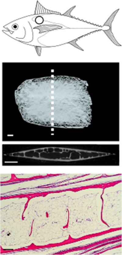
Scales of bigeye tuna (Thunnus obesus) are modified on certain parts of the body. Here on the fairing near the pectoral fin, scales are thickened with a three-dimensional bony shell supported by internal trabeculae-like struts. The space inside these thickened scales is filled with fat (adipocytes) and includes arterioles and venules.
Gonadal histology of box jellyfish (Cnidaria: Cubozoa) reveals variation between internal fertilizing species Alatina alata (Alatinidae) and Copula sivickisi (Tripedaliidae)
- Pages: 841-856
- First Published: 23 March 2018
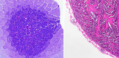
The gonadal ultrastructure of two species of Cubozoa exhibit different modes of internal fertilization. Copula sivickisi testes (left) with a distinct gametogenic differentiation gradient. Alatina alata testes (right) lack this differentiation gradient.