Label-free volumetric imaging of conjunctival collecting lymphatics ex vivo by optical coherence tomography lymphangiography
Corresponding Author
Peijun Gong
Optical+Biomedical Engineering Laboratory, Department of Electrical, Electronic and Computer Engineering, The University of Western Australia, Perth, WA, Australia
Correspondence
Peijun Gong, Optical+Biomedical Engineering Laboratory, Department of Electrical, Electronic and Computer Engineering, The University of Western Australia, 35 Stirling Highway, Perth WA 6009, Australia.
Email: [email protected]
Search for more papers by this authorDao-Yi Yu
Centre for Ophthalmology and Visual Science, The University of Western Australia, Perth, WA, Australia
Lions Eye Institute, Nedlands, WA, Australia
Search for more papers by this authorQiang Wang
Optical+Biomedical Engineering Laboratory, Department of Electrical, Electronic and Computer Engineering, The University of Western Australia, Perth, WA, Australia
Search for more papers by this authorPaula K. Yu
Centre for Ophthalmology and Visual Science, The University of Western Australia, Perth, WA, Australia
Lions Eye Institute, Nedlands, WA, Australia
Search for more papers by this authorKarol Karnowski
Optical+Biomedical Engineering Laboratory, Department of Electrical, Electronic and Computer Engineering, The University of Western Australia, Perth, WA, Australia
Search for more papers by this authorMorgan Heisler
Biomedical Optics Research Group, School of Engineering Science, Simon Fraser University, Burnaby, BC, Canada
Search for more papers by this authorAshley Francke
Biomedical Optics Research Group, School of Engineering Science, Simon Fraser University, Burnaby, BC, Canada
Search for more papers by this authorDong An
Centre for Ophthalmology and Visual Science, The University of Western Australia, Perth, WA, Australia
Lions Eye Institute, Nedlands, WA, Australia
Search for more papers by this authorMarinko V. Sarunic
Biomedical Optics Research Group, School of Engineering Science, Simon Fraser University, Burnaby, BC, Canada
Search for more papers by this authorDavid D. Sampson
Optical+Biomedical Engineering Laboratory, Department of Electrical, Electronic and Computer Engineering, The University of Western Australia, Perth, WA, Australia
University of Surrey, Guildford, Surrey, UK
Search for more papers by this authorCorresponding Author
Peijun Gong
Optical+Biomedical Engineering Laboratory, Department of Electrical, Electronic and Computer Engineering, The University of Western Australia, Perth, WA, Australia
Correspondence
Peijun Gong, Optical+Biomedical Engineering Laboratory, Department of Electrical, Electronic and Computer Engineering, The University of Western Australia, 35 Stirling Highway, Perth WA 6009, Australia.
Email: [email protected]
Search for more papers by this authorDao-Yi Yu
Centre for Ophthalmology and Visual Science, The University of Western Australia, Perth, WA, Australia
Lions Eye Institute, Nedlands, WA, Australia
Search for more papers by this authorQiang Wang
Optical+Biomedical Engineering Laboratory, Department of Electrical, Electronic and Computer Engineering, The University of Western Australia, Perth, WA, Australia
Search for more papers by this authorPaula K. Yu
Centre for Ophthalmology and Visual Science, The University of Western Australia, Perth, WA, Australia
Lions Eye Institute, Nedlands, WA, Australia
Search for more papers by this authorKarol Karnowski
Optical+Biomedical Engineering Laboratory, Department of Electrical, Electronic and Computer Engineering, The University of Western Australia, Perth, WA, Australia
Search for more papers by this authorMorgan Heisler
Biomedical Optics Research Group, School of Engineering Science, Simon Fraser University, Burnaby, BC, Canada
Search for more papers by this authorAshley Francke
Biomedical Optics Research Group, School of Engineering Science, Simon Fraser University, Burnaby, BC, Canada
Search for more papers by this authorDong An
Centre for Ophthalmology and Visual Science, The University of Western Australia, Perth, WA, Australia
Lions Eye Institute, Nedlands, WA, Australia
Search for more papers by this authorMarinko V. Sarunic
Biomedical Optics Research Group, School of Engineering Science, Simon Fraser University, Burnaby, BC, Canada
Search for more papers by this authorDavid D. Sampson
Optical+Biomedical Engineering Laboratory, Department of Electrical, Electronic and Computer Engineering, The University of Western Australia, Perth, WA, Australia
University of Surrey, Guildford, Surrey, UK
Search for more papers by this authorAbstract
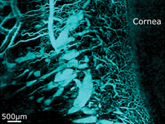
We employ optical coherence tomography (OCT) and optical coherence microscopy (OCM) to study conjunctival lymphatics in porcine eyes ex vivo. This study is a precursor to the development of in vivo imaging of the collecting lymphatics for potentially guiding and monitoring glaucoma filtration surgery. OCT scans at 1300 nm and higher-resolution OCM scans at 785 nm reveal the lymphatic vessels via their optical transparency. Equivalent signal characteristics are also observed from blood vessels largely free of blood (and devoid of flow) in the ex vivo conjunctiva. In our lymphangiography, vessel networks were segmented by compensating the depth attenuation in the volumetric OCT/OCM signal, projecting the minimum intensity in two dimensions and thresholding to generate a three-dimensional vessel volume. Vessel segmentation from multiple locations of a range of porcine eyes (n = 21) enables visualization of the vessel networks and indicates the varying spatial distribution of patent lymphatics. Such visualization provides a new tool to investigate conjunctival vessels in tissue ex vivo without need for histological tissue processing and a valuable reference on vessel morphology for the in vivo label-free imaging studies of lymphatics to follow.
Supporting Information
| Filename | Description |
|---|---|
| jbio201800070-sup-0001-VideoS1.mp4MPEG-4 video, 8.7 MB | Video S1 Volumetric imaging of conjunctival vessels in Sample 1. |
| jbio201800070-sup-0002-VideoS2.mp4MPEG-4 video, 8.2 MB | Video S2 Volumetric imaging of conjunctival vessels in Sample 4. |
Please note: The publisher is not responsible for the content or functionality of any supporting information supplied by the authors. Any queries (other than missing content) should be directed to the corresponding author for the article.
REFERENCES
- 1M. A. Swartz, Adv. Drug Deliv. Rev. 2001, 50, 3.
- 2D. Negrini, A. Moriondo, J. Physiol. 2011, 589, 2927.
- 3F. Zhang, G. Niu, G. Lu, X. Chen, Mol. Imaging Biol. 2011, 13, 599.
- 4T. F. O'Donnell, J. C. Rasmussen, E. M. Sevick-Muraca, J. Vasc. Surg. Venous Lymphat. Disord. 2017, 5, 261.
- 5B. Zhu, E. M. Sevick-Muraca, Br. J. Radiol. 2015, 88, 20140547.
- 6L. L. Munn, T. P. Padera, Microvasc. Res. 2014, 96, 55.
- 7E. I. Galanzha, M. S. Kokoska, E. V. Shashkov, J. W. Kim, V. V. Tuchin, V. P. Zharov, J. Biophotonics 2009, 2, 528.
- 8D. Y. Yu, W. H. Morgan, X. Sun, E. N. Su, S. J. Cringle, P. K. Yu, P. House, W. Guo, X. Yu, Prog. Retin. Eye Res. 2009, 28, 303.
- 9H. A. Quigley, A. T. Broman, Br. J. Ophthalmol. 2006, 90, 262.
- 10C. L. Chen, R. K. Wang, Biomed. Opt. Express 2017, 8, 1056.
- 11R. A. Leitgeb, R. M. Werkmeister, C. Blatter, L. Schmetterer, Prog. Retin. Eye Res. 2014, 41, 26.
- 12B. J. Vakoc, R. M. Lanning, J. A. Tyrrell, T. P. Padera, L. A. Bartlett, T. Stylianopoulos, L. L. Munn, G. J. Tearney, D. Fukumura, R. K. Jain, B. E. Bouma, Nat. Med. 2009, 15, 1219.
- 13R. K. Wang, S. Hurst, Opt. Express 2007, 15, 11402.
- 14P. Gong, S. Es'haghian, F. M. Wood, D. D. Sampson, R. A. McLaughlin, Exp. Dermatol. 2016, 25, 722.
- 15P. Gong, S. Es'haghian, K. A. Harms, A. Murray, S. Rea, B. F. Kennedy, F. M. Wood, D. D. Sampson, R. A. McLaughlin, J. Biophotonics 2016, 9, 626.
- 16D. M. Sampson, P. Gong, D. An, M. Menghini, A. Hansen, D. A. Mackey, D. D. Sampson, F. K. Chen, Invest. Ophthalmol. Vis. Sci. 2017, 58, 3065.
- 17P. Gong, S. Es'Haghian, K. A. Harms, A. Murray, S. Rea, F. M. Wood, D. D. Sampson, R. A. McLaughlin, Biomed. Opt. Express 2016, 7, 4886.
- 18S. Yousefi, J. Qin, Z. Zhi, R. K. Wang, J. Biomed. Opt. 2013, 18, 086004.
- 19P. Li, Y. Sun, S. Hariri, Z. Zhou, Y. Inamoto, S. J. Lee, T. T. Shen, R. K. Wang, Quant. Imaging Med. Surg. 2015, 5, 163.
- 20Y. M. Liew, R. A. McLaughlin, P. Gong, F. M. Wood, D. D. Sampson, J. Biomed. Opt. 2013, 18, 061213.
- 21A. Zhang, Q. Zhang, R. K. Wang, Biomed. Opt. Express 2015, 6, 4130.
- 22M. Zhang, T. S. Hwang, J. P. Campbell, S. T. Bailey, D. J. Wilson, D. Huang, Y. Jia, Biomed. Opt. Express 2016, 7, 816.
- 23J. M. Schmitt, A. Knüttel, R. F. Bonner, Appl. Opt. 1993, 32, 6032.
- 24L. Scolaro, R. A. McLaughlin, B. R. Klyen, B. A. Wood, P. D. Robbins, C. M. Saunders, S. L. Jacques, D. D. Sampson, Opt. Express 2012, 3, 366.
- 25P. Gong, R. A. McLaughlin, Y. M. Liew, P. R. T. Munro, F. M. Wood, D. D. Sampson, J. Biomed. Opt. 2014, 19, 021111.
- 26A. Curatolo, M. Villiger, D. Lorenser, P. Wijesinghe, A. Fritz, B. F. Kennedy, D. D. Sampson, Opt. Lett. 2016, 41, 21.
- 27J. M. Lauweryns, L. Boussauw, Z. Zellforsch. Mikrosk. Anat. 1973, 143, 149.
- 28X. Zhang, Q. Li, B. Liu, H. Zhou, H. Wang, Z. Zhang, M. Xiang, Z. Han, H. Zou, Invest. Ophthalmol. Vis. Sci. 2011, 52, 7787.
- 29J. Horstmann, H. Schulz-Hildebrandt, F. Bock, S. Siebelmann, E. Lankenau, G. Hüttmann, P. Steven, C. Cursiefen, Invest. Ophthalmol. Vis. Sci. 2017, 58, 5880.
- 30R. R. Allingham, K. F. Damji, S. Freedman, S. E. Moroi, G. Shafranov, M. B. Shields, Shields' Textbook of Glaucoma, Lippincott Williams & Wilkins, Philadelphia, PA 2005.
- 31K. Nouri-Mahdavi, L. Brigatti, M. Weitzman, J. Caprioli, Ophthalmology 1995, 102, 1760.
- 32A. Louveau, I. Smirnov, T. J. Keyes, J. D. Eccles, S. J. Rouhani, J. D. Peske, N. C. Derecki, D. Castle, J. W. Mandell, K. S. Lee, T. H. Harris, J. Kipnis, Nature 2015, 523, 337.