Volume 11, Issue 4 e201700029
FULL ARTICLE

TiO2-coated fluoride nanoparticles for dental multimodal optical imaging

Ana K.S. Braz

Ana K.S. Braz

Laboratory of Biomedical Optics and Imaging, Federal University of Pernambuco, Recife, Pernambuco, Brazil

Department of Operative Dentistry, University of Pernambuco, Recife, Pernambuco, Brazil

Search for more papers by this author
Diógenes S. Moura

Diógenes S. Moura

Laboratory of Biomedical Optics and Imaging, Federal University of Pernambuco, Recife, Pernambuco, Brazil

Colégio de Aplicação, Federal University of Pernambuco, Recife, Pernambuco, Brazil

Search for more papers by this author
Anderson S.L. Gomes

Anderson S.L. Gomes

Department of Physics, Federal University of Pernambuco, Recife, Pernambuco, Brazil

Search for more papers by this author
Tymish Y. Ohulchanskyy

Tymish Y. Ohulchanskyy

The Institute for Lasers, Photonics and Biophotonics, University at Buffalo, Buffalo, New York

Key Laboratory of Optoelectronic Devices and Systems of Ministry of Education and Guangdong Province, College of Optoelectronic Engineering, Shenzhen University, Shenzhen, China

Search for more papers by this author
Guanying Chen

Guanying Chen

The Institute for Lasers, Photonics and Biophotonics, University at Buffalo, Buffalo, New York

Search for more papers by this author
Maixian Liu

Maixian Liu

The Institute for Lasers, Photonics and Biophotonics, University at Buffalo, Buffalo, New York

Search for more papers by this author
Jossana Damasco

Jossana Damasco

The Institute for Lasers, Photonics and Biophotonics, University at Buffalo, Buffalo, New York

Search for more papers by this author
Renato E. de Araujo

Renato E. de Araujo

Laboratory of Biomedical Optics and Imaging, Federal University of Pernambuco, Recife, Pernambuco, Brazil

Search for more papers by this author
Paras N. Prasad

Corresponding Author

Paras N. Prasad

The Institute for Lasers, Photonics and Biophotonics, University at Buffalo, Buffalo, New York

Key Laboratory of Optoelectronic Devices and Systems of Ministry of Education and Guangdong Province, College of Optoelectronic Engineering, Shenzhen University, Shenzhen, China

Correspondence

Paras N. Prasad, The Institute for Lasers, Photonics and Biophotonics, University at Buffalo, Buffalo, NY. Email: [email protected]

Search for more papers by this author
First published: 13 July 2017
Citations: 8
Funding information Air Force Office of Scientific Research, Grant/Award number: FA9550-12-1-0450; PRONEX/CNPq/FACEPE Center of Excellence program; CNPq; CAPES

Abstract

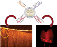
Core-shell nanostructures associated with photonics techniques have found innumerous applications in diagnostics and therapy. In this work, we introduce a novel core-shell nanostructure design that serves as a multimodal optical imaging contrast agent for dental adhesion evaluation. This nanostructure consists of a rare-earth-doped (NaYF4:Yb 60%, Tm 0.5%)/NaYF4 particle as the core (hexagonal prism, ~51 nm base side length) and the highly refractive TiO2 material as the shell (~thickness of 15 nm). We show that the TiO2 shell provides enhanced contrast for optical coherence tomography (OCT), while the rare-earth-doped core upconverts excitation light from 975 nm to an emission peaked at 800 nm for photoluminescence imaging. The OCT and the photoluminescence wide-field images of human tooth were demonstrated with this nanoparticle core-shell contrast agent. In addition, the described core-shell nanoparticles (CSNps) were dispersed in the primer of a commercially available dental bonding system, allowing clear identification of dental adhesive layers with OCT. We evaluated that the presence of the CSNp in the adhesive induced an enhancement of 67% scattering coefficient to significantly increase the OCT contrast. Moreover, our results highlight that the upconversion photoluminescence in the near-infrared spectrum region is suitable for image of deep dental tissue.

image

The full text of this article hosted at iucr.org is unavailable due to technical difficulties.