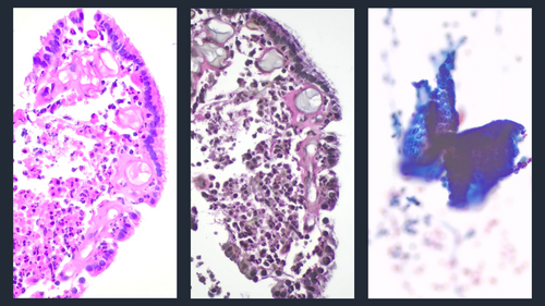
Well-Differentiated Adenocarcinoma With Papillary Architecture, Focal Residual Cilia, Apical Snouts, and CHEK2 and p53 Mutations
Funding: The author received no specific funding for this work.
ABSTRACT
Ciliated adenocarcinomas are rare non-terminal respiratory unit-type lung adenocarcinomas characterised by ciliated cells with nuclear atypia and glandular or papillary architecture. A few cases of ciliated adenocarcinoma of the lung have been reported. These adenocarcinomas are negative for TTF1 and have either KRAS (25%) or EGFR (9%) mutations. A case of a well-differentiated adenocarcinoma with papillary architecture, focal residual cilia, apical snouts, and CHEK2 and p53 mutations diagnosed at stage IV(T4NxM0) is reported. The article describes the cytomorphological features of this tumour, the potential pitfalls, diagnostic limitations, challenges identified in cytology specimens, and the necessary work-up for a definitive diagnosis. The differential diagnosis of papillary lesions of the lung and the clinical significance of CHEK2 mutations are also discussed in this article.
Graphical Abstract
Conflicts of Interest
The author declares no conflicts of interest.
Open Research
Data Availability Statement
The data that support the findings of this study are available from the corresponding author upon reasonable request.


