Volume 16, Issue 6 e202300002
RESEARCH ARTICLE

Depth-resolved attenuation mapping of the human ovary and fallopian tube using optical coherence tomography

Shuying Li

Shuying Li

Department of Biomedical Engineering, Washington University in St. Louis, 63130 St. Louis, Missouri, USA

Search for more papers by this author
Hongbo Luo

Hongbo Luo

Department of Electrical & Systems Engineering, Washington University in St. Louis, St. Louis, Missouri, 63130 USA

Search for more papers by this author
Sitai Kou

Sitai Kou

Department of Biomedical Engineering, Washington University in St. Louis, 63130 St. Louis, Missouri, USA

Search for more papers by this author
Ian S. Hagemann

Ian S. Hagemann

Department of Pathology & Immunology, Washington University School of Medicine, 63110 St. Louis, Missouri, USA

Department of Obstetrics & Gynecology, Washington University School of Medicine, 63110 St. Louis, Missouri, USA

Search for more papers by this author
Quing Zhu

Corresponding Author

Quing Zhu

Department of Biomedical Engineering, Washington University in St. Louis, 63130 St. Louis, Missouri, USA

Department of Electrical & Systems Engineering, Washington University in St. Louis, St. Louis, Missouri, 63130 USA

Department of Radiology, Washington University School of Medicine, 63110 St. Louis, Missouri, USA

Correspondence

Quing Zhu, Department of Biomedical Engineering, Washington University in St. Louis, 63130 St. Louis, MO, USA.

Email: [email protected]

Search for more papers by this author
First published: 14 March 2023
Citations: 1

Shuying Li and Hongbo Luo contributed equally to this study.

Abstract

Due to the lack of reliable early-diagnostic tools, most ovarian cancers are diagnosed at late stages. Although optical coherence tomography (OCT) has shown promise for identifying diseased ovaries and fallopian tubes at an earlier stage, previous studies either did not provide quantitative scattering mapping or simply used Beer's law to fit the scattering coefficients of each A-line. In this paper, we calculated the pixel-wise attenuation coefficients of ovaries and fallopian tubes in OCT images. Data from 73 freshly excised human ovaries and fallopian tubes from 36 patients have shown that statistical features are statistically different between cancerous ovaries, infundibula, and fimbriae and normal ones.image

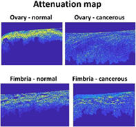
CONFLICT OF INTEREST STATEMENT

The authors declare no conflict of interest.

DATA AVAILABILITY STATEMENT

Data are available from the corresponding author upon request.

The full text of this article hosted at iucr.org is unavailable due to technical difficulties.