Immunofluorescence and histopathological assessment using ex vivo confocal laser scanning microscopy in lichen planus
This study has been conducted at the Dermatology and Allergy Department of University Hospital, LMU Munich, Germany.
[Correction added on 11 January, after first online publication: Ludwig-Maximilians-Universität München was added for Işın Sinem Bağcı.]
Abstract
Ex vivo confocal laser scanning microscopy (CLSM) provides rapid, high-resolution imaging, fluorescence detection and digital haematoxylin–eosin (H&E)-like staining. We aimed to assess the performance of ex vivo CLSM in identifying histomorphology and immunoreactivity in lichen planus (LP) and comparing its accuracy with conventional histopathology and direct immunofluorescence (DIF). Thirty-three sections of 17 LP patients stained with acridine orange (AO) and FITC-labelled anti-fibrinogen antibody and 21 control samples stained with AO were examined using ex vivo CLSM. Ex vivo CLSM was in perfect agreement with conventional histopathology in identifying interface dermatitis, vacuolar degeneration and band-like infiltration. ROC analysis showed that the presence of vacuolar degeneration, interface dermatitis and band-like infiltration was useful to distinguish LP sections from controls (p < .0001). The detection rates of fibrinogen deposition using DIF and in conclusion ex vivo CLSM were 93.8% and 62.5%, respectively. ex vivo CLSM enables histopathological and immunofluorescence examination in LP with the advantage of digital H&E-like staining.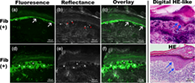
CONFLICT OF INTEREST
The Vivascope 2500 M-G4 device was provided by Mavig GmbH for the time of the study from January to May 2019. All authors declare that they have no financial or personal relationships that could be viewed as a potential conflict of interest.
Open Research
DATA AVAILABILITY STATEMENT
Research data are not shared