Cortex-wide microcirculation mapping with ultrafast large-field multifocal illumination microscopy
Zhenyue Chen and Quanyu Zhou contributed equally to this study.
Funding information: H2020 European Research Council, Grant/Award Number: ERC-2015-CoG-682379; H2020 Marie Skłodowska-Curie Actions, Grant/Award Number: 746430
Abstract
The recently introduced large-field multifocal illumination (LMI) fluorescence microscopy technique opened new possibilities for transcranial observations of mouse brain dynamics with a unique combination of capillary level resolution and centimeter-scale field-of-view (FOV). Here we report on a new acceleration scheme for LMI based on raster scan of a lattice pattern combined with a parallel camera exposure scheme, which attains 200 Hz frame rate over 12 × 12 mm2 FOV with 7.5 μm spatial resolution. We demonstrate real-time transcranial in vivo tracking of particles and imaging of microcirculation across the entire mouse cortex, thus corroborating the superb spatiotemporal resolution performance of LMI unattainable with other techniques. Potential applications include investigations into cerebrovascular function, cell tracking, as well as large-scale functional neuroimaging.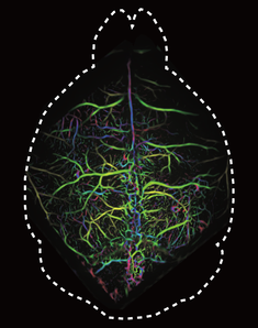
CONFLICTS OF INTEREST
The authors declare no potential conflict of interest.
Open Research
DATA AVAILABILITY STATEMENT
The data that support the findings of this study are available from the corresponding author upon reasonable request.