Peri-tumoural stroma collagen organization of invasive ductal carcinoma assessed by polarized light microscopy differs between OncotypeDX risk group
Mohammadali Khorasani and Alex Vitkin are senoir coauthors
Funding information: Canadian Institutes of Health Research, Grant/Award Number: PJT-156110; Natural Sciences and Engineering Research Council of Canada, Grant/Award Number: 410009540
Abstract
A commercially available genomic test, OncotypeDX has emerged as a useful postsurgical treatment guide for early stage breast cancer. Despite widespread clinical adoption, there remain logistical issues with its implementation. Collagenous stromal architecture has been shown to hold prognostic value that may complement OncotypeDX. Polarimetric analysis of breast cancer surgical samples allows for the quantification of collagenous stroma abundance and organization. We examine intratumoural collagen abundance and alignment along the tumor-host interface for 45 human samples of invasive ductal carcinoma categorized as low or higher risk by OncotypeDX. Furthermore, we probe the separatory power of collagen alignment patterns to classify unlabeled samples as low or higher OncotypeDX risk group using a linear discriminant (LD) model. No significant difference in mean collagen abundance was found between the two risk groups. However, collagen alignment along the tumor boundary was found to be significantly lower in higher risk samples. The LD model achieved a 71% total accuracy and 81% sensitivity to higher risk samples. Prognostic information extracted from the stromal morphology has potential to complement OncotypeDX as an easy-to-implement prescreening methodology.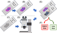
CONFLICT OF INTEREST
The authors declare no financial or commercial conflict of interest.
Open Research
DATA AVAILABILITY STATEMENT
The data that support the findings of this study are available from the corresponding author upon reasonable request.