Evaluation of corneal structures in myopic eyes more than twenty-two years after photorefractive keratectomy
Daniela Montorio
Department of Neurosciences, Reproductive Sciences and Dentistry, University of Naples Federico II, Naples, Italy
Search for more papers by this authorGilda Cennamo
Eye Clinic, Public Health Department, University of Naples Federico II, Naples, Italy
Search for more papers by this authorFeliciana Menna
Department of Neurosciences, Reproductive Sciences and Dentistry, University of Naples Federico II, Naples, Italy
Search for more papers by this authorPiero Donna
Department of Neurosciences, Reproductive Sciences and Dentistry, University of Naples Federico II, Naples, Italy
Search for more papers by this authorPasquale Napolitano
Department of Neurosciences, Reproductive Sciences and Dentistry, University of Naples Federico II, Naples, Italy
Search for more papers by this authorMaria Angelica Breve
Department of Neurosciences, Reproductive Sciences and Dentistry, University of Naples Federico II, Naples, Italy
Search for more papers by this authorUgo Fiore
Department of Management and Quantitative Studies, Parthenope University, Naples, Italy
Search for more papers by this authorGiovanni Cennamo
Department of Neurosciences, Reproductive Sciences and Dentistry, University of Naples Federico II, Naples, Italy
Search for more papers by this authorCorresponding Author
Nicola Rosa
Department of Medicine, Surgery and Dentistry, “Scuola Medica Salernitana”, University of Salerno, Salerno, Italy
Correspondence
Nicola Rosa, Department of Medicine, Surgery and Dentistry, “Scuola Medica Salernitana,” University of Salerno, Salerno, Italy.
Email: [email protected]
Search for more papers by this authorDaniela Montorio
Department of Neurosciences, Reproductive Sciences and Dentistry, University of Naples Federico II, Naples, Italy
Search for more papers by this authorGilda Cennamo
Eye Clinic, Public Health Department, University of Naples Federico II, Naples, Italy
Search for more papers by this authorFeliciana Menna
Department of Neurosciences, Reproductive Sciences and Dentistry, University of Naples Federico II, Naples, Italy
Search for more papers by this authorPiero Donna
Department of Neurosciences, Reproductive Sciences and Dentistry, University of Naples Federico II, Naples, Italy
Search for more papers by this authorPasquale Napolitano
Department of Neurosciences, Reproductive Sciences and Dentistry, University of Naples Federico II, Naples, Italy
Search for more papers by this authorMaria Angelica Breve
Department of Neurosciences, Reproductive Sciences and Dentistry, University of Naples Federico II, Naples, Italy
Search for more papers by this authorUgo Fiore
Department of Management and Quantitative Studies, Parthenope University, Naples, Italy
Search for more papers by this authorGiovanni Cennamo
Department of Neurosciences, Reproductive Sciences and Dentistry, University of Naples Federico II, Naples, Italy
Search for more papers by this authorCorresponding Author
Nicola Rosa
Department of Medicine, Surgery and Dentistry, “Scuola Medica Salernitana”, University of Salerno, Salerno, Italy
Correspondence
Nicola Rosa, Department of Medicine, Surgery and Dentistry, “Scuola Medica Salernitana,” University of Salerno, Salerno, Italy.
Email: [email protected]
Search for more papers by this authorAbstract
The aim of this study is to evaluate corneal epithelial thickness (CET), corneal densitometry (CD) in 84 myopic eyes (57 patients) more than 22 years after photorefractive keratectomy, using anterior segment-optical coherence tomography (AS-OCT) and Scheimpflug imaging system. The CET was significantly higher in all operated eyes than in unoperated eyes in central sector. A statistically significant increase in CD in corneal anterior layer of central sector was shown in groups of operated eyes with greater ablation depth respect to unoperated eyes. While there was no significant difference in CD between the operated eyes groups with lower ablation depth and unoperated eyes. A significant trend toward higher values in anterior CD with deeper ablations in central sector was found. These noninvasive imaging techniques allow to better understand the corneal remodeling process after photoablation and to monitor the patients over time.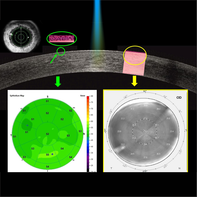
CONFLICT OF INTEREST
The authors declare no potential conflict of interest.
REFERENCES
- 1B. C. Ang, R. C. Foo, E. W. Lim, M. M. Tan, G. K. Nah, L. S. Thean, C. W. Tan, P. S. Zhao, J. Cataract. Refract. Surg. 2016, 42, 710.
- 2G. Cennamo, N. Rosa, M. A. Breve, M. di Grazia, J. Refract. Surg. 2003, 19, 438.
- 3N. Rosa, G. Cennamo, M. Rinaldi, J. Refract. Surg. 2001, 17, 129.
- 4G. Cennamo, N. Rosa, E. Guida, A. Del Prete, A. Sebastiani, J. Refract. Corneal Surg. 1994, 10, 137.
- 5G. Ambrosio, G. Cennamo, R. De Marco, L. Loffredo, N. Rosa, A. Sebastiani, J. Refract. Corneal Surg. 1994, 10, 129.
- 6N. Rosa, G. Cennamo, A. Del Prete, B. Pastena, A. Sebastiani, Ophthalmologica 1995, 209, 17.
- 7J. V. Jester, T. Møller-Pedersen, J. Huang, C. M. Sax, W. T. Kays, H. D. Cavangh, W. M. Petroll, J. Piatigorsky, J. Cell Sci. 1999, 112, 613.
- 8F. Poyales, N. Garzón, J. Mendicute, I. Illarramendi, P. Caro, O. Jáñez, F. Argüeso, A. López, Eye 2017, 31, 1647.
- 9J. Hou, Y. Wang, Y. Lei, X. Zheng, Y. Zhang, J. Ophthalmol. 2016, 2016, 8582362.
- 10X. Chen, A. Stojanovic, Y. Liu, Y. Chen, Y. Zhou, T. P. Utheim, J. Refract. Surg. 2015, 31, 446.
- 11Y. Yang, J. Hong, S. X. Deng, J. Xu, Invest. Ophthalmol. Vis. Sci. 2014, 55, 5032.
- 12N. Garzón, F. Poyales, I. Illarramendi, J. Mendicute, Ó. Jáñez, P. Caro, A. López, F. Argüeso, Int. Ophthalmol. 2017, 37, 1263.
- 13D. Montorio, G. Cennamo, M. A. Breve, U. Fiore, M. Reibaldi, V. Brescia Morra, G. Cennamo, J. Biophotonics 2020, 13, e201900095.
- 14A. M. Otri, U. Fares, M. A. Al-Aqaba, H. S. Dua, Ophthalmology 2012, 119, 501.
- 15S. Ní Dhubhghaill, J. J. Rozema, S. Jongenelen, I. Ruiz Hidalgo, N. Zakaria, M. J. Tassignon, Invest. Ophthalmol. Vis. Sci. 2014, 55, 162.
- 16N. Rosa, G. Cennamo, A. Pasquariello, F. Maffulli, A. Sebastiani, Ophthalmology 1996, 103, 1130.
- 17N. Rosa, A. Iura, M. Romano, G. Verolino, A. Romano, J. Refract. Surg. 2002, 18, 449.
- 18R. R. Wilcox, Introduction to Robust Estimation and Hypothesis Testing, 4th ed., Elsevier, Amsterdam, The Netherlands 2017.
- 19J. C. Erie, Trans. Am. Ophthalmol. Soc. 2003, 101, 293.
- 20M. R. Sedaghat, H. Momeni-Moghaddam, M. Gazanchian, D. Z. Reinstein, T. J. Archer, J. B. Randleman, S. R. Hosseini, G. Nouri-Hosseini, J. Refract. Surg. 2019, 35, 632.
- 21A. Ivarsen, W. Fledelius, J. Ø. Hjortdal, Invest. Ophthalmol. Vis. Sci. 2009, 50, 2061.
- 22C. A. Gauthier, D. Epstein, B. A. Holden, B. Tengroth, P. Fagerholm, H. Hamberg-Nyström, R. Sievert, J. Refract. Surg. 1995, 11, 113.
- 23S. V. Patel, J. C. Erie, J. W. McLaren, W. M. Bourne, J. Refract. Surg. 2007, 23, 385.
- 24S. E. Wilson, Q. Li, J. Weng, P. A. Barry-Lane, J. V. Jester, Q. Liang, R. J. Wordinger, Invest. Ophthalmol. Vis. Sci. 1996, 37, 1582.
- 25R. R. Mohan, Q. Liang, W. J. Kim, M. C. Helena, F. Baerveldt, S. E. Wilson, Exp. Eye Res. 1997, 65, 575.
- 26R. R. Mohan, W. J. Kim, R. R. Mohan, L. Chen, S. E. Wilson, Invest. Ophthalmol. Vis. Sci. 1998, 39, 2626.
- 27J. C. Erie, J. W. McLaren, D. O. Hodge, W. M. Bourne, Trans. Am. Ophthalmol. Soc. 2005, 103, 56.
- 28A. Micera, A. Lambiase, I. Puxeddu, L. Aloe, B. Stampachiacchiere, F. Levi-Schaffer, S. Bonini, S. Bonini, Exp. Eye Res. 2006, 83, 747.
- 29T. Møller-Pedersen, H. D. Cavanagh, W. M. Petroll, J. V. Jester, Curr. Eye Res. 1998, 17, 736.
- 30J. V. Jester, W. M. Petroll, H. D. Cavanagh, Prog. Ret. Eye Res. 1999, 18, 311.
- 31J. D. Zieske, Curr. Opin. Ophthalmol. 2001, 12, 237.
- 32H. Qi, Y. Chen, X. Zhu, Zhonghua Yan Ke Za Zhi 2001, 37, 87.
- 33G. Cennamo, A. Intravaja, D. Boccuzzi, G. Marotta, G. Cennamo, J. Refract. Surg. 2008, 24, 145.
- 34N. Rosa, M. Borrelli, M. De Bernardo, M. Lanza, Cornea 2011, 30, 130.
- 35N. Rosa, M. De Bernardo, S. Iaccarino, M. Lanza, Semin. Ophthalmol. 2015, 30, 328.
- 36T. Møller-Pedersen, H. D. Cavanagh, W. M. Petroll, J. V. Jester, Ophthalmology 2000, 107, 1235.
- 37T. Møller-Pedersen, Exp. Eye Res. 2004, 78, 553.
- 38K. M. Meek, C. Knupp, Prog. Retin. Eye Res. 2015, 49, 1.
- 39M. C. Corbett, J. I. Prydal, S. Verma, K. M. Oliver, M. Pande, J. Marshall, Ophthalmology 1996, 103, 1366.
- 40A. A. Torricelli, A. Santhanam, J. Wu, V. Singh, S. E. Wilson, Exp. Eye Res. 2016, 142, 110.
- 41T. Møller-Pedersen, H. F. Li, W. M. Petroll, H. D. Cavanagh, J. V. Jester, Invest. Ophthalmol. Vis. Sci. 1998, 39, 487.
- 42T. Møller-Pedersen, H. D. Cavanagh, W. M. Petroll, J. V. Jester, Cornea 1998, 17, 627.
- 43M. Boulze-Pankert, R. Dariel, L. Hoffart, J. Refract. Surg. 2016, 32, 788.
- 44G. Savini, J. Huang, M. Lombardo, S. Serrao, D. Schiano-Lomoriello, S. Venanzio, P. Ducoli, J. Refract. Surg. 2016, 32, 20.
- 45G. Cennamo, R. Forte, B. Aufiero, A. La Rana, J. Cataract. Refract. Surg. 2011, 37, 1502.
- 46S. Patel, J. McLaren, W. Bourne, Invest. Ophthalmol. Vis. Sci. 2001, 42, 333.
- 47J. W. McLaren, W. M. Bourne, S. V. Patel, Invest. Ophthalmol. Vis. Sci. 2010, 51, 1918.
- 48J. I. Prydal, F. Franc, J. Marshall, Eye 1998, 12, 337.
- 49S. Patel, J. Marshall, F. W. Fitzke 3rd., J. Refract. Surg. 1995, 11, 100.
- 50L. Xi, C. Zhang, Y. He, BMC Ophthalmol. 2018, 18, 209.
- 51L. M. Coelho, R. O. Sieiro, Int. Ophthalmol. 2019, 39, 341.
- 52A. Shalaby, G. B. Kaye, H. V. Gimbel, J. Refract. Surg. 2009, 25, S93.
- 53E. M. Hofmeister, F. M. Bishop, S. E. Kaupp, S. C. Schallhorn, J. Cataract. Refract. Surg. 2013, 39, 1358.
- 54S. Adib-Moghaddam, S. Soleyman-Jahi, G. Tefagh, S. Tofighi, M. A. Grentzelos, G. D. Kymionis, J. Refract. Surg. 2018, 34, 400.
- 55R. M. Werkmeister, A. Alex, S. Kaya, A. Unterhuber, B. Hofer, J. Riedl, M. Bronhagl, M. Vietauer, D. Schmidl, T. Schmoll, G. Garhöfer, W. Drexler, R. A. Leitgeb, M. Groeschl, L. Schmetterer, Invest. Ophthalmol. Vis. Sci. 2013, 54, 5578.
Citing Literature