Effect of tissue staining in quantitative phase imaging
Sungbea Ban
Department of Biomedical Engineering, Ulsan National Institute of Science and Technology (UNIST), Ulsan, Republic of Korea
Search for more papers by this authorEunjung Min
Rowland Institute at Harvard University, Cambridge, Massachusetts
Search for more papers by this authorYujin Ahn
Department of Biomedical Engineering, Ulsan National Institute of Science and Technology (UNIST), Ulsan, Republic of Korea
Search for more papers by this authorCorresponding Author
Gabriel Popescu
Quantitative Light Imaging Laboratory, Department of Electrical and Computer Engineering, Beckman Institute for Advanced Science and Technology, University of Illinois at Urbana-Champaign, Urbana, Illinois
Correspondence
Gabriel Popescu, Quantitative Light Imaging Laboratory, Department of Electrical and Computer Engineering, Beckman Institute for Advanced Science and Technology, University of Illinois at Urbana-Champaign, Urbana, IL.
Email: [email protected]
Woonggyu Jung, Department of Biomedical Engineering, Ulsan National Institute of Science and Technology (UNIST), Ulsan, Republic of Korea.
Email: [email protected]
Search for more papers by this authorCorresponding Author
Woonggyu Jung
Department of Biomedical Engineering, Ulsan National Institute of Science and Technology (UNIST), Ulsan, Republic of Korea
Correspondence
Gabriel Popescu, Quantitative Light Imaging Laboratory, Department of Electrical and Computer Engineering, Beckman Institute for Advanced Science and Technology, University of Illinois at Urbana-Champaign, Urbana, IL.
Email: [email protected]
Woonggyu Jung, Department of Biomedical Engineering, Ulsan National Institute of Science and Technology (UNIST), Ulsan, Republic of Korea.
Email: [email protected]
Search for more papers by this authorSungbea Ban
Department of Biomedical Engineering, Ulsan National Institute of Science and Technology (UNIST), Ulsan, Republic of Korea
Search for more papers by this authorEunjung Min
Rowland Institute at Harvard University, Cambridge, Massachusetts
Search for more papers by this authorYujin Ahn
Department of Biomedical Engineering, Ulsan National Institute of Science and Technology (UNIST), Ulsan, Republic of Korea
Search for more papers by this authorCorresponding Author
Gabriel Popescu
Quantitative Light Imaging Laboratory, Department of Electrical and Computer Engineering, Beckman Institute for Advanced Science and Technology, University of Illinois at Urbana-Champaign, Urbana, Illinois
Correspondence
Gabriel Popescu, Quantitative Light Imaging Laboratory, Department of Electrical and Computer Engineering, Beckman Institute for Advanced Science and Technology, University of Illinois at Urbana-Champaign, Urbana, IL.
Email: [email protected]
Woonggyu Jung, Department of Biomedical Engineering, Ulsan National Institute of Science and Technology (UNIST), Ulsan, Republic of Korea.
Email: [email protected]
Search for more papers by this authorCorresponding Author
Woonggyu Jung
Department of Biomedical Engineering, Ulsan National Institute of Science and Technology (UNIST), Ulsan, Republic of Korea
Correspondence
Gabriel Popescu, Quantitative Light Imaging Laboratory, Department of Electrical and Computer Engineering, Beckman Institute for Advanced Science and Technology, University of Illinois at Urbana-Champaign, Urbana, IL.
Email: [email protected]
Woonggyu Jung, Department of Biomedical Engineering, Ulsan National Institute of Science and Technology (UNIST), Ulsan, Republic of Korea.
Email: [email protected]
Search for more papers by this authorAbstract
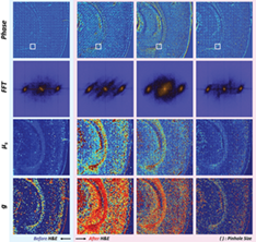
Quantitative phase imaging (QPI) is an emerging modality, which enables the identification of abnormalities in tissue based on optical properties. QPI can be applied to any biological specimen due to its label-free imaging capability, but its use in stained tissue is unclear. Here, we study the variability of QPI with the staining dye. Several tissues such as brain, heart and lung were stained with hematoxylin and eosin, and their optical properties compared at 550 and 730 nm. Our results showed that phase and scattering coefficients varied when QPI was used at the absorption wavelength of the staining dye. We also found that the variation of optical properties was dependent on tissue morphology.
REFERENCES
- 1V. Kumar, A. K. Abbas, J. C. Aster, S. L. Robbins, Robbins Basic Pathology, Elsevier Saunders, Amsterdam, 2013.
- 2B. Bhaduri, C. Edwards, H. Pham, R. J. Zhou, T. H. Nguyen, L. L. Goddard, G. Popescu, Adv. Opt. Photonics 2014, 6, 57.
- 3G. Popescu, Quantitative Phase Imaging of Cells and Tissues, McGraw-Hill, New York, NY 2011.
- 4M. G. Shan, M. E. Kandel, G. Popescu, Opt. Express 2017, 25, 1573.
- 5E. J. Min, S. B. Ban, Y. Y. Wang, S. C. Bae, G. Popescu, C. Best-Popescu, W. G. Jung, Biomed. Opt. Express 2017, 8, 1763.
- 6E. Min, M. E. Kandel, C. J. Ko, G. Popescu, W. Jung, C. Best-Popescu, Sci. Rep. 2016, 6, 39667.
- 7G. Di Caprio, M. A. Gioffre, N. Saffioti, S. Grilli, P. Ferraro, R. Puglisi, D. Balduzzi, A. Galli, G. Coppola, IEEE J. Sel. Top. Quantum Electron. 2010, 16, 833.
- 8F. Merola, L. Miccio, P. Memmolo, G. Di Caprio, A. Galli, R. Puglisi, D. Balduzzi, G. Coppola, P. Netti, P. Ferraro, Lab. Chip 2013, 13, 4512.
- 9G. Coppola, G. Di Caprio, M. Wilding, P. Ferraro, G. Esposito, L. Di Matteo, R. Dale, G. Coppola, B. Dale, Zygote 2014, 22, 446.
- 10P. Memmolo, G. Di Caprio, C. Distante, M. Paturzo, R. Puglisi, D. Balduzzi, A. Galli, G. Coppola, P. Ferraro, Opt. Express 2011, 19, 23215.
- 11P. Memmolo, M. Iannone, M. Ventre, P. A. Netti, A. Finizio, M. Paturzo, P. Ferraro, Opt. Express 2012, 20, 28485.
- 12P. Memmolo, L. Miccio, A. Finizio, P. A. Netti, P. Ferraro, Opt. Lett. 2014, 39, 2759.
- 13B. Kemper, G. von Bally, Appl. Opt. 2008, 47, A52.
- 14M. K. Kim, Principles and Techniques of Digital Holographic Microscopy, Vol. 1, SPIE, California, USA, 2010, p. 51.
- 15K. Lee, K. Kim, J. Jung, J. Heo, S. Cho, S. Lee, G. Chang, Y. Jo, H. Park, Y. Park, Sensors (Basel) 2013, 13, 4170.
- 16P. Marquet, C. Depeursinge, P. J. Magistretti, Neurophotonics 2014, 1, 020901.
- 17A. A. Evans, B. Bhaduri, G. Popescu, A. J. Levine, Proc. Natl. Acad. Sci. U.S.A. 2017, 114, 2865.
- 18G. Popescu, Y. Park, N. Lue, C. Best-Popescu, L. Deflores, R. R. Dasari, M. S. Feld, K. Badizadegan, Am. J. Phys. Cell Phys. 2008, 295, C538.
- 19H. V. Pham, C. Edwards, L. L. Goddard, G. Popescu, Appl. Opt. 2013, 52, A97.
- 20G. Popescu, L. P. Deflores, J. C. Vaughan, K. Badizadegan, H. Iwai, R. R. Dasari, M. S. Feld, Opt. Lett. 2004, 29, 2503.
- 21X. Mo, B. Kemper, P. Langehanenberg, A. Vollmer, J. Xie, G. V. Bally, Application of Color Digital Holographic Microscopy for Analysis of Stained Tissue Sections, Vol. 7367, SPIE, California, USA, 2009, p. 6.
- 22P. Lenz, D. Bettenworth, P. Krausewitz, M. Bruckner, S. Ketelhut, G. von Bally, D. Domagk, B. Kemper, Integr. Biol. (Camb) 2013, 5, 624.
- 23D. Bettenworth, A. Bokemeyer, C. Poremba, N. S. Ding, S. Ketelhut, P. Lenz, B. Kemper, Histol. Histopathol. 2017, 33, 11937.
- 24M. E. Kandel, S. Sridharan, J. Liang, Z. Luo, K. Han, V. Macias, A. Shah, R. Patel, K. Tangella, A. Kajdacsy-Balla, G. Guzman, G. Popescu, J. Biomed. Opt. 2017, 22, 66016.
- 25M. Lee, E. Lee, J. Jung, H. Yu, K. Kim, J. Yoon, S. Lee, Y. Jeong, Y. Park, Sci. Rep. 2016, 6, 31034.
- 26S. Sridharan, V. Macias, K. Tangella, A. Kajdacsy-Balla, G. Popescu, Sci. Rep. 2015, 5, 9976.
- 27P. Wang, R. Bista, R. Bhargava, R. E. Brand, Y. Liu, Opt. Lett. 2010, 35, 2840.
- 28Z. Wang, K. Tangella, A. Balla, G. Popescu, J. Biomed. Opt. 2011, 16, 116017.
- 29S. Ban, E. Min, S. Baek, H. M. Kwon, G. Popescu, W. Jung, Biomed. Opt. Express 2018, 9, 921.
- 30P. Wang, R. K. Bista, W. E. Khalbuss, W. Qiu, S. Uttam, K. Staton, L. Zhang, T. A. Brentnall, R. E. Brand, Y. Liu, J. Biomed. Opt. 2010, 15, 066028.
- 31G. Popescu, T. Ikeda, R. R. Dasari, M. S. Feld, Opt. Lett. 2006, 31, 775.
- 32Y. Jang, J. Jang, Y. Park, Opt. Express 2012, 20, 9673.
- 33C. Edwards, A. Arbabi, G. Popescu, L. L. Goddard, Light Sci. Appl. 2012, 1, e30.
- 34C. C. Wu, T. M. Liu, T. Y. Wei, L. Xin, Y. C. Li, L. S. Lee, C. K. Chang, J. L. Tang, S. S. Yang, T. H. Wei, Opt. Express 2010, 18, 22637.
- 35Z. Wang, H. Ding, G. Popescu, Opt. Lett. 2011, 36, 1215.