Current status, pitfalls and future directions in the diagnosis and therapy of lymphatic malformation
Ravi W. Sun
Department of Otolaryngology–Head and Neck Surgery, University of Arkansas for Medical Sciences, Little Rock, Arkansas
Arkansas Children's Hospital, Little Rock, Arkansas
Search for more papers by this authorValery V. Tuchin
Research-Educational Institute of Optics and Biophotonics, Saratov National Research State University, Saratov, Russia
Institute of Precision Mechanics and Control, Russian Academy of Sciences, Saratov, Russia
Laboratory of Femtomedicine, ITMO University, St. Petersburg, Russia
Search for more papers by this authorVladimir P. Zharov
Department of Otolaryngology–Head and Neck Surgery, University of Arkansas for Medical Sciences, Little Rock, Arkansas
Arkansas Nanomedicine Center, University of Arkansas for Medical Sciences, Little Rock, Arkansas
Search for more papers by this authorCorresponding Author
Ekaterina I. Galanzha
Department of Otolaryngology–Head and Neck Surgery, University of Arkansas for Medical Sciences, Little Rock, Arkansas
Arkansas Nanomedicine Center, University of Arkansas for Medical Sciences, Little Rock, Arkansas
Laboratory of Lymphatic Research, Diagnosis and Therapy (LLDT), University of Arkansas for Medical Sciences, Little Rock, Arkansas
Correspondence
Gresham T. Richter, Department of Otolaryngology, University of Arkansas for Medical Sciences, Arkansas Children's Hospital, 800 Marshall St., S3109, Little Rock, AR 72207.Email: [email protected]
Ekaterina Galanzha, Department of Otolaryngology, University of Arkansas for Medical Sciences, Laboratory of Lymphatic Research, Diagnosis, and Therapy (LLDT), 904 Stephens Spine Center, Mail Slot # 543, 4104 Outpatient Circle, Little Rock, AR 72205. Email: [email protected]
Search for more papers by this authorCorresponding Author
Gresham T. Richter
Department of Otolaryngology–Head and Neck Surgery, University of Arkansas for Medical Sciences, Little Rock, Arkansas
Arkansas Children's Hospital, Little Rock, Arkansas
Correspondence
Gresham T. Richter, Department of Otolaryngology, University of Arkansas for Medical Sciences, Arkansas Children's Hospital, 800 Marshall St., S3109, Little Rock, AR 72207.Email: [email protected]
Ekaterina Galanzha, Department of Otolaryngology, University of Arkansas for Medical Sciences, Laboratory of Lymphatic Research, Diagnosis, and Therapy (LLDT), 904 Stephens Spine Center, Mail Slot # 543, 4104 Outpatient Circle, Little Rock, AR 72205. Email: [email protected]
Search for more papers by this authorRavi W. Sun
Department of Otolaryngology–Head and Neck Surgery, University of Arkansas for Medical Sciences, Little Rock, Arkansas
Arkansas Children's Hospital, Little Rock, Arkansas
Search for more papers by this authorValery V. Tuchin
Research-Educational Institute of Optics and Biophotonics, Saratov National Research State University, Saratov, Russia
Institute of Precision Mechanics and Control, Russian Academy of Sciences, Saratov, Russia
Laboratory of Femtomedicine, ITMO University, St. Petersburg, Russia
Search for more papers by this authorVladimir P. Zharov
Department of Otolaryngology–Head and Neck Surgery, University of Arkansas for Medical Sciences, Little Rock, Arkansas
Arkansas Nanomedicine Center, University of Arkansas for Medical Sciences, Little Rock, Arkansas
Search for more papers by this authorCorresponding Author
Ekaterina I. Galanzha
Department of Otolaryngology–Head and Neck Surgery, University of Arkansas for Medical Sciences, Little Rock, Arkansas
Arkansas Nanomedicine Center, University of Arkansas for Medical Sciences, Little Rock, Arkansas
Laboratory of Lymphatic Research, Diagnosis and Therapy (LLDT), University of Arkansas for Medical Sciences, Little Rock, Arkansas
Correspondence
Gresham T. Richter, Department of Otolaryngology, University of Arkansas for Medical Sciences, Arkansas Children's Hospital, 800 Marshall St., S3109, Little Rock, AR 72207.Email: [email protected]
Ekaterina Galanzha, Department of Otolaryngology, University of Arkansas for Medical Sciences, Laboratory of Lymphatic Research, Diagnosis, and Therapy (LLDT), 904 Stephens Spine Center, Mail Slot # 543, 4104 Outpatient Circle, Little Rock, AR 72205. Email: [email protected]
Search for more papers by this authorCorresponding Author
Gresham T. Richter
Department of Otolaryngology–Head and Neck Surgery, University of Arkansas for Medical Sciences, Little Rock, Arkansas
Arkansas Children's Hospital, Little Rock, Arkansas
Correspondence
Gresham T. Richter, Department of Otolaryngology, University of Arkansas for Medical Sciences, Arkansas Children's Hospital, 800 Marshall St., S3109, Little Rock, AR 72207.Email: [email protected]
Ekaterina Galanzha, Department of Otolaryngology, University of Arkansas for Medical Sciences, Laboratory of Lymphatic Research, Diagnosis, and Therapy (LLDT), 904 Stephens Spine Center, Mail Slot # 543, 4104 Outpatient Circle, Little Rock, AR 72205. Email: [email protected]
Search for more papers by this authorAbstract
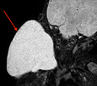
Lymphatic malformations are complex congenital vascular lesions composed of dilated, abnormal lymphatic channels of varying size that can result in significant esthetic and physical impairment due to relentless growth. Lymphatic malformations comprised of micro-lymphatic channels (microcystic) integrate and infiltrate normal soft tissue, leading to a locally invasive mass. Ultrasonography and magnetic resonance imaging assist in the diagnosis but are unable to detect microvasculature present in microcystic lymphatic malformations. In this review, we examine existing tools and elaborate on alternative diagnostic methods in assessing lymphatic malformations. In particular, photoacoustics, low-toxicity nanoparticles and optical clearing can overcome existing challenges in the examination of lymphatic channels in vivo. In combination with photothermal scanning and flow cytometry, Photoacoustic techniques may provide a versatile tool for lymphatic-related clinical applications, potentially leading to a single diagnostic and therapeutic platform to overcome limitations in current imaging techniques and permit targeted theranostics of microcystic lymphatic malformations.
Supporting Information
| Filename | Description |
|---|---|
| jbio201700124-sup-0001-author-biographies.docxapplication/docx, 812.5 KB | Author Biographies |
Please note: The publisher is not responsible for the content or functionality of any supporting information supplied by the authors. Any queries (other than missing content) should be directed to the corresponding author for the article.
REFERENCES
- 1G. M. Legiehn, M. K. Heran, Semin Intervent Radiol. 2010, 27, 209.
- 2J. A. Perkins, S. C. Manning, R. M. Tempero, M. J. Cunningham, J. L. Edmonds Jr, F. A. Hoffer, M. A. Egbert, Otolaryngol. Head Neck 2010, 142, 789.
- 3P. Brouillard, L. Boon, M. Vikkula, J. Clin. Invest. 2014, 124, 898.
- 4G. T. Richter, A. B. Friedman, Int. J. Pediatr. 2012, 2012, 645678.
- 5V. L. Luks, N. Kamitaki, M. P. Vivero, W. Uller, R. Rab, J. V. M. G. Bovée, K. L. Rialon, C. J. Guevara, A. I. Alomari, A. K. Greene, S. J. Fishman, H. P. W. Kozakewich, R. A. Maclellan, J. B. Mulliken, R. Rahbar, S. A. Spencer, C. C. Trenor III., J. Upton, D. Zurakowski, J. A. Perkins, A. Kirsh, J. T. Bennett, W. B. Dobyns, K. C. Kurek, M. L. Warman, S. A. McCarroll, R. Murillo, J. Pediatr. 2015, 166, 1048.
- 6Y. Samuels, Z. Wang, A. Bardelli, N. Silliman, J. Ptak, S. Szabo, H. Yan, A. Gazdar, S. M. Powell, G. J. Riggins, J. K. Willson, S. Markowitz, K. W. Kinzler, B. Vogelstein, V. E. Velculescu, Science 2004, 304, 554.
- 7J. B. Mulliken, J. Glowacki, Plast. Reconstr. Surg. 1982, 69, 412.
- 8G. T. Richter, J. Y. Suen, Head and Neck Vascular Anomalies, Plural Publishing Inc., San Diego, CA 2015, p. 229.
- 9L. M. de Serres, K. C. Y. Sie, M. A. Richardson, Arch. Otolaryngol. Head Neck Surg. 1995, 121, 577.
- 10S. Belov, Semin. Vasc. Surg. 1993, 6, 219.
- 11P. K. Upputuri, K. Sivasubramanian, C. S. K. Mark, M. Pramanik, Biomed. Res. Int. 2015, 2015, 783983.
- 12S. Banerji, J. Ni, S. X. Wang, S. Casper, J. Su, R. Tammi, M. Jones, D. G. Jackson, J. Cell Biol. 1999, 144, 789.
- 13S. Breiteneder-Geleff, A. Soleiman, H. Kowalski, R. Horvat, G. Amann, E. Kriehuber, K. Diem, W. Weninger, E. Tschachler, K. Aliatalo, D. Kerjaschki, Am. J. Pathol. 1999, 154, 385.
- 14A. Kaipainen, J. Korhonen, T. Mustomen, V. W. van Hinsbergh, G. H. Fang, D. Dumont, M. Breitman, K. Alitalo, Proc. Natl. Acad. Sci. U. S. A. 1995, 92, 3566.
- 15C. Galambos, L. Nodit, Pediatr. Dev. Pathol. 2005, 8, 181.
- 16E. C. Castro, C. Galambos, Pediatr. Dev. Pathol. 2009, 12, 187.
- 17A. Florez-Vargas, S. O. Vargas, L. V. Debelenko, A. R. Perez-Atayde, T. Archibald, H. P. Kozakewich, D. Zurakowski, Lymphology 2008, 41, 103.
- 18E. I. Galanzha, V. P. Zharov, Methods 2012, 57, 280.
- 19V. P. Zharov, E. I. Galanzha, V. V. Tuchin, Proc. SPIE. 2004, 5320, 185.
- 20E. I. Galanzha, E. V. Shashkov, T. Kelly, J.-W. Kim, L. Yang, Z. P. Zharov, Nat. Nanotechnol. 2009, 4, 855.
- 21L. V. Wang, Nat. Photonics. 2009, 3, 503.
- 22L. V. Wang, S. Hu, Science 2012, 335, 1458.
- 23S. Mallidi, G. P. Luke, S. Emelianov, Trends Biotechnol. 2011, 29, 213.
- 24D. Razansky, A. Buehler, V. Ntziachristos, Nat. Protoc. 2011, 6, 1121.
- 25V. P. Zharov, E. I. Galanzha, V. V. Tuchin, Opt. Lett. 2005, 30, 628.
- 26V. V. Tuchin, A. Tárnok, V. P. Zharov, J. Biophotonics 2009, 2, 457.
- 27E. I. Galanzha, E. V. Shashkov, P. Spring, J. Y. Suen, V. P. Zharov, Cancer Res. 2009, 69, 7926.
- 28E. I. Galanzha, E. V. Shashkov, V. V. Tuchin, V. P. Zharov, Cytometry 2008, 73, 884.
- 29E. I. Galanzha, V. P. Zharov, Cancers (Basel). 2013, 5, 1691.
- 30E. I. Galanzha, M. S. Kokoska, E. V. Shashkov, J.-W. Kim, V. V. Tuchin, V. P. Zharov, J. Biophotonics 2009, 2, 528.
- 31D. A. Nedosekin, M. A. Juratli, M. Sarimollaoglu, C. L. Moore, N. J. Rusch, M. S. Smeltzer, V. P. Zharov, E. I. Galanzha, J. Biophotonics 2013, 6, 523.
- 32J. B. Dixon, S. T. Greiner, A. A. Gashev, G. L. Cote, J. E. Moore, D. C. Zawieja, Microcirculation 2006, 13, 597.
- 33M. Foldi, E. Foldi, S. Kubik, Textbook of Lymphology, Urban & Fisher Munchen, Jena, GR 2003.
- 34E. I. Galanzha, V. V. Tuchin, V. P. Zharov, World J. Gastroenterol. 2007, 13, 192.
- 35W. Olszewski, A. Tarnok, Cytometry 2008, 73A, 1111.
- 36E. I. Galanzha, E. V. Shashkov, M. S. Kokoska, J. A. Myhill, V. P. Zharov, Laser Surg Med. 2008, S20, 81.
- 37Y. J. Lee, E. J. Jeong, H. W. Song, C. G. Ahn, H. W. Noh, J. Y. Sim, D. H. Song, M. Y. Jeon, S. Lee, H. Kim, M. Zhang, B. K. Kim, J. Biomed. Opt. 2017, 22, 91513.
- 38Y. A. Menyaev, K. A. Carey, D. A. Nedosekin, M. Sarimollaoglu, E. I. Galanzha, J. S. Stumhofer, V. P. Zharov, Biomed. Opt. Express 2016, 7, 3643.
- 39J.-W. Kim, E. I. Galanzha, E. V. Shashkov, H.-M. Moon, V. P. Zharov, Nat. Nanotechnol. 2009, 4, 688.
- 40Z. Cheng, A. Al Zaki, J. Z. Hui, V. R. Muzykantov, A. Tsourkas, Science 2012, 338, 903.
- 41A. de la Zerda, J. W. Kim, E. I. Galanzha, S. S. Gambhir, V. P. Zharov, Contrast Media Mol. Imaging 2011, 6, 346.
- 42X. Yang, E. W. Stein, S. Ashkenazi, L. V. Wang, Wiley Interdiscip. Rev. Nanomed. Nanobiotechnol. 2009, 1, 360.
- 43C. W. Wei, J. Xia, I. Pelivanov, C. Jia, S. W. Huang, X. Hu, X. Gao, M. O'Donnell, J. Biophotonics 2013, 6, 513.
- 44W. Li, X. Chen, Nanomedicine (Lond.) 2015, 10, 299.
- 45C. Gao, Z. J. Deng, D. Peng, Y. S. Jin, Y. Ma, Y. Y. Li, Y. K. Zhu, J. Z. Xi, J. Tian, Z. F. Dai, C. H. Li, X. L. Liang, Cancer Biol. Med. 2016, 13, 349.
- 46S. Mallidi, S. Kim, A. Karpiouk, P. P. Joshi, K. Sokolov, S. Emelianov, Photoacoustics 2015, 3, 26.
- 47G. Balasundaram, C. J. Ho, K. Li, W. Driessen, U. S. Dinish, C. L. Wong, V. Ntziachristos, B. Liu, M. Olivo, Int. J. Nanomedicine 2015, 10, 387.
- 48K. T. Nguyen, Y. Zhao, Acc. Chem. Res. 2015, 48, 3016.
- 49D. R. McCormack, K. Bhattacharyya, R. Kannan, K. Katti, J. A. Viator, Lasers Surg. Med. 2011, 43, 333.
- 50Y. Wang, E. M. Strohm, Y. Sun, Z. Wang, Y. Zheng, Z. Wang, M. C. Kolios, Biomed. Opt. Express 2016, 7, 4125.
- 51C. Di Guglielmo, J. De Lapuente, C. Porredon, D. Ramos-López, J. Sendra, M. Borràs, J. Nanosci. Nanotechnol. 2012, 12, 6185.
- 52V. P. Zharov, Nat. Photonics. 2011, 5, 110.
- 53G. Ravizzini, B. Turkbey, T. Barrett, H. Kobayashi, P. L. Choyke, Wiley Interdiscip. Rev. 2009, 1, 610.
- 54D. Pan, X. Cai, C. Yalaz, A. Senpan, K. Omanakuttan, S. A. Wickline, L. V. Wang, G. M. Lanza, ACS Nano 2012, 6, 1260.
- 55R. Madru, T. A. Tran, J. Axelsson, C. Ingvar, A. Bibic, F. Ståhlberg, L. Knutsson, S. E. Strand, Am. J. Nucl. Med. Mol. Imaging 2013, 4, 60.
- 56V. V. Tuchin, Rivista Del. Nuovo Cimento. 2014, 37, 375.
- 57V. V. Tuchin, I. L. Maksimova, D. A. Zimnyakov, I. L. Kon, A. H. Mavlutov, A. A. Mishin, J. Biomed. Opt. 1997, 2, 401.
- 58V. V. Tuchin, Optical Clearing of Tissues and Blood, PM 154, SPIE Press, Bellingham, WA 2006, p. 254.
- 59V. V. Tuchin, Tissue Optics: Light Scattering Methods and Instruments for Medical Diagnostics. PM 254, 3 rd ed., SPIE Press, Bellingham, WA 2015, p. 419.
- 60K. V. Larin, M. G. Ghosn, A. N. Bashkatov, E. A. Genina, N. A. Trunina, V. V. Tuchin, IEEE J. Select. Tops. Quant. Electr. 2012, 18, 1244.
- 61D. Zhu, K. V. Larin, Q. Luo, V. V. Tuchin, Laser Photonics Rev. 2013, 7, 732.
- 62E. A. Genina, A. N. Bashkatov, Y. P. Sinichkin, I. Y. Yanina, V. V. Tuchin, J. Biomed, Photon. Eng. 2015, 1, 22.
- 63D. Zhu, B. Choi, E. Genina, V. V. Tuchin, J. Biomed. Opt. 2016, 21, 081201.
- 64E. A. Genina, A. N. Bashkatov, Y. P. Sinichkin, I. Y. Yanina, V. V. Tuchin, Optical Clearing of Tissues: Benefits for Biology, Medical Diagnostics, and Phototherapy, Chapter 10 in Optical Biomedical Diagnostics. Methods. PM263, 2 nd ed., Vol. 2, SPIE Press, Bellingham, WA 2016, p. 565.
- 65E. A. Susaki, H. R. Ueda, Cell Chem. Biol. 2016, 23, 137.
- 66J. Seo, M. Choe, S.-Y. Kim, Mol. Cells 2016, 39, 439.
- 67A. Azaripour, T. Lagerweij, C. Scharfbillig, A. E. Jadczak, B. Willershausen, C. J. F. Van Noordenr, Histochem. Cytochem. 2016, 51, 9.
- 68L. Silvestri, I. Costantini, L. Sacconi, F. S. Pavone, J. Biomed. Opt. 2016, 21, 081205.
- 69E. Song, H. Seo, K. Choe, Y. Hwang, J. Ahn, S. Ahn, P. Kim, Biomed. Opt. Exp. 2015, 6, 4154.
- 70C. G. Rylander, O. F. Stumpp, T. E. Milner, N. J. Kemp, J. M. Mendenhall, K. R. Diller, A. J. Welch, Biomed. Opt. 2006, 11, 041117.
- 71D. K. Tuchina, R. Shi, A. N. Bashkatov, E. A. Genina, D. Zhu, Q. Luo, V. V. Tuchin, J. Biophotonics 2015, 8, 332.
- 72J. M. Hirshburg, K. M. Ravikumar, W. Hwang, A. T. Yeh, J. Biomed. Opt. 2010, 15, 055002.
- 73W. Feng, R. Shi, N. Ma, D. K. Tuchina, V. V. Tuchin, D. Zhu, J. Biomed. Opt. 2016, 21, 081207.
- 74Y. A. Menyaev, D. A. Nedosekin, M. Sarimollaoglu, M. A. Juratli, E. I. Galanzha, V. V. Tuchin, V. P. Zharov, Biomed. Opt. Exp. 2013, 4, 3030.
- 75Y. Zhou, J. Yao, L. V. Wang, Opt. Lett. 2013, 38, 2592.
- 76Y. Liu, X. Yang, D. Zhu, R. Shi, Q. Luo, Opt. Lett. 2013, 38, 4236.
- 77X. Yang, Y. Liu, D. Zhu, R. Shi, Q. Luo, Opt. Exp. 2014, 22, 1094.
- 78Q. Zhao, L. Li, Q. Li, X. Jiang, Q. Ren, X. Chai, C. Zhou, J. Biomed. Opt. 2014, 19, 36019.
- 79A. Liopo, R. Su, D. A. Tsyboulski, A. A. Oraevsky, J. Biomed. Opt. 2016, 21, 081208.
- 80X. Yang, Y. Zhang, K. Zhao, Y. Zhao, Y. Liu, H. Gong, Q. Luo, D. Zhu, IEEE Trans. Med. Imag. 2016, 35, 1903.
- 81C.-H. Liu, M. Singh, J. Li, Z. Han, C. Wu, S. Wang, R. Idugboe, R. Raghunathan, E. N. Sobol, V. V. Tuchin, M. D. Twa, K. V. Larin, Modern Technol. Med. 2015, 7, 44.
- 82Y. Tanaka, A. Kubota, M. Yamato, T. Okano, K. Nishida, Biomaterials 2011, 32, 1080.
- 83Y. Ding, J. Wang, Z. Fan, D. Wei, R. Shi, Q. Luo, D. Zhu, X. Wei, Biomed. Opt. Exp. 2013, 4, 2518.
- 84 F. S. Pavone, P. J. Campagnola Eds., Second Harmonic Generation Imaging, CRC Press, Taylor & Francis Group, Boca Raton, London, New York 2014.
- 85B. Lloyd-Lewis, F. M. Davis, O. B. Harris, J. R. Hitchcock, F. C. Lourenco, M. Pasche, C. J. Watson, Breast Cancer Res. 2016, 18.
- 86Y. Cui, X. Wang, W. Ren, J. Liu, J. Irudayaraj, ACS Nano 2016, 10, 3132.
- 87R. Graaff, J. G. Aarnoudse, J. R. Zijp, P. M. A. Sloot, F. F. M. de Mul, J. Greve, M. H. Koelink, Appl. Opt. 1992, 31, 1370.
- 88K. V. Berezin, K. N. Dvoretskiy, M. L. Chernavina, V. V. Nechaev, A. M. Likhter, I. T. Shagautdinova, E. Y. Stepanovich, O. N. Grechukhina, V. V. Tuchin, Proc. SPIE. 2017, 10336, 103360J.
- 89Z. Li, H. Chen, F. Zhou, H. Li, W. R. Chen, J. Innov. Opt. Health Sci. 2017, 11, 1750011.
- 90R. M. Nolan, S. G. Adie, M. Marjanovic, E. J. Chaney, F. A. South, G. L. Monroy, N. D. Shemonski, S. J. Erickson-Bhatt, R. L. Shelton, A. J. Bower, D. G. Simpson, K. A. Cradock, Z. G. Liu, P. S. Ray, S. A. Boppart, BMC Cancer 2016, 16, 144.
- 91H. Yu, P. Lee, Y. Ju Jo, K. R. Lee, V. V. Tuchin, Y. Jeong, Y. K. Park, J. Biomed. Opt. 2016, 21, 121510.
- 92A. P. Popov, E. V. Khaydukov, A. V. Bykov, V. A. Semchishen, V. V. Tuchin, Proc. SPIE 2015, 9540, 95400B.
- 93I. Gabay, K. G. Subramanian, C. Martin, M. Yildirima, V. V. Tuchin, A. Ben-Yakar, Proc. SPIE, 2016, 9707, 97070X-1-8.
- 94P. J. D. Whiteside, C. Qian, N. Golda, H. K. Hunt, Lasers Surg. Med. 2017.
- 95I. B. Pathan, C. M. Setty, Trop. J. Pharmaceut. Res. 2009, 8, 173.
- 96S. Giwa, J. K. Lewis, L. Alvarez, R. Langer, A. E. Roth, G. M. Church, J. F. Markmann, D. H. Sachs, A. Chandraker, J. A. Wertheim, M. Rothblatt, E. S. Boyden, E. Eidbo, W. P. A. Lee, B. Pomahac, G. Brandacher, D. M. Weinstock, G. Elliott, D. Nelson, J. P. Acker, K. Uygun, B. Schmalz, B. P. Weegman, A. Tocchio, G. M. Fahy, K. B. Storey, B. Rubinsky, J. Bischof, J. A. W. Elliott, T. K. Woodruff, G. J. Morris, U. Demirci, K. G. M. Brockbank, E. J. Woods, R. N. Ben, J. G. Baust, D. Gao, B. Fuller, Y. Rabin, C. C. Kravitz, M. J. Taylor, M. Toner, Nat. Biotechnol. 2017, 35, 530.
- 97A. H. Lusic, M. W. Grinstaff, Chem. Rev. 2103, 113, 1.