Villoglandular Carcinoma of the Uterine Cervix: Clinic-Cytopathological-Histological Features of a Rare Case With Brief Review of the Literature
Neelam Sood and Ruchika Gupta are equally contributed to this study.
Funding: The authors received no specific funding for this work.
ABSTRACT
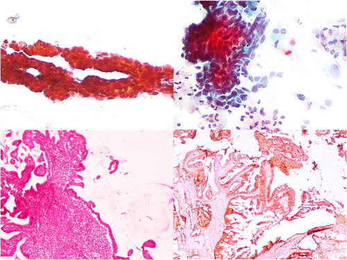
Villoglandular adenocarcinoma (VGA) of the cervix is a rare tumour with very few reports of cytological diagnosis on a cervical smear. Since the usual prognosis of VGA is more favourable than that of a conventional cervical adenocarcinoma, a pre-operative diagnosis is essential for appropriate therapeutic decisions. We report the clinical, cytological, and histopathological features of a case of VGA in an elderly woman. A 60-year-old female presented with postmenopausal bleeding and was found to have a hypertrophied cervix. Conventional smears from the cervix demonstrated features of atypical glandular cells with a villoglandular configuration. Endocervical cell nuclei were enlarged with overlapping, hyperchromasia and mild to moderate anisonucleosis. The cytological impression of villoglandular carcinoma was confirmed on subsequent hysterectomy and histopathology. Hence, cytopathologists need to be aware of the entity of villoglandular carcinoma of the cervix, its relatively bland nuclear features, and the diagnostic clues to allow for an accurate diagnosis on cervical smear. An accurate pre-operative diagnosis can assist the gynaecologists in arriving at appropriate therapeutic and prognostic decisions.
Graphical Abstract
Villoglandular carcinoma of the cervix is a rare subtype of cervical adenocarcinoma that can be recognised on cytology by three-dimensional papillary fragments and relatively bland nuclear features. Knowledge of this entity is imperative for an accurate pre-operative diagnosis and appropriate management.
Conflicts of Interest
The authors declare no conflicts of interest.
Open Research
Data Availability Statement
All data generated or analysed during this study are included in this article.


