FNA Diagnosis of Adult Rhabdomyoma of the Tongue and Floor of the Mouth
Funding: The authors received no specific funding for this work.
ABSTRACT
Adult rhabdomyoma is a rare benign tumour of skeletal muscle origin and is rarely reported on fine needle aspiration cytology. This tumour has a preference for males older than 40 years of age. The most common sites include the parapharyngeal space, larynx, salivary glands, mouth, and soft tissue of the head and neck region. Fine needle aspiration cytology (FNAC), a less invasive alternative diagnostic method than incisional biopsy, can accurately diagnose this benign neoplasm and avoid aggressive treatment. To the best of our knowledge, only 11 cases of adult rhabdomyoma are diagnosed on FNAC. We herein report one such rare case of adult rhabdomyoma of the floor of the mouth in a 39-year-old male diagnosed with FNAC, emphasising its utility in making a definite diagnosis of adult rhabdomyoma.
Graphical Abstract
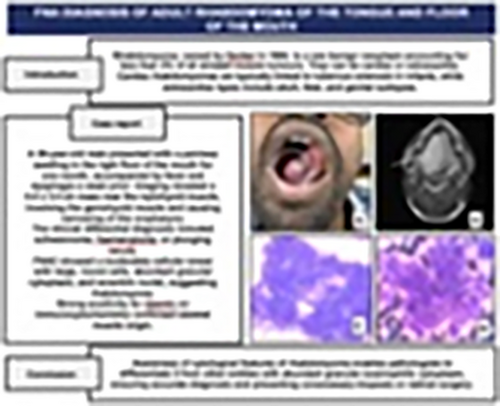
Awareness of rhabdomyoma and its distinct morphological features enables pathologists to differentiate it from other entities with abundant granular eosinophilic cytoplasm, ensuring accurate diagnosis and preventing unnecessary biopsies or radical surgical interventions.
Rhabdomyoma is a slow-growing mass occurring in the head and neck region. A definite diagnosis can be made in FNA cytology with the awareness of the classical morphological features (tumor cells in cohesive clusters, abundant granular eosinophilic cytoplasm without atypia and mitosis), obviating the need for a biopsy or frozen section. Immunocytochemistry can be performed on the smears, and immunohistochemistry on the cell blocks if available. Preoperative diagnosis is necessary to avoid aggressive or unnecessary radical surgery.
Conflicts of Interest
The authors declare no conflicts of interest.
Open Research
Data Availability Statement
Data sharing not applicable to this article as no datasets were generated or analysed during the current study.


