Prostate cancer tissue classification by multiphoton imaging, automated image analysis and machine learning
Egleidson F. A. Gomes
Departamento de Física, Instituto de Ciências Exatas, Universidade Federal de Minas Gerais, Belo Horizonte, MG, Brazil
Search for more papers by this authorEduardo Paulino Junior
Departamento de Anatomia Patológica e Medicina Legal, Faculdade de Medicina, Universidade Federal de Minas Gerais, Belo Horizonte, MG, Brazil
Search for more papers by this authorMário F. R. de Lima
Laboratório Analys Patologia, Belo Horizonte, MG, Brazil
Search for more papers by this authorLuana A. Reis
Departamento de Física, Instituto de Ciências Exatas, Universidade Federal de Minas Gerais, Belo Horizonte, MG, Brazil
Search for more papers by this authorGiovanna Paranhos
Departamento de Física, Instituto de Ciências Exatas, Universidade Federal de Minas Gerais, Belo Horizonte, MG, Brazil
Search for more papers by this authorMarcelo Mamede
Departamento Anatomia e Imagem, Faculdade de Medicina, Universidade Federal de Minas Gerais, Belo Horizonte, MG, Brazil
Search for more papers by this authorFrancis G. J. Longford
University of Southampton, Southampton, UK
Search for more papers by this authorCorresponding Author
Ana Maria de Paula
Departamento de Física, Instituto de Ciências Exatas, Universidade Federal de Minas Gerais, Belo Horizonte, MG, Brazil
Correspondence
Ana Maria de Paula, Departamento de Física, Instituto de Ciências Exatas, Universidade Federal de Minas Gerais, 31270-901 Belo Horizonte, MG, Brazil.
Email: [email protected]
Search for more papers by this authorEgleidson F. A. Gomes
Departamento de Física, Instituto de Ciências Exatas, Universidade Federal de Minas Gerais, Belo Horizonte, MG, Brazil
Search for more papers by this authorEduardo Paulino Junior
Departamento de Anatomia Patológica e Medicina Legal, Faculdade de Medicina, Universidade Federal de Minas Gerais, Belo Horizonte, MG, Brazil
Search for more papers by this authorMário F. R. de Lima
Laboratório Analys Patologia, Belo Horizonte, MG, Brazil
Search for more papers by this authorLuana A. Reis
Departamento de Física, Instituto de Ciências Exatas, Universidade Federal de Minas Gerais, Belo Horizonte, MG, Brazil
Search for more papers by this authorGiovanna Paranhos
Departamento de Física, Instituto de Ciências Exatas, Universidade Federal de Minas Gerais, Belo Horizonte, MG, Brazil
Search for more papers by this authorMarcelo Mamede
Departamento Anatomia e Imagem, Faculdade de Medicina, Universidade Federal de Minas Gerais, Belo Horizonte, MG, Brazil
Search for more papers by this authorFrancis G. J. Longford
University of Southampton, Southampton, UK
Search for more papers by this authorCorresponding Author
Ana Maria de Paula
Departamento de Física, Instituto de Ciências Exatas, Universidade Federal de Minas Gerais, Belo Horizonte, MG, Brazil
Correspondence
Ana Maria de Paula, Departamento de Física, Instituto de Ciências Exatas, Universidade Federal de Minas Gerais, 31270-901 Belo Horizonte, MG, Brazil.
Email: [email protected]
Search for more papers by this authorAbstract
Prostate carcinoma, a slow-growing and often indolent tumour, is the second most commonly diagnosed cancer among men worldwide. The prognosis is mainly based on the Gleason system through prostate biopsy analysis. However, new treatment and monitoring strategies depend on a more precise diagnosis. Here, we present results by multiphoton imaging for prostate tumour samples from 120 patients that allow to obtain quantitative parameters leading to specific tumour aggressiveness signatures. An automated image analysis was developed to recognise and quantify stromal fibre and neoplastic cell regions in each image. The set of metrics was able to distinguish between non-neoplastic tissue and carcinoma areas by linear discriminant analysis and random forest with accuracy of 89% ± 3%, but between Gleason groups of only 46% ± 6%. The reactive stroma analysis improved the accuracy to 65% ± 5%, clearly demonstrating that stromal parameters should be considered as additional criteria for a more accurate diagnosis.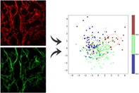
CONFLICT OF INTEREST STATEMENT
The authors declare no potential conflicts of interest.
Open Research
DATA AVAILABILITY STATEMENT
The data that support the findings of this study are available from the corresponding author upon reasonable request.
REFERENCES
- 1J. N. Ouellette, C. R. Drifka, K. B. Pointer, Y. Liu, T. J. Lieberthal, W. John Kao, J. S. Kuo, A. G. Loeffler, K. W. Eliceiri, Bioengineering 2021, 8, 17.
- 2X. Catteau, P. Simon, M. Jondet, M. Vanhaeverbeek, J.-C. Noël, PLoS One 2019, 14, e0210263.
- 3G. Ayala, J. A. Tuxhorn, T. M. Wheeler, A. Frolov, P. T. Scardino, M. Ohori, M. Wheeler, J. Spitler, D. R. Rowley, Clin. Cancer Res. 2003, 9, 4792.
- 4G. Falzon, S. Pearson, R. Murison, Phys. Med. Biol. 2008, 53, 6641.
- 5P. P. Provenzano, K. W. Eliceiri, J. M. Campbell, D. R. Inman, J. G. White, P. J. Keely, BMC Med. 2006, 4, 38.
- 6X. Han, R. M. Burke, M. L. Zettel, P. Tang, E. B. Brown, Opt. Express 2008, 16, 1846.
- 7P. P. Provenzano, D. R. Inman, K. W. Eliceiri, J. G. Knittel, L. Yan, C. T. Rueden, J. G. White, P. J. Keely, BMC Med. 2008, 6, 11.
- 8A. E. Tuer, S. Krouglov, N. Prent, R. Cisek, D. Sandkuijl, K. Yasufuku, B. C. Wilson, V. Barzda, J. Phys. Chem. B 2011, 115, 12759.
- 9P. Campagnola, Anal. Chem. 2011, 83, 3224.
- 10M. W. Conklin, J. C. Eickhoff, K. M. Riching, C. A. Pehlke, K. W. Eliceiri, P. P. Provenzano, A. Friedl, P. J. Keely, Am. J. Pathol. 2011, 178, 1221.
- 11V. Ajeti, O. Nadiarnykh, S. M. Ponik, P. J. Keely, K. W. Eliceiri, P. J. Campagnola, Biomed. Opt. Express 2011, 2, 2307.
- 12K. Burke, P. Tang, E. B. Brown, J. Biomed. Opt. 2012, 18, 031106.
10.1117/1.JBO.18.3.031106 Google Scholar
- 13A. Ghazaryan, H. F. Tsai, G. Hayrapetyan, W.-L. Chen, Y.-F. Chen, M.-Y. Jeong, C.-S. Kim, S.-J. Chen, C.-Y. Dong, J. Biomed. Opt. 2012, 18, 031105.
10.1117/1.JBO.18.3.031105 Google Scholar
- 14K. Zhang, C. A. Corsa, S. M. Ponik, J. L. Prior, D. Piwnica-Worms, K. W. Eliceiri, P. J. Keely, G. D. Longmore, Nat. Cell Biol. 2013, 15, 677.
- 15J. S. Bredfeldt, Y. Liu, C. A. Pehlke, M. W. Conklin, J. M. Szulczewski, D. R. Inman, P. J. Keely, R. D. Nowak, T. R. Mackie, K. W. Eliceiri, J. Biomed. Opt. 2014, 19, 016007.
- 16J. Adur, V. B. Pelegati, A. A. de Thomaz, M. O. Baratti, D. B. Almeida, L. A. L. A. Andrade, F. Bottcher-Luiz, H. F. Carvalho, C. L. Cesar, PLoS One 2012, 7, e47007.
- 17J. Adur, V. B. Pelegati, A. A. de Thomaz, M. O. Baratti, L. A. L. A. Andrade, H. F. Carvalho, F. Bottcher-Luiz, C. L. Cesar, J. Biophotonics 2014, 7, 37.
- 18W. Xiufeng, G. Chen, L. Jianping, W. Zhu, J. Qiu, J. Chen, S. Xie, S. Zhuo, J. Yan, PLoS One 2013, 8, e65933.
- 19M. Bizzarri, A. Cucina, BioMed Res. Int. 2014, 2014, 934038.
10.1155/2014/934038 Google Scholar
- 20Y. K. Tao, D. Shen, Y. Sheikine, O. O. Ahsen, H. H. Wang, D. B. Schmolze, N. B. Johnson, J. S. Brooker, A. E. Cable, J. L. Connolly, J. G. Fujimoto, Proc. Natl. Acad Sci. 2014, 111, 15304.
- 21A. Brabrand, I. I. Kariuki, M. J. Engstrøm, O. A. Haugen, L. A. Dyrnes, B. O. Åsvold, M. B. Lilledahl, A. M. Bofin, APIMIS 2015, 123, 1.
- 22W. J. Tan, J. Yan, S. Xu, A. A. Thike, B. H. Bay, H. Yu, M.-H. Tan, P. H. Tan, J. Clin. Pathol. 2015, 68, 1033.
- 23K. Tilbury, P. J. Campagnola, Perspect. Med Chem. 2015, 7, 21.
- 24A. Golaraei, L. Kontenis, R. Cisek, D. Tokarz, S. J. Done, B. C. Wilson, V. Barzda, Biomed. Opt. Express 2016, 7, 4054.
- 25M. S. Hall, F. Alisafaei, E. Ban, X. Feng, C.-Y. Hui, V. B. Shenoy, M. Wu, Proc. Natl. Acad. Sci. 2016, 113, 14043.
- 26K. Wang, W. Fei, B. R. Seo, C. Fischbach, W. Chen, L. Hsu, D. Gourdon, Matrix Biol. 2017, 60–61, 86.
- 27A. Case, B. K. Brisson, A. C. Durham, S. Rosen, J. Monslow, E. Buza, P. Salah, J. Gillem, G. Ruthel, S. Veluvolu, V. Kristiansen, E. Puré, D. C. Brown, K. U. Sørenmo, S. W. Volk, PLoS One 2017, 12, e0180448.
- 28M. W. Conklin, R. E. Gangnon, B. L. Sprague, L. Van Gemert, J. M. Hampton, K. W. Eliceiri, J. S. Bredfeldt, Y. Liu, N. Surachaicharn, P. A. Newcomb, A. Friedl, P. J. Keely, A. Trentham-Dietz, Cancer Epidemiol. Biomarkers Prev. 2018, 27, 138.
- 29R. A. Natal, J. Vassallo, G. R. Paiva, V. B. Pelegati, G. O. Barbosa, G. R. Mendonça, C. Bondarik, S. F. Derchain, H. F. Carvalho, C. S. Lima, C. L. Cesar, L. O. Sarian, Tumor Biol. 2018, 40, 1010428318770953.
10.1177/1010428318770953 Google Scholar
- 30R. A. Natal, G. R. Paiva, V. B. Pelegati, L. Marenco, C. A. Alvarenga, R. F. Vargas, S. F. Derchain, L. O. Sarian, C. Franchet, C. L. Cesar, et al., Sci. Rep. 2019, 9, 7715.
- 31S. L. Best, Y. Liu, A. Keikhosravi, C. R. Drifka, K. M. Woo, G. S. Mehta, M. Altwegg, T. N. Thimm, M. Houlihan, J. S. Bredfeldt, E. Jason Abel, W. Huang, K. W. Eliceiri, BMC Cancer 2019, 19, 490.
- 32L. Gole, J. Yeong, J. C. T. Lim, K. H. Ong, H. Han, A. A. Thike, Y. C. Poh, S. Yee, J. Iqbal, W. Hong, B. Lee, W. Yu, P. H. Tan, Breast Cancer Res. 2020, 22, 42.
- 33Y. Ling, C. Li, K. Feng, S. Palmer, P. L. Appleton, S. Lang, D. McGloin, Z. Huang, G. Nabi, J. Biophotonics 2016, 10, 911.
- 34A. M. Garcia, F. L. Magalhães, J. S. Soares, E. P. Junior, M. F. R. de Lima, M. Mamede, A. M. de Paula, Biomed. Phys. Eng. Express 2017, 4, 025026.
10.1088/2057-1976/aaa379 Google Scholar
- 35J. I. Epstein, M. J. Zelefsky, D. D. Sjoberg, J. B. Nelson, L. Egevad, C. Magi-Galluzzi, A. J. Vickers, A. V. Parwani, V. E. Reuter, S. W. Fine, J. A. Eastham, P. Wiklund, M. Han, C. A. Reddy, J. P. Ciezki, T. Nyberg, E. A. Klein, Eur. Urol. 2016, 69, 428.
- 36I. P. Pavlova, S. S. Nair, D. Lundon, S. Sobotka, R. Roshandel, P.-J. Treacy, P. Ratnani, R. Brody, J. I. Epstein, G. E. Ayala, N. Kyprianou, A. K. Tewari, J. Personalized Med. 2021, 11, 1061.
- 37L. A. Reis, A. P. V. Garcia, E. F. A. Gomes, F. G. J. Longford, J. G. Frey, G. D. Cassali, A. M. de Paula, Biomed. Opt. Express 2020, 11, 6413.
- 38F. G. J. Longford, PyFibre: Python Fibrous Image Analysis Toolkit (Version 2.1.1). https://github.com/franklongford/PyFibre, 2022.
- 39A. M. Stein, D. A. Vader, L. M. Jawerth, D. A. Weitz, L. M. Sander, J. Microsc. 2008, 232, 463.
- 40Y. Sato, S. Nakajima, N. Shiraga, H. Atsumi, S. Yoshida, T. Koller, G. Gerig, R. Kikinis, Med. Image Anal. 1998, 2, 143.
- 41J. S. Bredfeldt, Y. Liu, M. W. Conklin, P. J. Keely, T. R. Mackie, K. W. Eliceiri, J. Pathol. Informatics 2014, 5, 28.
- 42Y. Liu, A. Keikhosravi, G. S. Mehta, C. R. Drifka, K. W. Eliceiri, in Fibrosis: Methods in Molecular Biology, Vol. 1627 (Ed: L. Rittié), Springer, New York, NY 2017, p. 429.
- 43D. Sculley, Proc. 19th Int. Conf. World Wide Web, 2010, 1177–1178.
- 44A. M. Martínez, A. C. Kak, IEEE Trans. Pattern Anal. Mach. Intell. 2001, 23, 228.
- 45K. Fukunaga, Statistical pattern recognition, World Scientific, Singapore 1993.
- 46B. Dai, R. Chen, S. Zhu, W. Zhang, In: 2018 Int. Symp. Comput. Consumer Control (IS3C), 2018, 449–452.
- 47B. Jahne, Digital image processing, Vol. 4, Springer, Berlin 2005.
- 48F. J. Ávila, O. del Barco, J. M. Bueno, J. Biomed. Opt. 2015, 20, 086001.
- 49F. J. Ávila, O. del Barco, J. M. Bueno, J. Biomed. Opt. 2016, 21, 066015.
- 50F. J. Ávila, O. Del Barco, J. M. Bueno, J. Opt. 2017, 19, 105301.
10.1088/2040-8986/aa825d Google Scholar
- 51F. J. Ávila, J. M. Bueno, Appl. Opt. 2015, 54, 9848.
- 52R. Rezakhaniha, A. Agianniotis, J. T. C. Schrauwen, A. Griffa, D. Sage, C. V. C. vd Bouten, F. N. Van de Vosse, M. Unser, N. Stergiopulos, Biomechan. Model. Mechanobiol. 2012, 11, 461.
- 53L. Egevad, B. Delahunt, D. M. Berney, D. G. Bostwick, J. Cheville, E. Comperat, A. J. Evans, S. W. Fine, D. J. Grignon, P. A. Humphrey, J. Hörnblad, K. A. Iczkowski, J. G. Kench, G. Kristiansen, K. R. M. Leite, C. Magi-Galluzzi, J. K. McKenney, J. Oxley, C.-C. Pan, H. Samaratunga, J. R. Srigley, H. Takahashi, L. D. True, T. Tsuzuki, T. van der Kwast, M. Varma, M. Zhou, M. Clements, Histopathology 2018, 73, 8.
- 54D. J. Lomas, H. U. Ahmed, Nat. Rev. Clin. Oncol. 2020, 17, 372.
Citing Literature