Volume 13, Issue 10 e202000245
FULL ARTICLE

Optical coherence tomography angiography for mapping cerebral microvasculature based on normalized differentiation analysis

Jiang Zhu

Corresponding Author

Jiang Zhu

Key Laboratory of the Ministry of Education for Optoelectronic Measurement Technology and Instrument, Beijing Information Science and Technology University, Beijing, China

Beijing Laboratory of Biomedical Testing Technology and Instruments, Beijing Information Science and Technology University, Beijing, China

Correspondence

Jiang Zhu and Lianqing Zhu, Key Laboratory of the Ministry of Education for Optoelectronic Measurement Technology and Instrument, Beijing Information Science and Technology University, Beijing 100192, China.

Email: [email protected] (J. Z.) and [email protected] (L. Z.)

Search for more papers by this author
Jianting Liu

Jianting Liu

Key Laboratory of the Ministry of Education for Optoelectronic Measurement Technology and Instrument, Beijing Information Science and Technology University, Beijing, China

Beijing Laboratory of Biomedical Testing Technology and Instruments, Beijing Information Science and Technology University, Beijing, China

Search for more papers by this author
Lianqing Zhu

Corresponding Author

Lianqing Zhu

Key Laboratory of the Ministry of Education for Optoelectronic Measurement Technology and Instrument, Beijing Information Science and Technology University, Beijing, China

Beijing Key Laboratory of Optoelectronic Measurement Technology, Beijing Information Science and Technology University, Beijing, China

Correspondence

Jiang Zhu and Lianqing Zhu, Key Laboratory of the Ministry of Education for Optoelectronic Measurement Technology and Instrument, Beijing Information Science and Technology University, Beijing 100192, China.

Email: [email protected] (J. Z.) and [email protected] (L. Z.)

Search for more papers by this author
Chongyang Wang

Chongyang Wang

Key Laboratory of the Ministry of Education for Optoelectronic Measurement Technology and Instrument, Beijing Information Science and Technology University, Beijing, China

Beijing Laboratory of Biomedical Testing Technology and Instruments, Beijing Information Science and Technology University, Beijing, China

Search for more papers by this author
Fan Fan

Fan Fan

Beijing Laboratory of Biomedical Testing Technology and Instruments, Beijing Information Science and Technology University, Beijing, China

Search for more papers by this author
Qiang Yang

Qiang Yang

Beijing Laboratory of Biomedical Testing Technology and Instruments, Beijing Information Science and Technology University, Beijing, China

Search for more papers by this author
Fan Zhang

Fan Zhang

Beijing Laboratory of Biomedical Testing Technology and Instruments, Beijing Information Science and Technology University, Beijing, China

Search for more papers by this author
First published: 08 July 2020
Citations: 3

Funding information: Key Research Cultivation Project of Beijing Information Science and Technology University for Promoting University Connotative Development and Improving Research Level, Grant/Award Number: 2019KYNH205; National Natural Science Foundation of China, Grant/Award Number: 61975019; Research Project of Beijing Municipal Education Commission, Grant/Award Number: KZ202011232050; Start-up Foundation of Beijing Information Science and Technology University, Grant/Award Number: 5029011104

Abstract

Optical coherence tomography angiography (OCTA) is a label-free, noninvasive biomedical imaging modality for mapping microvascular networks and quantifying blood flow velocities in vivo. Simple computation and fast processing are critical for the OCTA in some applications. Herein, we report on a normalized differentiation method for mapping cerebral microvasculature with the advantages of simple analysis and high image quality, benefitting from computation of differentiation and characteristics of normalization. Normalized differentiation values are validated to have a nearly linear relationship with flow velocities in a range using a flow phantom. The measurements in a rat cerebral cortex show that the OCTA based on the normalized differentiation analysis can generate microvascular images with high quality and monitor spatiotemporal dynamics of blood flow with simple computation and fast processing before and after localized ischemia induced by arterial occlusion.image

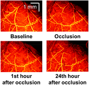
CONFLICT OF INTEREST

The authors declare no financial or commercial conflict of interest.

The full text of this article hosted at iucr.org is unavailable due to technical difficulties.