Rapid identification of human ovarian cancer in second harmonic generation images using radiomics feature analyses and tree-based pipeline optimization tool
Guangxing Wang
School of Science, Jimei University, Xiamen, Fujian, China
Key Laboratory of OptoElectronic Science and Technology for Medicine of Ministry of Education & Fujian Provincial Key Laboratory of Photonics Technology, Fujian Normal University, Fuzhou, Fujian, China
Guangxing Wang and Yang Sun contributed equally to this study.
Search for more papers by this authorCorresponding Author
Yang Sun
Department of Gynecology, Fujian Cancer Hospital, Affiliated Cancer Hospital of Fujian Medical University, Fuzhou, Fujian, China
Guangxing Wang and Yang Sun contributed equally to this study.
Correspondence
Yang Sun, Yang Sun, Department of Gynecology, Fujian Cancer Hospital, Affiliated Cancer Hospital of Fujian Medical University, Fuzhou, Fujian, China.
Email: [email protected]
Shuangmu Zhuo, School of Science, Jimei University, Xiamen, Fujian, China.
Email: [email protected]
Search for more papers by this authorYouting Chen
Department of Hepatopancreatobiliary Surgery, the First Affiliated Hospital, Fujian Medical University, Fuzhou, China
Search for more papers by this authorQiqi Gao
Department of Gynecology, Fujian Cancer Hospital, Affiliated Cancer Hospital of Fujian Medical University, Fuzhou, Fujian, China
Search for more papers by this authorDongqing Peng
School of Science, Jimei University, Xiamen, Fujian, China
Search for more papers by this authorHongxin Lin
Key Laboratory of OptoElectronic Science and Technology for Medicine of Ministry of Education & Fujian Provincial Key Laboratory of Photonics Technology, Fujian Normal University, Fuzhou, Fujian, China
Search for more papers by this authorZhenlin Zhan
Key Laboratory of OptoElectronic Science and Technology for Medicine of Ministry of Education & Fujian Provincial Key Laboratory of Photonics Technology, Fujian Normal University, Fuzhou, Fujian, China
Search for more papers by this authorZhiyi Liu
State Key Laboratory of Modern Optical Instrumentation, College of Optical Science and Engineering, Zhejiang University, Hangzhou Zhejiang, China
Search for more papers by this authorCorresponding Author
Shuangmu Zhuo
School of Science, Jimei University, Xiamen, Fujian, China
Key Laboratory of OptoElectronic Science and Technology for Medicine of Ministry of Education & Fujian Provincial Key Laboratory of Photonics Technology, Fujian Normal University, Fuzhou, Fujian, China
Correspondence
Yang Sun, Yang Sun, Department of Gynecology, Fujian Cancer Hospital, Affiliated Cancer Hospital of Fujian Medical University, Fuzhou, Fujian, China.
Email: [email protected]
Shuangmu Zhuo, School of Science, Jimei University, Xiamen, Fujian, China.
Email: [email protected]
Search for more papers by this authorGuangxing Wang
School of Science, Jimei University, Xiamen, Fujian, China
Key Laboratory of OptoElectronic Science and Technology for Medicine of Ministry of Education & Fujian Provincial Key Laboratory of Photonics Technology, Fujian Normal University, Fuzhou, Fujian, China
Guangxing Wang and Yang Sun contributed equally to this study.
Search for more papers by this authorCorresponding Author
Yang Sun
Department of Gynecology, Fujian Cancer Hospital, Affiliated Cancer Hospital of Fujian Medical University, Fuzhou, Fujian, China
Guangxing Wang and Yang Sun contributed equally to this study.
Correspondence
Yang Sun, Yang Sun, Department of Gynecology, Fujian Cancer Hospital, Affiliated Cancer Hospital of Fujian Medical University, Fuzhou, Fujian, China.
Email: [email protected]
Shuangmu Zhuo, School of Science, Jimei University, Xiamen, Fujian, China.
Email: [email protected]
Search for more papers by this authorYouting Chen
Department of Hepatopancreatobiliary Surgery, the First Affiliated Hospital, Fujian Medical University, Fuzhou, China
Search for more papers by this authorQiqi Gao
Department of Gynecology, Fujian Cancer Hospital, Affiliated Cancer Hospital of Fujian Medical University, Fuzhou, Fujian, China
Search for more papers by this authorDongqing Peng
School of Science, Jimei University, Xiamen, Fujian, China
Search for more papers by this authorHongxin Lin
Key Laboratory of OptoElectronic Science and Technology for Medicine of Ministry of Education & Fujian Provincial Key Laboratory of Photonics Technology, Fujian Normal University, Fuzhou, Fujian, China
Search for more papers by this authorZhenlin Zhan
Key Laboratory of OptoElectronic Science and Technology for Medicine of Ministry of Education & Fujian Provincial Key Laboratory of Photonics Technology, Fujian Normal University, Fuzhou, Fujian, China
Search for more papers by this authorZhiyi Liu
State Key Laboratory of Modern Optical Instrumentation, College of Optical Science and Engineering, Zhejiang University, Hangzhou Zhejiang, China
Search for more papers by this authorCorresponding Author
Shuangmu Zhuo
School of Science, Jimei University, Xiamen, Fujian, China
Key Laboratory of OptoElectronic Science and Technology for Medicine of Ministry of Education & Fujian Provincial Key Laboratory of Photonics Technology, Fujian Normal University, Fuzhou, Fujian, China
Correspondence
Yang Sun, Yang Sun, Department of Gynecology, Fujian Cancer Hospital, Affiliated Cancer Hospital of Fujian Medical University, Fuzhou, Fujian, China.
Email: [email protected]
Shuangmu Zhuo, School of Science, Jimei University, Xiamen, Fujian, China.
Email: [email protected]
Search for more papers by this authorFunding information: Incubation program of Fujian Health and Family Planning Department for Young and Mid-aged Backbones, Grant/Award Number: 2018-ZQN-44; Joint Funds of Fujian Provincial Health and Education Research, Grant/Award Number: 2019-WJ-21; National Natural Science Foundation of China, Grant/Award Numbers: 81771881, 61905214; Natural Science Foundation of Fujian Province, Grant/Award Numbers: 2018J07004, 2018J01416; Zhejiang Provincial Natural Science Foundation of China, Grant/Award Number: LR20F050001; Fundamental Research Funds for the Central Universities, Grant/Award Number: 2019QNA5004
Abstract
Ovarian cancer is currently one of the most common cancers of the female reproductive organs, and its mortality rate is the highest among all types of gynecologic cancers. Rapid and accurate classification of ovarian cancer plays an important role in the determination of treatment plans and prognoses. Nevertheless, the most commonly used classification method is based on histopathological specimen examination, which is time-consuming and labor-intensive. Thus, in this study, we utilize radiomics feature extraction methods and the automated machine learning tree-based pipeline optimization tool (TOPT) for analysis of 3D, second harmonic generation images of benign, malignant and normal human ovarian tissues, to develop a high-efficiency computer-aided diagnostic model. Area under the receiver operating characteristic curve values of 0.98, 0.96 and 0.94 were obtained, respectively, for the classification of the three tissue types. Furthermore, this approach can be readily applied to other related tissues and diseases, and has great potential for improving the efficiency of medical diagnostic processes.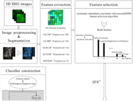
Supporting Information
| Filename | Description |
|---|---|
| jbio202000050-sup-0001-Supinfo.docxWord 2007 document , 1.5 MB | Appendix S1. Supporting Information. |
Please note: The publisher is not responsible for the content or functionality of any supporting information supplied by the authors. Any queries (other than missing content) should be directed to the corresponding author for the article.
REFERENCES
- 1W. Chen, R. Zheng, P. D. Baade, S. Zhang, H. Zeng, F. Bray, A. Jemal, X. Q. Yu, J. He, CA Cancer J. Clin. 2016, 66, 115.
- 2F. Bray, J. Ferlay, I. Soerjomataram, R. L. Siegel, L. A. Torre, A. Jemal, CA Cancer J. Clin. 2018, 68, 394.
- 3T. Kaku, S. Ogawa, Y. Kawano, Y. Ohishi, H. Kobayashi, T. Hirakawa, H. Nakano, Med. Electron Microsc. 2003, 36, 9.
- 4E. L. Moss, J. Hollingworth, T. M. Reynolds, J. Clin. Pathol. 2005, 58, 308.
- 5N. Bharwani, R. H. Reznek, A. G. Rockall, Eur. J. Radiol. 2011, 78, 41.
- 6N. Roberts, D. Magee, Y. Song, K. Brabazon, M. Shires, D. Crellin, N. M. Orsi, R. Quirke, P. Quirke, D. Treanor, Am. J. Pathol. 2012, 180, 1835.
- 7P. J. Campagnola, C. Y. Dong, Laser Photonics Rev. 2011, 5, 13.
- 8P. J. Campagnola, L. M. Loew, Nat. Biotechnol. 2003, 21, 1356.
- 9C. Xiyi, N. Oleg, P. Sergey, P. J. Campagnola, Nat. Protoc. 2012, 7, 654.
- 10B. Kathleen, T. Ping, B. Edward, J. Biomed. Opt. 2012, 18, 31106.
- 11R. Lacomb, O. Nadiarnykh, P. J. Campagnola, Biophys. J. 2008, 94, 4504.
- 12M. Kochueva, V. Dudenkova, S. Kuznetsov, A. Varlamova, A. Maslennikova, J. Biomed. Opt. 2018, 23, 1.
- 13G. Thomas, O. Christophe, L. G. Yann, G. Luc, T. Bruno, E. Frédérick, D. Yoann, B. Georges, G. Dominique, Opt. Express 2010, 18, 25794.
- 14J. M. Watson, P. F. Rice, S. L. Marion, M. A. Brewer, J. R. Davis, J. J. Rodriguez, U. Urs, P. B. Hoyer, J. K. Barton, J. Biomed. Opt. 2012, 17, 0760021.
- 15C. Ricciardelli, R. J. Rodgers, Semin. Reprod. Med. 2006, 24, 270.
- 16L. Philippe, R. V. Emmanuel, L. Ralph, C. Sara, R. G. P. M. Stiphout, G. Van, C. M. L. Patrick, G. Zegers, B. Robert, D. A. Ronald, Eur. J. Cancer 2007, 43, 441.
- 17H. J. Aerts, E. R. Velazquez, R. T. Leijenaar, C. Parmar, P. Grossmann, S. Carvalho, J. Bussink, R. Monshouwer, B. Haibe-Kains, D. Rietveld, Nat. Commun. 2014, 5, 4006.
- 18H. Arimura, M. Soufi, K. Ninomiya, H. Kamezawa, M. Yamada, Radiol. Phys. Technol. 2018, 11, 365.
- 19S. Huang, B. L. Franc, R. J. Harnish, G. Liu, D. Mitra, T. P. Copeland, V. A. Arasu, J. Kornak, E. F. Jones, S. C. Behr, Npj Breast Cancer. 2018, 4, 24.
- 20Y. Q. Huang, C. H. Liang, L. He, J. Tian, C.-s. Liang, X. Chen, Z.-l. Ma, Z.-Y. Liu, J. Clin. Oncol. 2016, 34, 2157.
- 21T. W. Sawyer, J. W. Koevary, F. P. Rice, C. C. Howard, O. J. Austin, D. C. Connolly, K. Q. Cai, J. K. Barton, J. Biomed. Opt. 2019, 24, 096010.
- 22W. Huang, S. Tu, Medical Phys. Ther. 2016, 43, 3377.
- 23T. T. Le, W. Fu, J. H. Moore, Bioinformatics 2020, 36, 250.
- 24P. Hanchuan, L. Fuhui, D. Chris, IEEE Trans. Pattern. Anal. Mach. Intell. 2005, 27, 1226.
- 25H. Lin, L. Lin, G. Wang, N. Zuo, Z. Zhan, S. Xie, G. Chen, J. Chen, S. Zhuo, Biomed. Opt. Express 2018, 9, 3783.
- 26X. Zheng, N. Zuo, H. Lin, L. Zheng, M. Ni, G. Wu, J. Chen, S. Zhuo, Surg. Endosc. 2020, 34, 408.
- 27P. Coupé, M. Munz, J. V. Manjón, E. S. Ruthazer, D. L. Collins, Med. Image Anal. 2012, 16, 849.
- 28V. G. Jjm, A. Fedorov, C. Parmar, A. Hosny, N. Aucoin, V. Narayan, B. T. Rgh, J. C. Fillion-Robin, S. Pieper, A. Hjwl, Cancer Res. 2017, 77, e104.
- 29R. M. Haralick, K. Shanmugam, I. H. Dinstein, IEEE Trans. Syst. Man Cybern. 1973, SMC-3, 610.
10.1109/TSMC.1973.4309314 Google Scholar
- 30G. Thibault, J. Angulo, F. Meyer, IEEE Trans. Biomed. Eng. 2014, 61, 630.
- 31M. M. Galloway, Comput Graph Image Proce. 1975, 4, 172.
10.1016/S0146-664X(75)80008-6 Google Scholar
- 32M. Amadasun, R. King, IEEE Transactions Systems Man & Cybernetics, IEEE, Piscataway, Vol. 19, 1989, p. 1264.
- 33C. Sun, W. G. Wee, Comput. Vis. Graph. Image Proce. 1983, 23, 341.
- 34M. Avanzo, J. Stancanello, N. I. El, Phys. Med. 2017, 38, 122.
- 35R. S. Olson, R. J. Urbanowicz, P. C. Andrews, N. A. Lavender, L. C. Kidd, J. H. Moore in Automating Biomedical Data Science Through Tree-Based Pipeline Optimization, Vol. (Ed.^Eds.: Squillero, Giovanni and Burelli, Paolo), Springer International Publishing, Porto, 2016.
10.1007/978-3-319-31204-0_9 Google Scholar
- 36T. Chen, C. Guestrin in Xgboost: A scalable tree boosting system, Vol. (Ed.^Eds.: Balaji Krishnapuram, Mohak Shah), ACM, New York, 2016, pp. 785–794.
- 37F. Pedregosa, G. Varoquaux, A. Gramfort, V. Michel, B. Thirion, O. Grisel, M. Blondel, P. Prettenhofer, R. Weiss, V. Dubourg, J. Mach. Learn. Res. 2011, 12, 2825.
- 38F.-A. Fortin, D. Rainville, G. François-Michel, P. Marc-André, G. Marc, Christ. J. Mach. Learn. Res. 2012, 13, 2171.
- 39L. A. Torre, B. Trabert, C. E. DeSantis, K. D. Miller, G. Samimi, C. D. Runowicz, M. M. Gaudet, A. Jemal, R. L. Siegel, CA Cancer J. Clin. 2018, 68, 284.
- 40Z. Momenimovahed, A. Tiznobaik, S. Taheri, H. Salehiniya, Int. J. Womens Health 2019, 11, 287.
- 41R. P. Mecham, Curr. Protoc. Cell Biol. 2012, 57, 10.11. 11.
10.1002/0471143030.cb1001s57 Google Scholar
- 42I. G. Schauer, A. K. Sood, S. Mok, J. Liu, Neoplasia 2011, 13, 393.
- 43K. B. Tilbury, K. R. Campbell, K. W. Eliceiri, S. M. Salih, M. Patankar, P. J. Campagnola, BMC Cancer 2017, 17, 102.
- 44H. Bao, A. Boussioutas, R. Jeremy, S. Russell, M. Gu, Opt. Express 2010, 18, 1255.
- 45Y. Zhang, M. L. Akins, K. Murari, J. Xi, M.-J. Li, K. Luby-Phelps, M. Mahendroo, X. Li, Proc. Natl. Acad. Sci. 2012, 109, 12878.
- 46J. Adur, V. B. Pelegati, A. A. de Thomaz, M. O. Baratti, L. A. Andrade, H. F. Carvalho, F. Bottcher-Luiz, C. L. Cesar, J. Biophotonics 2014, 7, 37.
- 47B. Wen, K. R. Campbell, K. Tilbury, O. Nadiarnykh, M. A. Brewer, M. Patankar, V. Singh, K. W. Eliceiri, P. J. Campagnola, Sci. Rep. 2016, 6, 35734.
Citing Literature