Optical effects on the surrounding structure during quantitative analysis using indocyanine green videoangiography: A phantom vessel study
Abstract
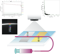
Various reports have been published regarding quantitative evaluations of intraoperative fluorescent intensity studies using indocyanine green (ICG) with videoangiography (VAG). The effects of scattering and point-spread functions (PSF) on quantitative ICG-VAG evaluations have not been investigated. Clinically, when ICG is administered through the peripheral vein, it reaches the tissue intra-arterially. To achieve more reliable intraoperative quantitative intensity evaluations, we examined the impact of high-intensity structures on close areas. The study was conducted using a phantom model and surgical fluorescent microscope. A region of interest (ROI) was created for the vessel model and another ROI was created within 3 cm of that. With an ROI of 6.8 mm in the vessel phantom model, 10% intensity was confirmed, even though there was no fluorescent structure. Intensity decreased gradually as the ROI moved further from the vessel model. Our study results suggest that the presence of a high-intensity structure and the size of the ROI may affect quantitative intensity evaluations using ICG-VAG. Results of linear regression analysis indicate that the relationship of intensity (Y) and distance (X) is as follows: Y(real/A) = 29 Exp(−0.062X) + 164.3 Exp(−1.81X). The optical effect should be considered when performing an intraoperative intensity study with a surgical microscope.