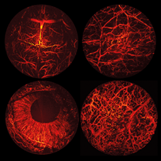
Portable optical-resolution photoacoustic microscopy for volumetric imaging of multiscale organisms
Tian Jin
School of Physical Electronics, University of Electronic Science and Technology of China, Chengdu, China
Search for more papers by this authorHeng Guo
School of Physical Electronics, University of Electronic Science and Technology of China, Chengdu, China
Search for more papers by this authorLei Yao
School of Physical Electronics, University of Electronic Science and Technology of China, Chengdu, China
Center for Information in Biomedicine, University of Electronic Science and Technology of China, Chengdu, China
Search for more papers by this authorHuikai Xie
Department of Electrical and Computer Engineering, University of Florida, Gainesville, Florida
Search for more papers by this authorCorresponding Author
Huabei Jiang
School of Physical Electronics, University of Electronic Science and Technology of China, Chengdu, China
Center for Information in Biomedicine, University of Electronic Science and Technology of China, Chengdu, China
Department of Medical Engineering, University of South Florida, Tampa, Florida
Correspondence
Huabei Jiang, Department of Medical Engineering, University of South Florida, Tampa, FL 33620. Email: [email protected]
Lei Xi, School of Physical Electronics, University of Electronic Science and Technology of China, Chengdu, 610054, China. Email: [email protected]
Search for more papers by this authorCorresponding Author
Lei Xi
School of Physical Electronics, University of Electronic Science and Technology of China, Chengdu, China
Center for Information in Biomedicine, University of Electronic Science and Technology of China, Chengdu, China
Correspondence
Huabei Jiang, Department of Medical Engineering, University of South Florida, Tampa, FL 33620. Email: [email protected]
Lei Xi, School of Physical Electronics, University of Electronic Science and Technology of China, Chengdu, 610054, China. Email: [email protected]
Search for more papers by this authorTian Jin
School of Physical Electronics, University of Electronic Science and Technology of China, Chengdu, China
Search for more papers by this authorHeng Guo
School of Physical Electronics, University of Electronic Science and Technology of China, Chengdu, China
Search for more papers by this authorLei Yao
School of Physical Electronics, University of Electronic Science and Technology of China, Chengdu, China
Center for Information in Biomedicine, University of Electronic Science and Technology of China, Chengdu, China
Search for more papers by this authorHuikai Xie
Department of Electrical and Computer Engineering, University of Florida, Gainesville, Florida
Search for more papers by this authorCorresponding Author
Huabei Jiang
School of Physical Electronics, University of Electronic Science and Technology of China, Chengdu, China
Center for Information in Biomedicine, University of Electronic Science and Technology of China, Chengdu, China
Department of Medical Engineering, University of South Florida, Tampa, Florida
Correspondence
Huabei Jiang, Department of Medical Engineering, University of South Florida, Tampa, FL 33620. Email: [email protected]
Lei Xi, School of Physical Electronics, University of Electronic Science and Technology of China, Chengdu, 610054, China. Email: [email protected]
Search for more papers by this authorCorresponding Author
Lei Xi
School of Physical Electronics, University of Electronic Science and Technology of China, Chengdu, China
Center for Information in Biomedicine, University of Electronic Science and Technology of China, Chengdu, China
Correspondence
Huabei Jiang, Department of Medical Engineering, University of South Florida, Tampa, FL 33620. Email: [email protected]
Lei Xi, School of Physical Electronics, University of Electronic Science and Technology of China, Chengdu, 610054, China. Email: [email protected]
Search for more papers by this authorAbstract
Photoacoustic microscopy (PAM) provides a fundamentally new tool for a broad range of studies of biological structures and functions. However, the use of PAM has been largely limited to small vertebrates due to the large size/weight and the inconvenience of the equipment. Here, we describe a portable optical-resolution photoacoustic microscopy (pORPAM) system for 3-dimensional (3D) imaging of small-to-large rodents and humans with a high spatiotemporal resolution and a large field of view. We show extensive applications of pORPAM to multiscale animals including mice and rabbits. In addition, we image the 3D vascular networks of human lips, and demonstrate the feasibility of pORPAM to observe the recovery process of oral ulcer and cancer-associated capillary loops in human oral cavities. This technology is promising for broad biomedical studies from fundamental biology to clinical diseases.

Supporting Information
| Filename | Description |
|---|---|
| jbio201700250-sup-0001-author-biographies.docxapplication/docx, 184 KB | Author Biographies |
| jbio201700250-sup-0002-AppendixS1.docxWord document, 584.7 KB |
Figure S1. The layout of the CUPAM configuration. CFT, cylindrically focused transducer; DAQ, data acquisition card; GVS, galvanometer scanner; OL, objective; PC, Personal computer; PH, pinhole; R, rotator; SL, scan lens; SMF, single-mode fiber Figure S2. The photographs of the mouse in the experiments of (A) brain and (B) ear imaging |
| jbio201700250-sup-0003-MovieS1.mp43.7 MB | Movie S1. Scanning mechanism of pORPAM. |
| jbio201700250-sup-0004-MovieS2.mp41.1 MB | Movie S2. The volume rendering of a mouse brain imaged by the low-resolution mode in different views and cross-sectional slices at different depths. |
| jbio201700250-sup-0005-MovieS3.mp41.1 MB | Movie S3. The volume rendering of a mouse brain imaged by the high-resolution mode in different views and cross-sectional slices at different depths. |
| jbio201700250-sup-0006-MovieS4.mp43.1 MB | Movie S4. Volumetric rendering and slices of the vascular network at different depths in a human lip. |
Please note: The publisher is not responsible for the content or functionality of any supporting information supplied by the authors. Any queries (other than missing content) should be directed to the corresponding author for the article.
REFERENCES
- 1V.Ntziachristos, Nat. Methods 2010, 7, 603.
- 2V.Ntziachristos, J.Ripoll, L. V.Wang, R.Weissleder, Nat. Biotechnol. 2005, 23, 313.
- 3A. Oraevsky, A. Karabutov, in Biomedical Photonics Handbook (T. Vo-Dinh, USA, 2003), p. 3401–3434.
- 4A.Taruttis, V.Ntziachristos, Nat. Photonics 2015, 9, 219.
- 5P.Beard, Interface Focus 2011, 1, 602.
- 6L. V.Wang, Nat. Photonics 2009, 3, 503.
- 7R.Kruger, R.Lam, D.Reinecke, S.Del Rio, R.Doyle, Med. Phys. 2010, 37, 6096.
- 8R.Ma, S.Söntges, S.Shoham, V.Ntziachristos, D.Razansky, Biomed. Opt. Exp. 2012, 3, 1724.
- 9H.Estrada, J.Turner, M.Kneipp, D.Razansky, Laser Phys. Lett. 2014, 11, 045601.
- 10A.Taruttis, G.Dam, V.Ntziachristos, Cancer Res. 2015, 75, 1548.
- 11A. P.Jathoul, A. P. Jathoul, J. Laufer, O. Ogunlade, B. Treeby, B. Cox, E. Zhang, P. Johnson, A. R. Pizzey, B. Philip, T. Marafioti, M. F. Lythgoe, R. B. Pedley, M. A. Pule, P. Beard, Nat. Photonics 2015, 9, 239.
- 12M.Kircher, A. Zerda, J. V. Jokerst, C. L. Zavaleta, P. J. Kempen, E. Mittra, K. Pitter, R. Huang, C. Campos, F. Habte, R. Sinclair, C. W. Brennan, I. K. Mellinghoff, E. C. Holland, S. S. Gambhir, Nat. Med. 2012, 18, 829.
- 13S.Manohar, S. E.Vaartjes, J. C. G.van Hespen, J. M.Klaase, F. M.van den Engh, W.Steenbergen, T. G.van Leeuwen, Opt. Express 2007, 15, 12277.
- 14X.Deánben, G. Sela, A. Lauri, M. Kneipp, V. Ntziachristos, G. Westmeyer, S. Shoham, D. Razansky, Light Sci. Appl. 2016, 5.
- 15S.Ermilov, T. Khamapirad, A. Conjusteau, M. H. Leonard, R. Lacewell, K. Mehta, T. Miller, A. A. Oraevsky, J. Biomed. Opt. 2009, 14, 024007.
- 16H.Zhang, K.Maslov, G.Stoica, L. V.Wang, Nat. Biotechnol. 2006, 24, 848.
- 17S.Hu, K.Maslov, L. V.Wang, Opt. Lett. 2011, 36, 1134.
- 18H.Wang, X.Yang, Y.Liu, B.Jiang, Q.Luo, Opt. Express 2013, 21, 24210.
- 19W.Song, W. Zheng, R. Liu, R. Lin, H. Huang, X. Gong, S. Yang, R. Zhang, L. Song, Biomed. Opt. Express 2014, 12, 4235.
- 20L.Lin, P. Zhang, S. Xu, J. Shi, L. Li, J. Yao, L. Wang, J. Zou, L. V. Wang, J. Biomed. Opt. 2017, 22, 41002.
- 21J.Kim, C.Lee, K.Park, G.Lim, C.Kim, Sci. Rep. 2015, 5, 7932.
- 22P.Hajireza, W.Shi, A.Forbrich, P.Shao, R.Zemp, Proc. SPIE 2012, 8223, 82230I-1.
10.1117/12.931287 Google Scholar
- 23Y.Yuan, S.Yang, D.Xing, Appl. Phys. Lett. 2012, 100, 023702.
- 24H.Kang, S.Lee, E.Lee, S.Kim, T.Lee, Biomed. Opt. Express 2015, 6, 4650.
- 25L.Li, C. Yeh, S. Hu, L. Wang, B. T. Soetikno, R. Chen, Q. Zhou, K. K. Shung, K. I. Maslov, L. V. Wang, Opt. Lett. 2014, 39, 2117.
- 26B.Rao, L.Li, K.Maslov, L.Wang, Opt. Lett. 2010, 35, 1521.
- 27W.Qi, T.Jin, J.Rong, H.Jiang, L.Xi, J. Biophotonics 2017, 10, 950.
- 28J.Takano, T.Yakushiji, I.Kamiyama, T.Nomura, A.Katakura, N.Takano, T.Shibahara, Int. J. Oral Maxillofac. Surg. 2010, 39, 208.
- 29P.Hajireza, A.Forbrich, R.Zemp, Biomed. Opt. Express 2014, 5, 539.
- 30S.Hu, K.Maslov, V.Tsytsarev, L. V.Wang, J. Biomed. Opt. 2009, 14, 040503.
- 31L.Wang, K.Maslov, L. V.Wang, Proc. Natl. Acad. Sci. U.S.A. 2013, 110, 5759.
- 32Z.Luo, P.Wang, A.Zhang, G.Zuo, Y.Zheng, Y.Huang, PLoS One 2015, 10, e0116076.
- 33H.Inoue, M. Kaga, H. Ikeda, C. Sato, H. Sato, H. Minami, E. G. Santi, B. Hayee, N. Eleftheriadis, Ann. Gastroenterol. 2015, 28, 41.
- 34S.Oladipipo, S. Hu, A. Santeford, J. Yao, J. Kovalski, R. Shohet, K. Maslov, L. V. Wang, J. Arbeit, Blood 2011, 117, 4142.
- 35P.Hai, Y. Zhou, R. Zhang, J. Ma, Y. Li, J. Shao, L. V. Wang, J. Biomed. Opt. 2017, 22, 41004.
- 36R.Cao, J.Li, B.Ning, N.Sun, T.Wang, Z.Zuo, S.Hu, NeuroImage 2017, 150, 77.
- 37W.Lu, Q.Huang, G.Ku, X.Wen, M.Zhou, D.Guzatov, P.Brecht, R.Su, A.Oraevsky, L. V.Wang, C.Li, Biomaterials 2010, 31, 2617.
- 38L.Xiang, L. Ji, T. Zhang, B. Wang, J. Yang, Q. Zhang, M. S. Jiang, J. Zhou, P. R. Carney, H. Jiang, Neuroimage 2013, 66, 240.
- 39A.Zerda, C. Zavaleta, S. Keren, S. Vaithilingam, S. Bodapati, Z. Liu, J. Levi, B.R. Smith, T. Ma, O. Oralkan, Z. Cheng, X. Chen, H. Dai, B. T. Khuri-Yakub, S. S. Gambhir, Nat. Nanotechnol. 2008, 3, 557.
- 40R. Lin, J. Chen, H. Wang, M. Yan, W. Zheng, L. Song, Quant. Imaging Med. Surg. 2015, 5, 23.


