Volume 11, Issue 4 e201700232
FULL ARTICLE

Perfusion-based fluorescence imaging method delineates diverse organs and identifies multifocal tumors using generic near-infrared molecular probes

Jessica Miller

Jessica Miller

Optical Radiology Laboratory, Mallinckrodt Institute of Radiology, Washington University School of Medicine, St. Louis, Missouri

Biomedical Engineering, Washington University in St. Louis, St. Louis, Missouri

Search for more papers by this author
Steven T. Wang

Steven T. Wang

Optical Radiology Laboratory, Mallinckrodt Institute of Radiology, Washington University School of Medicine, St. Louis, Missouri

Search for more papers by this author
Inema Orukari

Inema Orukari

Optical Radiology Laboratory, Mallinckrodt Institute of Radiology, Washington University School of Medicine, St. Louis, Missouri

Biomedical Engineering, Washington University in St. Louis, St. Louis, Missouri

Search for more papers by this author
Julie Prior

Julie Prior

Optical Radiology Laboratory, Mallinckrodt Institute of Radiology, Washington University School of Medicine, St. Louis, Missouri

Search for more papers by this author
Gail Sudlow

Gail Sudlow

Optical Radiology Laboratory, Mallinckrodt Institute of Radiology, Washington University School of Medicine, St. Louis, Missouri

Search for more papers by this author
Xinming Su

Xinming Su

Department of Medicine, Division of Oncology, Washington University School of Medicine, St. Louis, Missouri

Search for more papers by this author
Kexian Liang

Kexian Liang

Optical Radiology Laboratory, Mallinckrodt Institute of Radiology, Washington University School of Medicine, St. Louis, Missouri

Search for more papers by this author
Rui Tang

Rui Tang

Optical Radiology Laboratory, Mallinckrodt Institute of Radiology, Washington University School of Medicine, St. Louis, Missouri

Search for more papers by this author
Elizabeth M.C. Hillman

Elizabeth M.C. Hillman

Department of Biomedical Engineering, Columbia University, New York, New York

Search for more papers by this author
Katherine N. Weilbaecher

Katherine N. Weilbaecher

Department of Medicine, Division of Oncology, Washington University School of Medicine, St. Louis, Missouri

Search for more papers by this author
Joseph P. Culver

Joseph P. Culver

Optical Radiology Laboratory, Mallinckrodt Institute of Radiology, Washington University School of Medicine, St. Louis, Missouri

Biomedical Engineering, Washington University in St. Louis, St. Louis, Missouri

Search for more papers by this author
Mikhail Y. Berezin

Mikhail Y. Berezin

Optical Radiology Laboratory, Mallinckrodt Institute of Radiology, Washington University School of Medicine, St. Louis, Missouri

Department of Chemistry, Washington University in St. Louis, St. Louis, Missouri

Search for more papers by this author
Samuel Achilefu

Corresponding Author

Samuel Achilefu

Optical Radiology Laboratory, Mallinckrodt Institute of Radiology, Washington University School of Medicine, St. Louis, Missouri

Biomedical Engineering, Washington University in St. Louis, St. Louis, Missouri

Department of Biochemistry and Molecular Biophysics, Washington University School of Medicine, St. Louis, Missouri

Correspondence

Samuel Achilefu, Optical Radiology Laboratory, Mallinckrodt Institute of Radiology, Washington University School of Medicine, 4515 McKinley Ave, St. Louis, MO 63110.

Email: [email protected]

Search for more papers by this author
First published: 05 December 2017
Citations: 6
Funding information National Cancer Institute, Grant/Award numbers: R01 CA171651, R01 CA171651-05S1, P50 CA094056, U54CA199092, R01 CA208623; National Institutes of Health, Grant/Award numbers: R01 EB008111, S10 OD016237, S10 RR031625, S10 RR031621, S10 OD020129; Alvin J. Siteman Cancer Research Fund, Grant/Award number: 11-FY16-01; Department of Defense Breast Cancer Research Program, Grant/Award number: W81XWH-16-1-0286; National Institute of Biomedical Imaging and Bioengineering, Grant/Award numbers: R01 EB008111, NIBIB R01 EB007276

Abstract

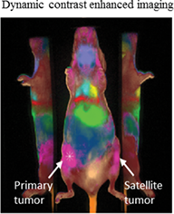
Rapid detection of multifocal cancer without the use of complex imaging schemes will improve treatment outcomes. In this study, dynamic fluorescence imaging was used to harness differences in the perfusion kinetics of near-infrared (NIR) fluorescent dyes to visualize structural characteristics of different tissues. Using the hydrophobic nontumor-selective NIR dye cypate, and the hydrophilic dye LS288, a high tumor-to-background contrast was achieved, allowing the delineation of diverse tissue types while maintaining short imaging times. By clustering tissue types with similar perfusion properties, the dynamic fluorescence imaging method identified secondary tumor locations when only the primary tumor position was known, with a respective sensitivity and specificity of 0.97 and 0.75 for cypate, and 0.85 and 0.81 for LS288. Histological analysis suggests that the vasculature in the connective tissue that directly surrounds the tumor was a major factor for tumor identification through perfusion imaging. Although the hydrophobic dye showed higher specificity than the hydrophilic probe, use of other dyes with different physical and biological properties could further improve the accuracy of the dynamic imaging platform to identify multifocal tumors for potential use in real-time intraoperative procedures.

image

The full text of this article hosted at iucr.org is unavailable due to technical difficulties.