Optical emission of 223Radium: in vitro and in vivo preclinical applications
Abstract
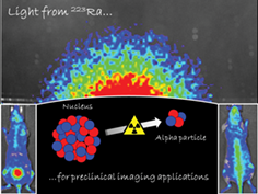
223Radium (223Ra) is widely used in nuclear medicine to treat patients with osseous metastatic prostate cancer. In clinical practice 223Ra cannot be imaged directly; however, gamma photons produced by its short-lived daughter nuclides can be captured by conventional gamma cameras. In this work, we show that 223Ra and its short-lived daughter nuclides can be detected with optical imaging techniques. The light emission of 223Ra was investigated in vitro using different setups in order to clarify the mechanism of light production. The results demonstrate that the luminescence of the 223Ra chloride solution, usually employed in clinical treatments, is compatible with Cerenkov luminescence having an emission spectrum that is almost indistinguishable from CR one. This study proves that luminescence imaging can be successfully employed to detect 223Ra in vivo in mice by imaging whole body 223Ra biodistribution and more precisely its uptake in bones.