Multicontrast endomyocardial imaging by single-channel high-resolution cross-polarization optical coherence tomography
Xinwen Yao
Department of Electrical Engineering, Columbia University, New York, New York
Search for more papers by this authorYu Gan
Department of Electrical Engineering, Columbia University, New York, New York
Search for more papers by this authorYuye Ling
Department of Electrical Engineering, Columbia University, New York, New York
Search for more papers by this authorCharles C. Marboe
Department of Pathology and Cell Biology, Columbia University Medical Center, New York, New York
Search for more papers by this authorCorresponding Author
Christine P. Hendon
Department of Electrical Engineering, Columbia University, New York, New York
Correspondence
Christine P. Hendon, Department of Electrical Engineering, Columbia University, 500 West 120th Street, Mudd 1310, New York, NY 10027.
Email: [email protected]
Search for more papers by this authorXinwen Yao
Department of Electrical Engineering, Columbia University, New York, New York
Search for more papers by this authorYu Gan
Department of Electrical Engineering, Columbia University, New York, New York
Search for more papers by this authorYuye Ling
Department of Electrical Engineering, Columbia University, New York, New York
Search for more papers by this authorCharles C. Marboe
Department of Pathology and Cell Biology, Columbia University Medical Center, New York, New York
Search for more papers by this authorCorresponding Author
Christine P. Hendon
Department of Electrical Engineering, Columbia University, New York, New York
Correspondence
Christine P. Hendon, Department of Electrical Engineering, Columbia University, 500 West 120th Street, Mudd 1310, New York, NY 10027.
Email: [email protected]
Search for more papers by this authorAbstract
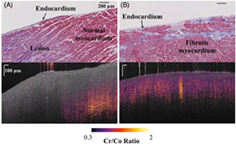
A single-channel high-resolution cross-polarization (CP) optical coherence tomography (OCT) system is presented for multicontrast imaging of human myocardium in one-shot measurement. The intensity and functional contrasts, including the ratio between the cross- and co-polarization channels as well as the cumulative retardation, are reconstructed from the CP-OCT readout. By comparing the CP-OCT results with histological analysis, it is shown that the system can successfully delineate microstructures in the myocardium and differentiate the fibrotic myocardium from normal or ablated myocardium based on the functional contrasts provided by the CP-OCT system. The feasibility of using A-line profiles from the 2 orthogonal polarization channels to identify fibrotic myocardium, normal myocardium and ablated lesion is also discussed.
REFERENCES
- 1M.Nathan, L. C.Ying, C.Pierre, B.David, L.João, J. Am. Coll. Cardiol. 2011, 57, 891.
- 2M.Ciulla, R.Paliotti, D. B.Hess, E.Tjahja, S. E.Campbell, F.Magrini, K. T.Weber, J. Am. Soc. Echocardiogr. 1997, 10(6), 657.
- 3M.Nahrendorf, C.Badea, L. W.Hedlund, J.-L.Figueiredo, D. E.Sosnovik, G. A.Johnson, R.Weissleder, Am. J. Phys. Heart Circ. Phys. 2007, 292, H3172.
- 4L. T.Cooper, K. L.Baughman, A. M.Feldman, A.Frustaci, M.Jessup, U.Kuhl, G. N.Levine, J.Narula, R. C.Starling, J.Towbin, R.Virmani, Circulation 2007, 116, 2216.
- 5F.Saraiva, V.Matos, L.Gonçalves, M.Antunes, L.Providência, Transplant. Proc. 2011, 43, 1908.
- 6A. M.From, J. J.Maleszewski, C. S.Rihal, Mayo Clin. Proc. 2011, 86, 1095.
- 7C. P.Fleming, K. J.Quan, H.Wang, G.Amit, A. M.Rollins, Opt. Express 2010, 18, 3079.
- 8Y.Gan, C. P.Fleming, Biomed. Opt. Express 2013, 4, 2150.
- 9M.Cua, E.Lin, L.Lee, X.Sheng, K. S. K.Wong, G. F.Tibbits, M. F.Beg, M. V.Sarunic, J. Biomed. Opt. 2014, 19, 116007.
- 10X.Yao, Y.Gan, C. C.Marboe, C. P.Hendon, J. Biomed. Opt. 2016, 21, 061006.
- 11C. J.Goergen, H. H.Chen, S.Sakadžić, V. J.Srinivasan, D. E.Sosnovik, Phys. Rep. 2016, 4, e12894.
- 12C. J.Goergen, H.Radhakrishnan, S.Sakadžić, E. T.Mandeville, E. H.Lo, D. E.Sosnovik, V. J.Srinivasan, Opt. Lett. 2012, 37, 3882.
- 13C.Fan, G.Yao, Biomed. Opt. Express 2013, 4, 460.
- 14Y.Wang, G.Yao, Biomed. Opt. Express 2013, 4, 2540.
- 15S.Wang, A.Lopez, Y.Morikawa, G.Tao, J.Li, I.Larina, J.Martin, K.Larin, Biomed. Opt. Express 2014, 5, 1980.
- 16C. W.Sun, Y. M.Wang, L. S.Lu, C. W.Lu, I. J.Hsu, M. T.Tsai, C. C.Yang, Y. W.Kiang, C. C.Wu, J. Biomed. Opt. 2006, 11, 054016.
- 17C. P.Fleming, N.Rosenthal, A. M.Rollins, M.Arruda, J. Innovations Cardiac Rhythm Manage. 2011, 2, 199.
- 18L. M.Peterson, M. W.Jenkins, S.Gu, L.Barwick, M.Watanabe, A. M.Rollins, Biomed. Opt. Express 2012, 3, 3022.
- 19S.Wang, D. S.Lakomy, M. D.Garcia, A. L.Lopez, K. V.Larin, I. V.Larina, J. Biophotonics 2016, 9, 837.
- 20X.Fu, Z.Wang, H.Wang, Y. T.Wang, M. W.Jenkins, A. M.Rollins, Opt. Lett. 2014, 39, 5066.
- 21C.Mekkaoui, S.Huang, H. H.Chen, G.Dai, T. G.Reese, W. J.Kostis, A.Thiagalingam, P.Maurovich-Horvat, J. N.Ruskin, U.Hoffmann, M. P.Jackowski, D. E.Sosnovik, J. Cardiovasc. Magn. Reson. 2012, 14(1), 70.
- 22D. E.Sosnovik, R.Wang, G.Dai, T.Wang, E.Aikawa, M.Novikov, A.Rosenzweig, R. J.Gilbert, V. J.Wedeen, Circ. Cardiovasc. Imaging 2017, 10(9), 206.
- 23Y.Gan, X.Yao, D.Tsay, C. C.Marboe, C. P.Hendon, Proc. SPIE 2017, 10042, 1004207.
10.1117/12.2253200 Google Scholar
- 24I.Ahmad, A.Gribble, M.Ikram, M.Pop, A.Vitkin, J. Biophotonics 2016, 9, 750.
- 25N.Ghosh, M. F. G.Wood, S.-H.Li, R. D.Weisel, B. C.Wilson, R.-K.Li, I. A.Vitkin, J. Biophotonics 2009, 2, 145.
- 26M.-R.Tsai, Y.-W.Chiu, M. T.Lo, C.-K.Sun, J. Biomed. Opt. 2010, 15, 026002.
- 27T. P.Martin, G.Norris, G.McConnell, S.Currie, Int. J. Cardiovasc. Imaging 2013, 29, 1733.
- 28J. M.Schmitt, S. H.Xiang, Opt. Lett. 1998, 23, 1060.
- 29F. I.Feldchtein, G. V.Gelikonov, V. M.Gelikonov, R. R.Iksanov, R. V.Kuranov, A. M.Sergeev, N. D.Gladkova, M. N.Ourutina, J. A.Warren, D. H.Reitze, Opt. Express 1998, 3, 239.
- 30N.Gladkova, O.Streltsova, E.Zagaynova, E.Kiseleva, V.Gelikonov, G.Gelikonov, M.Karabut, K.Yunusova, O.Evdokimova, J. Biophotonics 2011, 4, 519.
- 31E.Gubarkova, M.Kirillin, V.Dudenkova, P.Timashev, S.Kotova, E.Kiseleva, L.Timofeeva, G.Belkova, A.Solovieva, A.Moiseev, G.Gelikonov, I.Fiks, F.Feldchtein, N.Gladkova, J. Biomed. Opt. 2016, 21, 126010.
- 32K.Rasmussen, C.Reilly, Y.Li, R. S.Jones, Biotechnol. Bioeng. 2016, 113, 198.
- 33C.Fan, Y.Wang, R. K.Wang, Opt. Express 2007, 15, 7950.
- 34C. K.Hitzenberger, E.Götzinger, M.Sticker, M.Pircher, A. F.Fercher, Biomed. Opt. Express 2001, 9, 780.
- 35Y.Ling, Y.Gan, X.Yao, C. P.Hendon, Opt. Lett. 2017, 42, 1333.
- 36R. P.Singh-Moon, C. C.Marboe, C. P.Hendon, Biomed. Opt. Express 2015, 6, 2494.
- 37H.Wang, T.Akkin, C.Magnain, R.Wang, J.Dubb, W. J.Kostis, M. A.Yaseen, A.Cramer, S.Sakadžić, D.Boas, Opt. Lett. 2016, 41, 2213.
- 38S.Fialovć, M.Augustin, M.Glösmann, T.Himmel, S.Rauscher, M.Gröger, M.Pircher, C. K.Hitzenberger, B.Baumann, Biomed. Opt. Express 2016, 7, 1479.
- 39C.Fan, G.Yao, Opt. Lett. 2012, 37, 1415.
- 40C.Fan, G.Yao, Opt. Express 2012, 20, 22360.
Citing Literature