Volume 10, Issue 6-7 pp. 854-861
Full Article

Comparative study of presurgical skin infiltration depth measurements of melanocytic lesions with OCT and high frequency ultrasound

Arthur Varkentin

Corresponding Author

Arthur Varkentin

Hannoversches Zentrum für Optische Technologien (HOT), Leibniz Universität Hannover, Nienburger Str. 17, 30167 Hannover, Germany

Corresponding author: e-mail: [email protected]

Search for more papers by this author
Mikhail Mazurenka

Mikhail Mazurenka

Hannoversches Zentrum für Optische Technologien (HOT), Leibniz Universität Hannover, Nienburger Str. 17, 30167 Hannover, Germany

Search for more papers by this author
Elias Blumenröther

Elias Blumenröther

Hannoversches Zentrum für Optische Technologien (HOT), Leibniz Universität Hannover, Nienburger Str. 17, 30167 Hannover, Germany

Search for more papers by this author
Merve Meinhardt-Wollweber

Merve Meinhardt-Wollweber

Hannoversches Zentrum für Optische Technologien (HOT), Leibniz Universität Hannover, Nienburger Str. 17, 30167 Hannover, Germany

Search for more papers by this author
Maik Rahlves

Maik Rahlves

Hannoversches Zentrum für Optische Technologien (HOT), Leibniz Universität Hannover, Nienburger Str. 17, 30167 Hannover, Germany

Search for more papers by this author
Sigrid M. C. Broekaert

Sigrid M. C. Broekaert

Universitätsmedizin Göttingen, Abteilung Dermatologie, Venerologie und Allergologie, Robert-Koch-Str. 40, 37075 Göttingen, Germany

Search for more papers by this author
Susanne Schäd-Trcka

Susanne Schäd-Trcka

Universitätsmedizin Rostock, Klinik und Poliklinik für Dermatologie und Venerologie, Strempelstr. 13, 18057 Rostock, Germany

Search for more papers by this author
Steffen Emmertinst

Steffen Emmertinst

Universitätsmedizin Rostock, Klinik und Poliklinik für Dermatologie und Venerologie, Strempelstr. 13, 18057 Rostock, Germany

Search for more papers by this author
Uwe Morgner

Uwe Morgner

Institut für Quantenoptik, Leibniz Universität Hannover, Welfengarten 1, 30167 Germany

Search for more papers by this author
Bernhard Roth

Bernhard Roth

Hannoversches Zentrum für Optische Technologien (HOT), Leibniz Universität Hannover, Nienburger Str. 17, 30167 Hannover, Germany

Search for more papers by this author
First published: 23 December 2016
Citations: 37

Abstract

A reliable, fast, and non-invasive determination of melanoma thickness in vivo is highly desirable for clinical dermatology as it may facilitate the identification of surgical melanoma margins, determine if a sentinel node biopsy should be performed or not, and reduce the number of surgical interventions for patients. In this work, optical coherence tomography (OCT) and high frequency ultrasound (HFUS) are evaluated for quantitative in vivo preoperative assessment of the skin infiltration depth of melanocytic tissue. Both methods allow non-invasive imaging of skin at similar axial resolution. Comparison with the Breslow lesion thickness obtained from histopathology revealed that OCT is slightly more precise in terms of thickness determination while HFUS has better contrast. The latter does not require image post-processing, as necessary for the OCT images. The findings of our pilot study suggest that non-invasive OCT and HFUS are able to determine the infiltration depth of lesions like melanocytic nevi or melanomas preoperatively and in vivo with a precision comparable to invasive histopathology measurements on skin biopsies. In future, to further strengthen our findings a statistically significant study comprising a larger amount of data is required which will be conducted in an extended clinical study in the next step.

figure

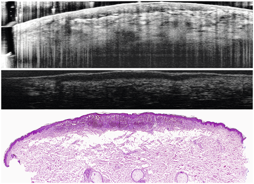
Comparison of optical coherence tomography and high frequency ultrasound B-Scans and a H&E stained histology of a melanocytic nevus.

The full text of this article hosted at iucr.org is unavailable due to technical difficulties.