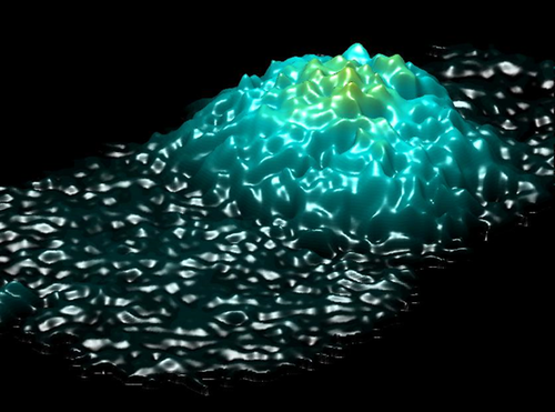
Investigating fibroblast cells under “safe” and “injurious” blue-light exposure by holographic microscopy
Alejandro Calabuig
National Council of Research, Institute of Applied Science & Intelligent Systems (ISASI) ’E. Caianiello', Via Campi Flegrei 34, 80078 Pozzuoli (NA), Italy
Department of Chemical, Materials and Industrial Production Engineering, University of Naples Federico II, P. le Tecchio 80, 80125 Napoli, Italy
Search for more papers by this authorMartina Mugnano
National Council of Research, Institute of Applied Science & Intelligent Systems (ISASI) ’E. Caianiello', Via Campi Flegrei 34, 80078 Pozzuoli (NA), Italy
Department of Chemical, Materials and Industrial Production Engineering, University of Naples Federico II, P. le Tecchio 80, 80125 Napoli, Italy
Search for more papers by this authorLisa Miccio
National Council of Research, Institute of Applied Science & Intelligent Systems (ISASI) ’E. Caianiello', Via Campi Flegrei 34, 80078 Pozzuoli (NA), Italy
Search for more papers by this authorCorresponding Author
Simonetta Grilli
- [email protected]
- +39 081 867 5305
National Council of Research, Institute of Applied Science & Intelligent Systems (ISASI) ’E. Caianiello', Via Campi Flegrei 34, 80078 Pozzuoli (NA), Italy
Corresponding author: e-mail: [email protected], Phone: +39 081 867 5305
Search for more papers by this authorPietro Ferraro
National Council of Research, Institute of Applied Science & Intelligent Systems (ISASI) ’E. Caianiello', Via Campi Flegrei 34, 80078 Pozzuoli (NA), Italy
Search for more papers by this authorAlejandro Calabuig
National Council of Research, Institute of Applied Science & Intelligent Systems (ISASI) ’E. Caianiello', Via Campi Flegrei 34, 80078 Pozzuoli (NA), Italy
Department of Chemical, Materials and Industrial Production Engineering, University of Naples Federico II, P. le Tecchio 80, 80125 Napoli, Italy
Search for more papers by this authorMartina Mugnano
National Council of Research, Institute of Applied Science & Intelligent Systems (ISASI) ’E. Caianiello', Via Campi Flegrei 34, 80078 Pozzuoli (NA), Italy
Department of Chemical, Materials and Industrial Production Engineering, University of Naples Federico II, P. le Tecchio 80, 80125 Napoli, Italy
Search for more papers by this authorLisa Miccio
National Council of Research, Institute of Applied Science & Intelligent Systems (ISASI) ’E. Caianiello', Via Campi Flegrei 34, 80078 Pozzuoli (NA), Italy
Search for more papers by this authorCorresponding Author
Simonetta Grilli
- [email protected]
- +39 081 867 5305
National Council of Research, Institute of Applied Science & Intelligent Systems (ISASI) ’E. Caianiello', Via Campi Flegrei 34, 80078 Pozzuoli (NA), Italy
Corresponding author: e-mail: [email protected], Phone: +39 081 867 5305
Search for more papers by this authorPietro Ferraro
National Council of Research, Institute of Applied Science & Intelligent Systems (ISASI) ’E. Caianiello', Via Campi Flegrei 34, 80078 Pozzuoli (NA), Italy
Search for more papers by this authorAbstract
The exposure to visible light has been shown to exert various biological effects, such as erythema and retinal degeneration. However, the phototoxicity mechanisms in living cells are still not well understood. Here we report a study on the temporal evolution of cell morphology and volume during blue light exposure. Blue laser irradiation is switched during the operation of a digital holography (DH) microscope between what we call here “safe” and “injurious” exposure (SE & IE). The results reveal a behaviour that is typical of necrotic cells, with early swelling and successive leakage of the intracellular liquids when the laser is set in the “injurious” operation. In the phototoxicity investigation reported here the light dose modulation is performed through the very same laser light source adopted for monitoring the cell's behaviour by digital holographic microscope. We believe the approach may open the route to a deep investigation of light-cell interactions, with information about death pathways and threshold conditions between healthy and damaged cells when subjected to light-exposure.
Supporting Information
| Filename | Description |
|---|---|
| jbio201500340-sup-0001-supp-information.pdfPDF document, 2.3 MB | Supporting Information |
| jbio201500340-sup-0002-SupplMovie1.aviAVI video, 2.2 MB | Supplementary Information - Movie 1 |
| jbio201500340-sup-0003-SupplMovie2.aviAVI video, 1.1 MB | Supplementary Information - Movie 2 |
| jbio201500340-sup-0004-SupplMovie3.aviAVI video, 2 MB | Supplementary Information - Movie 3 |
| jbio201500340-sup-0005-SupplMovie4.aviAVI video, 4.6 MB | Supplementary Information - Movie 4 |
| jbio201500340-sup-0006-SupplMovie5.aviAVI video, 7.8 MB | Supplementary Information - Movie 5 |
| jbio201500340-sup-0007-SupplMovie6.aviAVI video, 4.2 MB | Supplementary Information - Movie 6 |
Please note: The publisher is not responsible for the content or functionality of any supporting information supplied by the authors. Any queries (other than missing content) should be directed to the corresponding author for the article.
References
- 1P. B. Rottier and J. C. Van Der Leun, Br. J. Dermatol. 72, 256–260 (1960).
- 2N. Kollias and A. Baqer, Photochem. Photobiol. 39, 651–659 (1984).
- 3S. B. Porges, K. H. Kaidbey, and G. L. Grove, Photodermatology 5, 197–200 (1988).
- 4M. Mittelbrunn, R. Tejedor, H. de la Fuente, M. A. García-López, A. Ursa, P. F. Peñas, A. García-Díez, J. L. Alonso-Lebrero, J. P. Pivel, S. González, R. Gonzalez-Amaro, and F. Sánchez-Madrid, J. Invest. Dermatol. 130, 334–342 (2005).
- 5G. Monfrecola, S. Lembo, M. Cantelli, E. Ciaglia, L. Scarpato, G. Fabbrocini, and A. Balato, Biochimie 101, 252–255 (2014).
- 6S. Gottschalk, H. Estrada, O. Degtyaruk, J. Rebling, O. Klymenko, M. Rosemann, D. Razansky, P. B. Rottier, and J. C. Van Der Leun, Biomaterials 69, 38–44 (2015).
- 7N. N. Osborne, C. Núñez-Álvarez, and S. del Olmo-Aguado, Exp. Eye Re. 128, 8–14 (2014).
- 8I. Jaadane, P. Boulenguez, S. Chahory, S. Carré, M. Savoldelli, L. Jonet, F. Behar-Cohen, C. Martinsons, and A. Torriglia, Free Radic. Biol. Med. 84, 373–384.
- 9S. Orrenius, B. Zhivotovsky, and P. Nicotera, Nature 4, 552–565 (2003).
- 10P. Weerasinghe and L. M. Buja, Exp. Mol. Pathol. 93, 302–308 (2012).
- 11L. F. Barros, T. Kanaseki, R. Sabirov, S. Morishima, J. Castro, C. X. Bittner, E. Maeno, Y. Ando-Akatsuka, and Y. Okada, Cell Death Differ. 10, 687–697 (2003).
- 12H. Lecoeur, Exp. Cell Res. 277, 1–14 (2002).
- 13L. Zamai,
E. Falcieri,
G. Marhefka, and
M. Vitale,
Cytometry
23,
303–311
(1996).
10.1002/(SICI)1097-0320(19960401)23:4<303::AID-CYTO6>3.0.CO;2-H CAS PubMed Web of Science® Google Scholar
- 14T. Vanden Berghe, S. Grootjans, V. Goossens, Y. Dondelinger, D. V. Krysko, N. Takahashi, and P. Vandenabeele, Methods 61, 117–129 (2013).
- 15L. F. Barros, T. Hermosilla, and J. Castro, Comp. Biochem. Physiol. – A Mol. Integr. Physiol. 130, 401–409 (2001).
- 16G. Majno and I. Joris, Am. J. Pathol. 146, 3–15 (1995).
- 17W. Zong and C. B. Thompson, Genes Dev. 20, 1–15 (2006).
- 18N. Festjens, T. Vanden Berghe, and P. Vandenabeele, Biochim. Biophys. Acta 1757, 1371–1387 (2006).
- 19P. N. Unwin and P. D. Ennis, Nature 307, 609–613 (1984).
- 20P. N. Unwin and G. Zampighi, Nature 283, 545–549 (1980).
- 21E. Maeno, Y. Ishizaki, T. Kanaseki, A. Hazama, and Y. Okada, Proc. Natl. Acad. Sci. U.S.A. 97, 9487–9492 (2000).
- 22A. Tanaka, R. Tanaka, N. Kasai, S. Tsukada, T. Okajima, and K. Sumitomo, J. Struct. Biol. 191, 32–38 (2015).
- 23K. J. Chalut, J. H. Ostrander, M. G. Giacomelli, and A. Wax, Cancer Res. 69, 1199–1204 (2009).
- 24M. M. Comptont, J. S. Haskill, and J. A. Cidlowski, Endocrinology 122, 2158–2164 (1988).
- 25A. M. Petrunkina, E. Jebe, and E. Töpfer-Petersen, J. Cell. Physiol. 204, 508–521 (2005).
- 26T. Nabekura, S. Morishima, T. L. Cover, S. I. Mori, H. Kannan, S. Komune, and Y. Okada, Glia 41, 247–259 (2003).
- 27X. Yang, Y. Feng, Y. Liu, N. Zhang, W. Lin, Y. Sa, and X.-H. Hu, Biomed. Opt. Express 5, 2172–2183 (2014).
- 28B. Javidi, I. Moon, S. Yeom, and E. Carapezza, Opt. Express 13, 4492–4506 (2005).
- 29A. El Mallahi, C. Minetti, and F. Dubois, Appl. Opt. 52, 68–80 (2013).
- 30B. Rappaz, P. Marquet, E. Cuche, Y. Emery, C. Depeursinge, and P. J. Magistretti, Opt. Express 13, 9361–9373 (2005).
- 31B. Kemper, S. Kosmeier, P. Langehanenberg, G. von Bally, I. Bredebusch, W. Domschke, and J. Schnekenburger, J. Biomed. Opt. 12, 054009 (2014).
- 32M. Kemmler, M. Fratz, D. Giel, N. Saum, A. Brandenburg, and C. Hoffmann, J. Biomed. Opt. 12, 064002 (2014).
- 33G. Di Caprio, A. Galli, R. Puglisi, D. Balduzzi, G. Coppola, P. Netti, and P. Ferraro, P Digital holography as a method for 3D imaging and estimating the biovolume of motile cells F. Merola, L. Miccio, P. Memmolo, Lab on a Chip 13, 4512–4516 DOI: 10.1039/c3lc50515d (2013).
- 34G. Coppola, G. Di Caprio, M. Gioffré, R. Puglisi, D. Balduzzi, A. Galli, L. Miccio, M. Paturzo, S. Grilli, A. Finizio, and P. Ferraro, Digital self-referencing quantitative phase microscopy by wavefront folding in holographic image reconstruction, Opt. Lett. 35, 3390–3392 (2010).
- 35S. Grilli, P. Ferraro, S. De Nicola, A. Finizio, G. Pierattini, and R. Meucci, Opt. Express 9, 294–302 (2001).
- 36P. Memmolo, L. Miccio, V. Bianco, M. Patruzo, P. Ferraro, Diagnostic Tools for Lab-on-Chip Applications Based on Coherent Imaging Microscopy, Proc. of the IEEE 103, 4512 (2015).
- 37A. Calabuig, V. Micó, J. Garcia, Z. Zalevsky, and C. Ferreira, Opt. Lett. 36, 885–887 (2011).
- 38A. Calabuig, M. Matrecano, M. Paturzo, and P. Ferraro, Opt. Lett. 39, 2471–2474 (2014).
- 39C. Mann, L. Yu, C.-M. Lo, and M. Kim, Opt. Express 13, 8693–8698 (2005).
- 40F. Verpillat, F. Joud, P. Desbiolles, and M. Gross, Opt. Express 19, 26044 (2011).
- 41N. Pavillon, A. Benke, D. Boss, C. Moratal, J. Kühn, P. Jourdain, C. Depeursinge, P. J. Magistretti, and P. Marquet, J. Biophotonics 3, 432–436 (2010).
- 42N. Pavillon, J. Kühn, C. Moratal, P. Jourdain, C. Depeursinge, P. J. Magistretti, and P. Marquet, LoS One 7, e30912 (2012).
- 43A. Khmaladze, R. L. Matz, T. Epstein, J. Jasensky, M. M. Banaszak Holl, and Z. Chen, J. Struct. Biol. 178, 270–278 (2012).
- 44Z. El-Schich, A. Mölder, H. Tassidis, P. Härkönen, M. Falck Miniotis, and A. Gjörloff Wingren, J. Struct. Biol. 189, 207–212 (2015).
- 45M. F. Miniotis, A. Mukwaya, and A. Gjörloff Wingren, PLoS One 9, e106546 (2014).
- 46J. Balvan, A. Krizova, J. Gumulec, M. Raudenska, Z. Sladek, M. Sedlackova, P. Babula, M. Sztalmachova, R. Kizek, R. Chmelik, and M. Masarik, PLoS One 10, e0121674 (2015).
- 47Y. Kuse, K. Ogawa, K. Tsuruma, M. Shimazawa, and H. Hara, Sci. Rep. 4, 5223 (2014).
- 48P. N. Youssef, N. Sheibani, and D. M. Albert, Eye 25, 1 (2011).


