Nanotechnology-based CRISPR/Cas9 delivery system for genome editing in cancer treatment
Abstract
The clustered regularly interspaced short palindromic repeats (CRISPR)/CRISPR associated protein 9 (CRISPR/Cas9) systems initiate a revolution in genome editing, which have a significant potential for treating cancer. A significant amount of research has been conducted regarding genetic modification using CRISPR/Cas9 systems, and 33 clinical trials using ex vivo or in vivo CRISPR/Cas9 gene editing techniques have been carried out to treat cancer. Despite its potential advantages, the main obstacle to convert CRISPR/Cas9 technology into clinical genome editing applications is the safe and efficient transport of genetic material owing to various extra- and intracellular biological hurdles. We outline the characteristics of three forms of CRISPR/Cas9 cargos, plasmids, mRNA/sgRNA, and ribonucleoprotein (RNP) complexes in this review. The recent in vivo nanotechnology-based delivery techniques for these three categories to treat cancer are then reviewed. In the end, we outline the prerequisites for effective and secure in vivo CRISPR/Cas9 delivery in clinical contexts and discuss challenges with current nanocarriers. This review offers a thorough overview of the CRISPR/Cas9 nano-delivery system for the treatment of cancer, serving as a resource for the design and building of CRISPR/Cas9 delivery systems and offering fresh perspectives on the treatment of tumors.
1 INTRODUCTION
Despite significant advancements in treatment and diagnostic technology, cancer remains one of the leading causes of death and decreased life expectancy.1 Joint efforts to combat cancer are bearing fruit through surgery, radiotherapy, chemotherapy, and immunotherapy. However, malignancies that have displayed extensive metastasis and recurrence are lacking effective treatment. Seeking alternative cancer therapeutic approaches is therefore urgently needed.
Gene modification holds enormous potential for the treatment of many diseases since it allows for the manipulation of the cellular DNA sequences of living things.2-6 The advent of gene editing technology has resulted in significant advancements across multiple domains of tumor therapy, for example, expediting the creation and manufacturing of antibody therapeutics. It has particularly aided in the development of fresh approaches to tumor biological therapy, as seen by the invention of the “tumor cell normalization” editing technique and the revision of the immune cell adoptive therapy strategy. Genome modification technologies include three main categories: zinc finger structure nucleases (ZFN), transcription activator-like effector nucleases (TALEN), and clustered regularly interspaced short palindromic repeats (CRISPR)/CRISPR associated protein 9 (CRISPR/Cas9) technologies. CRISPR/Cas9 gene editing is targeted by a single guide RNA (sgRNA), in contrast to ZFN or TALEN gene editing, which recognizes distinct DNA sequences by modifying the zinc finger domains or amino acid modules of proteins.7 Building tools for mammalian gene editing can be done more quickly and at a lower cost using CRISPR/Cas9 because only the first 20 nucleotides of the sgRNA need to be changed to recognize different places. Besides, unlike ZFN and TALEN, which rely on protein and DNA binding, the targeting of CRISPR/Cas9 is more selective since it is dependent on the ribonucleotide complex. Furthermore, the mammalian genome contains a large number of protospacer adjacent motif (PAM) sequences that are identified by CRISPR/Cas9, meaning that the number of editable sites has expanded dramatically. As a result, the clinical applications of CRISPR/Cas9 technology are more widespread and easier to realize. In a word, the “genetic scissor” CRISPR/Cas9 is crucial to genome editing for medicinal uses owing to its versatility, simplicity, and high efficiency.4, 8-12
When the PAM sequence is present,13-17 sgRNA in this system accurately leads the Cas9 endonuclease to the target regions, where it causes DNA double strand breaks (DSBs), resulting in site-specific genomic change.18-21 Endogenous DNA repair can take place following the creation of a DSB via two primary genome editing pathways: nonhomologous end joining (NHEJ) or homology-directed repair (HDR).22-25 Gene knockouts could be achieved by the NHEJ pathway's random insertions and deletions (indels), while HDR can be used for selective gene repair or insertion utilizing a DNA donor.21, 23, 26 Beside genome editing, the CRISPR/Cas9 system can also be used for gene transcription regulation. The catalytically inactive dead Cas9 (dCas9) was created by mutating the catalytic motifs RuvC and HNH.27, 28 By fusing transcription repressors or activators into dCas9, gene suppression or activation could be achieved serving as CRISPR interference (CRISPRi) or CRISPR activation (CRISPRa) technologies, respectively.28-31
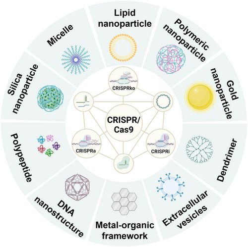
Given its promising potential in gene therapy, CRISPR/Cas9 has expanded rapidly in biomedical research of cancer treatment.24, 32-34 Different CRISPR/Cas9-mediated cancer therapy approaches, such as altering chemo-tolerance genes, changing metabolically relevant genes and tumor stem cell associated genes, as well as cancer immunotherapy, are well-established in diverse cancer types.32, 35-38 Although the discovery of CRISPR/Cas9 paved the way for simple, effective, and multiplex manipulation of genes, the off-target impacts, difficulties in targeting specific cells in vivo, and the successful transfer into the nuclei still remain the main obstacles keeping it from realizing its maximum capabilities.39, 40 Rational delivery systems and improvements in the CRISPR/Cas9 genome engineering technique may be able to solve these problems.41-43 Here, we outline current developments in nanotechnology-based strategies to deliver plasmids, mRNA/sgRNA, and ribonucleoprotein (RNP) complexes-based CRISPR/Cas9 systems against cancer (Figure 1). Firstly, we go over the mechanism of CRISPR/Cas9 gene editing technology. Then, we discuss various forms of CRISPR/Cas9 and their applications in treating cancer. We next aim at the rational design of non-vial nanocarriers for CRISPR/Cas9 delivery systems. In the end, we extend the development of CRISPR/Cas9 genome editing system to clinical applications and provide insights to address the complex therapeutic needs by nanotechnology-based delivery methods.
2 MECHANISM OF CRISPR/CAS9
CRISPR were found in Escherichia coli in 1987.44, 45 According to the different recognition and cutting mechanisms of endonucleases, CRISPR system can be classified into two types, including 16 subtypes.46-48 Among them, CRISPR system type II, namely CRISPR/Cas9, targeting a certain DNA sequence utilizing the Cas protein, shows good characteristics of gene editing and has primarily been used in mammalian cells.49-51 CRISPR RNA (crRNA), trans activating CRISPR RNA (tracrRNA) and Cas9 endonuclease are three important components of CRISPR/Cas9 system.15, 52, 53 The crRNA has a 20-nt protospacer sequence to direct Cas9 protein to a particular site, as well as contains an extra sequence to bind with tracrRNA via complementary pairing.54, 55 In addition to binding to crRNA, the tracrRNA has another functional component, which recruit Cas9 protein to form RNP complex.56, 57 Additionally, to simplify the system and improve the binding ability between Cas9 and target DNA, the crRNA and tracrRNA are connected as a sgRNA (Figure 2).58-60
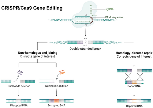
The 5′ end of crRNA specifically recognizes the target sequence and recognizes the “NGG” site of PAM, which is at downstream of the target DNA sequence.61-63 PAM sites usually exist as recognition sites,64 and more importantly, PAM sites are also recognition sites that distinguish the host's own DNA from foreign DNA to prevent autoimmunity.63, 65 There are six major domains in Cas9 endonuclease, among which HNH and RuvC are used to cleave the target gene sequence.66-68 The RuvC domain cleaves the non-complimentary strand of crRNA, whereas the HNH nuclease domain cleaves the complementary strand of crRNA.67-71 In the process of self-repair, NHEJ is used to introduce insertion/deletion (indel) of edited DNA strands bases to mutate DNA. NHEJ can initiate frameshift mutation or stop codons premature into target gene, thus achieving gene knockout.72, 73 In contrast to the NHEJ, which can straightly join damaged regions, HDR utilizes a donor DNA template to enable precise insertion.74, 75 Therefore, foreign artificial templates can be input for directional gene editing.76-78 Nevertheless, since HDR would only be functional in dividing cells, knock-in is far less effective than knockout.79
3 DERIVATIVES AND FORMS OF CRISPR/CAS9
By using the biological characteristics of Cas9 targeting specific DNA sequences under the guidance of sgRNA, scientists have further developed gene targeting activation and gene targeting inhibition tools based on dCas9, called CRISPRa and CRISPRi respectively (Figure 3).29, 80-83 By adding the amino acid alteration D10A to the RuvC nuclease domain and H840A to the NHN nuclease domain, respectively, dCas9 is rendered inactive in these two systems, but dCas9 still reserves the capability of binding DNA.84-86 The CRISPRi technique is initially only to spatially impede RNA polymerase and hinder target gene transcription.60, 85, 87 Subsequently, the inhibitory activity of dCas9 is enhanced by the addition of a transcriptional repressor domain including the KRAB (Krüppel-associated box) and the biparty repressor domain KRAB-MeCP2.88-92 CRISPRi has a number of advantages over shRNA and CRISPR knockout (CRISPR KO) in terms of its ability to suppress the production of the genes being targeted. As an illustration, the dCas9-KRAB CRISPRi method won't affect the genome sequence of target gene locations, unlike CRISPR KO and other conventional gene knockout technologies.93 Besides, high degrees of gene repression are frequently achieved by the dCas9-KRAB CRISPRi method with little to no off-targeting.94, 95 Allowing dCas9 to bind to different transcriptional activators can contribute to upregulated expression of the target genes, which is what CRISPRa.96, 97 Targeting promoter and enhancer regions through the fusion of dCas9 with transcriptional activators like VP64 and p65 can increase gene expression.27, 98 To better improve the level of target gene overexpression, dCas9 fusions need to recruit multiple activating structural domains. Accordingly, numerous systems have been created, involving multiple fusions (VPR, a triple fusion of VP64, p65 and Rta5), the use of protein scaffolds (e.g., SunTag system), and the use of RNA scaffolds (such as the Synergistic Activation Mediator complex).98-100 Applications of CRISPRa provide numerous benefits in terms of the potential to increase target gene expression.101 This technical strategy does not change the genetic sequence of target locus, in contrast to transgenic and gene editing technologies.102 Besides, even if precise information of the DNA sequence at the target location is required, prior knowledge of the target gene's natural regulatory mechanism is not necessary.103 Most notably, CRISPRa transcription typically results in extremely high levels of expression for genes.104

The above are derivatives of CRISPR/Cas9, while there are a total of three delivery forms regarding CRISPR/Cas9. RNP, a compound of Cas9 endonuclease and sgRNA, is the first kind. Delivering an RNP is the simplest method that does not need transcription or translation.105, 106 It begins genome editing as soon as it reaches a cell, resulting in high editing efficacy and minimized immunological responses, insertional mutagenesis, and off-target consequences.107-110 Nevertheless, this delivery form needs the highly active Cas9 protein to be purified.111 Besides, there are special needs when building delivery systems for RNP because of its distinctive properties that include complicated structure and charge property.112 In addition, the huge molecular weight makes Cas9 protein difficult to enter cells.
sgRNA with Cas9 mRNA delivery is the second method. Cas9 mRNA is typically synthesized in vitro using dsDNA templates with sequences obtained from CRISPR/Cas9 plasmid (pCas9). Nuclear entrance is not required for Cas9 mRNA to be translated into Cas9 protein, this may help to lessen off-target activities caused by temporary Cas9 protein production but could result in reduced genome editing efficiency.113, 114 Besides, this form only allows for brief Cas9 expression since mRNA is extremely unstable and susceptible to RNase destruction.115 Therefore, Cas9 mRNA delivery is severely hampered by the low stability of Cas9 mRNA.116
The third form is the plasmid encoding Cas9 endonuclease and sgRNA. DNA plasmid is more readily available, less expensive to manipulate, and generally more stable, therefore, the main method for delivering CRISPR components in laboratory environment is plasmid-based delivery. Longer expression time in cells is also needed through plasmid DNA-driven Cas9 production, which might be useful when persistent expression is needed for editing. There are several limitations, though. First, the huge size and negative charge reduce the possibility of pCas9 to be transported into cells, which demands delivery vehicles with substantial payload capacity. Second, pCas9 depending on nuclear entrance for DNA transcription makes genome editing less effective. Additionally, off-target effects are more likely to occur in the type of plasmid since Cas9 is expressed continuously in cells.114, 117 What's more, there is a chance of insertional mutagenesis with pCas9, and the big plasmid can also trigger immunological reactions.118, 119
The three types of CRISPR/Cas9 payloads have their own advantages and disadvantages. Nevertheless, regardless of the payload form, it is challenging for CRISPR/Cas9 to penetrate cells. Therefore, developing an effective nanotechnology strategy for multiple Cas9 components is essential. Nanocarriers, such as cationic lipid-based nanoparticles, cationic polymer/polypeptide-based nanoparticles, inorganic nanomaterials, DNA nanostructures, gold-based nanoparticles and exosomes or extracellular vesicles, are currently hopeful delivery methods for CRISPR/Cas9 systems. Therefore, next, we will overview the applications of different nanocarriers in terms of the different delivery forms of CRISPR/Cas9.
4 NANOTECHNOLOGY-BASED DELIVERY OF CRISPR/CAS9
4.1 Plasmid-based CRISPR/Cas9
It is well recognized that nanocarriers loaded with pCas9 are able to incorporate the CRISPR/Cas9 system into cells through a cellular internalization process, that begins with endocytic cell uptake and ends with endosomal escape. Besides, some nanocarriers can also enable the plasmid to enter the cell in a non-endocytosis manner without entering the lysosome, thus preventing it from being damaged.120-122 The plasmid reaches the nucleus after being released from the nanocarriers and begins transcription into mRNA and sgRNA. The Cas9 protein is then translated from the mRNA in the cytoplasm. Afterwards, sgRNA and the Cas9 protein combine to form the RNP complex. Finally, genome editing occurs as a result of the RNP complex being translocated into the nucleus. As a result of the strong negative charge as well as huge size of pCas9, most nanocarrier-based delivery strategies used positively charged materials to compress the plasmids into smaller size complexes by electrostatic interactions. Cationic lipids, PEI-based polymers, inorganic compounds, chitosan, protamine sulfate, and dendrimers are the most often employed positively charged substances.
4.1.1 Lipid-based nanotechnology
Lipid nanoparticles (LNP), the traditional nucleic acid delivery technology, has been the focus of in-depth research in recent years.123, 124 The cationic lipids interact with the negatively charged phosphate backbone of the DNA via electrostatic attraction, and they also promote cellular absorption by reacting with the negatively charged cell membranes. Significant research has produced LNPs as a delivery vector since the adoption of cationic lipids for cellular transfection.125-127 For example, Zhang and coworkers constructed a multifunctional LNP to deliver pCas9 targeting MutT homolog1 (MTH1) gene into nuclei of non-small cell lung cancer (NSCLC) cells.128 They pre-condensed plasmids by protamine, a protein with substantial positive charges and nuclear localization sequence (NLS), to create an adversely charged complex. Then the protein/DNA complex was cationic liposome-coated to avoid nuclease degradation in blood circulation, which was further modified with DSPE-PEG-HA to give the ability to actively target tumor cells. Results indicated that the enhanced cellular internalization and nuclei-targeting protamine caused higher transfection. The MTH1 gene disruption slowed the progression of NSCLC and liver metastasis was decreased while cell death in tumor tissue was promoted.
In further applications, LNP formulations have been modified to achieve targeted therapy, extended circulation time, elevated transfection efficiency, decreased toxicity, and enhanced particle stability. For example, to realize targeted therapy, Wang et al. have designed cationic lipid-assisted polymeric nanoparticles (CLANs) to deliver a pCas9 expressing sgRNA targeting the overhanging fusion region of the BCR-ABL gene (pCas9/gBCR-ABL) into chronic myeloid leukemia (CML) cells.129 CLANs were produced by a cationic lipid N,N-bis(2-hydroxyethyl)-N-methyl-N-(2-cholesteryloxycarbonyl aminoethyl) ammonium bromide (BHEM-Chol) to encapsulate plasmids in poly(ethylene glycol)-b-poly(lactic acid-co-glycolic acid) (PEG-PLGA). CLANs carrying pCas9/gBCR-ABL precisely damaged BCR-ABL gene while leaving BCR and ABL genes of normal cells intact, which increased the lifespan of CML mice and reduced the amount of CML cells gathering in the blood.
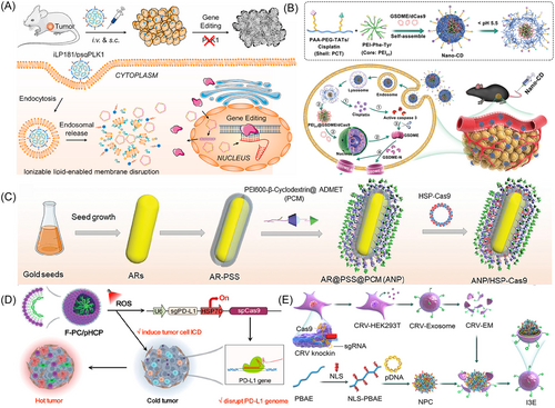
Wei and coauthors constructed another cationic LNP namely DOX-CB@lipo-pDNA-iRGD, to deliver the pCas9 expressing sgRNA targeting CD47 gene and boron drug to the nucleus.130 The cationic liposome was first prepared by DOTAP, DOPE, CHOL, and DSPE-PEG5000, and then combined with DOX-CB, a compound synthesized by doxorubicin (DOX) and carborane (CB), to utilize the nuclear tropism of DOX. To achieve tumor targeted delivery, the cationic liposome was further modified with the internalizing-RGD (iRGD), a homing peptide that bound to intern αvβ3/5 expressing on tumor cells and tumor blood vessel endothelial cells. On GL261 cells, the multifunctional nanoliposome DOX-CB@lipo-pDNA-iRGD demonstrated an increased percentage of genome recombination and transfection, which coupled boron neutron capture therapy (BNCT) with immune therapy that improved the prognosis for tumor-bearing mice, increased survival rates, and decreased tumor stemness. In another study,131 the cationic liposome was modified with R8-dGR (R8-dGR-Lip), which increased cell penetration by binding to integrin v3 and neuropilin-1 on tumor cells. The R8-dGR-Lip was used to complex pCas9 to decrease HIF-1α in pancreatic cancer and co-encapsulate paclitaxel (PTX). Contrasted to pegylated counterparts, R8-dGR-Lip improved the tumor dispersion, which promoted the cytotoxicity of PTX and reduced pancreatic cancer metastasis. Huang et al. created a novel ionizable LNP (iLNP), known as iLP181, for assessing the capability of transporting the pCas9 system targeting Polo-like kinase 1 (PLK1) (Figure 4A).132 By binding to ApoE, iLP181/psgPLK1 has been effectively incorporated by hepatoma carcinoma cells. Long-term modification of genes was carried out both in vivo and in vitro. Also, when compared to commercial Lipo2000, iLP181 LNPs showed robust endosomal escape, and dramatically slowed tumor growth in mice.
Since the plasmid needs to be transported in the nucleus to function, LNP was also modified to enhance the nucleation ability of pCas9. Lo et al. designed a nucleus directing peptide (R-peptide)-modified solid lipid nanoparticles (SLNs) to eliminate human antigen R (HuR), a protein that binds RNA and controls the stability of mRNA and protein translation in ways that affect the tumor's ability to survive, spread, invade, and drug resistance.137 The R-peptide is generated from the NLS of a human phosphatase with good deliverability, no immunogenicity, and minimal toxicity. Concurrently, the liposome was also modified with a pH-sensitive H-peptide and a P-peptide that aiming at the epithelial growth factor receptor on tumor cells, acquiring SLN-HPR. In comparison to commercially available transfection agents, SLN-HPR at pH 6.0 demonstrated a greater transfection efficiency. The nuclear directing R-peptide allowed pCas9 to enter the nucleus and downregulate HuR to inhibit tumor cells growth, metastasis, and resistance.
In conclusion, LNPs protect pCas9 from disintegration and immunological reactions in addition to allowing them to move to the nucleus and penetrate the cellular membrane barrier. Thus, LNPs are safe and potent CRISPR/Cas9 plasmids delivery vehicle and possess the advantage of easy preparation. Furthermore, functional lipid alterations such as cell targeting modification might introduce new varieties of efficient CRISPR/Cas9 plasmids delivery techniques, presenting good prospect for LNPs in CRISPR/Cas9 plasmid delivery.
4.1.2 Polymer polyethylenimine (PEI)-based nanotechnology
PEI, the gold standard, can transfer genes including both large plasmids and short nucleic acids into cells. PEI also promotes endosome escape through the “proton sponge effect” to facilitate escape of payloads which are internalized into the cell. Dioleoylphosphatidylethanolamine (PE)-modifying PEI2000 was utilized to condense the Cas9 nuclease plasmid (hCas9) or the Cas9 D10A nickase plasmid (hCas9-D10A).138 High molecular weight PEI is another option besides low molecular weight PEI, which shows higher transfection efficiency. Branched PEI 25 kDa was used by Ryu et al. to condense pCas9 into a nanocomplex, and successfully delivered it into Neuro2a cells.139
Compared to low molecular weight PEI, high molecular weight PEI often exhibits better transfection effectiveness, however, often has higher cytotoxicity. PEI with low molecular weight has the advantage of low cytotoxicity, but to improve its transfection efficiency, many studies have introduced special groups or cross-linked them to enhance transfection.140-144 Dahlman et al. modified low molecular weight PEI (PEI600) with alkyl chains to realize effective siRNA deliver.145 Guan et al. synthesized fluorocarbon (FC) modified PEI600 for that purpose.146 More recently, deoxycholic acid (DA) was used to decorate PEI1.8K by the Karimi group.147, 148 Compared to PEI1.8K, PEI1.8K-DA had a greater transfection efficiency. Later, this group further modified PEI1.8K with arginine containing disulfide bonds,149 which improved the membrane permeability, nuclear localization, and plasmid release. Additionally, in comparison with the polymeric gold standard transfection agent, this nanocarrier is capable of transfecting pCas9 in a variety of cells, involving difficult-to-transfect primary cells (HUVECs), cancer cells (HeLa), and neuronal cells (PC-12). In addition to arginine, phenylalanine (Phe) and tyrosine (Tyr) were also used to modify low molecular weight PEI. Gong's group synthesized PEI1.8K-Phe-Tyr to condense CRISPR/dCas9 plasmid. It was determined that the transfection efficiency was significantly higher than PEI25K in vitro.150 In the further utilization, they described a Nano-CRISPR scaffold (Nano-CD) for cellular pyroptosis-related immune therapy (Figure 4B).133 Nano-CD was constructed by coating the PEI1.8K-Phe-Tyr/CRISPR/dCas9 with a multifunctional copolymer, which was created by covalently attaching the drugs cisplatin and TAT to the backbone of PEGylated polyacrylic acid (PAA). Potent tumor pyroptosis was accurately and efficiently activated attributable to the cooperation of gasdermin E (GSDME) protein and caspase-3 stimulation brought on by cisplatin, subsequently inverting the immune suppressing tumor microenvironment (TME) and boosting the anticancer immunological cycle as beneficial feedback.
Gong's group introduced fluorinated groups on PEI to obtain PFs that can load nucleic acids.151, 152 Fluorinated PEI can enhance the affinity of the pCas9 with cell membrane, promote its passage through cell membrane and endosomal-lysosomal membrane, and accelerate endosomal-lysosomal escape, thus enhancing transfection efficiency. For example, PF10K was reacted with the heptafluorobutyric anhydride for obtaining the fluorinate-decorated PEI (PFn).153 PF34 with averages 5.4 fluorinate per PEI was chosen to condense pCas9 aiming at PD-L1 and CD47 due to the higher transfection performance with tiny mass value. Then the polyplex was encapsulated into a multistage sensitive nanocomplex (MUSE), which not only prolonged blood circulation and tumor targeting, but also improved cellular uptake and endosomal escape. The MUSE with MT-CRISPR/Cas9 loaded shown outstanding PD-L1 and CD47 disrupting effectiveness in cancerous cells based on these benefits. It has also been shown that in tumor models, the MUSE stimulated powerful adaptive and innate antitumor immune responses and produced a durable immunological memory response, greatly inhibiting tumor development and enhanced survival with nearly undetectable off-target delivery effect. Additionally, to produce the inner core, Qi et al. synthesized fluorinated PEI1.8K, which they then complexed with the plasmids.154 Then, a tumor selective response module was coated on the core, which was composed of hyaluronic acid and TME-sensitive peptides (TMSP), obtaining the HPT-PFs nanoparticles. Plasmids for both the generation of IL-12 and the knockout of CD47 were simultaneously delivered using HPT-PFs. Don't eat me signals (CD47) expression was lost in tumor cells, and tumor cells produced IL-12 at levels higher than 500 ng/mL. Combining CD47 deletion with IL-12 production significantly reduced tumor growth in melanoma-bearing mouse models by dramatically increasing M1-tumor-associated macrophage (TAMs) and secreting cytokines associated with inflammation. In another study, Huang et al. also synthesized a fluorinated PEI1.8K (PEI1.8K-F) and modified with PEG side chains for protection.155 In combination with PT-based chemotherapy, the reducible Pt(IV) prodrug was copolymerized with PEI1.8K-F. The resulting PtUTP-F nanoplatform was utilized to condense a CRISPR/dCas9 system targeting Cancer/testis antigen 45 (CT45). High transfection efficiency was achieved thanks to the outstanding cellular internalization, endo/lysosomal escape, and gene release characteristics, which were made by the hydrophobicity of fluorination and the proton sponge effect of PEI1.8K. PtUTP-F/dCas9-CT45 is able to induce CT45 expression for protein phosphatase 4C (PP4C) action restriction that blocks the DNA restoration process, increasing the response to Pt(II) drugs for specialized A2780 cancer treatment.
In addition to leveraging the higher charge density of PEI, many studies have designed responsive carriers to help achieve lysosomal escape. Yang et al. created a photoswitched CRISPR/Cas9 system for PD-L1 gene disruption.156 This system was constructed by complexing the photoactivated self-degradable PEI derivative PPCe with plasmid (pX330/sgPD-L1). After being exposed to 660 nm laser irradiation, PPCe could be activated and self-degraded to launch the pX330/sgPD-L1 in the cytoplasm, which promoted endosomal escape through the photochemical internalization (PCI) effect. As a result, PPCe acts as an optogenetic trigger in cancerous cells and cancerous stem cells to efficiently disrupt PD-L1 at the level of the genome, which effectively activated the T cell-regulated antitumor response.
Pu et al. grafted the semiconducting polymer with PEI600 brushes with 1O2-cleavable linkers and PEG2000 to generate a near-infrared regulated pCas9 delivery nano-vector (pSPN).157 This is the first NIR photo regulatable CRISPR/Cas9 nanocarrier. Under NIR light irradiation, pSPN was spontaneously cleaved to release the pCas9, thus initiating the gene editing process. In mice and live cells, respectively, this nanoplatform allowed for a 15- and 1.8-fold improvement in the expression of repaired genes compared with nonirradiated controls. PEI can also be combined with other materials to increase cell uptake. Zhang's group conjugated PEI with magnetic nanoparticle (MNP@PEI) to condense pCas9 targeting MutL homolog 1 (MLH1, MPM), and further coated with lipids (LMPM).158 Though an external magnetic field, LMPM showed an improved cellular internalization. By downregulating the expression of MLH1 and targeting the DNA repair pathway when an outside magnetic field was utilized, LMPM was able to increase the immunogenicity of tumor cells and intensify the immunological response against the tumor. In vitro and in vivo studies demonstrated LMPM coupled with a magnetic field and α-PD1 therapies was able to effectively limit cancer development and additionally enhancing CD8+ T cells penetration to the TME.
4.1.3 Inorganic materials-based nanotechnology
In addition to protonatable polymers, inorganic materials were also commonly used in pCas9 delivery because of superior cell uptake and lysosomal escape capabilities. For instance, a promising platform for bio-applications has arisen in the form of metal organic frameworks (MOFs), which are made up of metal ions or clusters coupled to organic ligand linkages.159, 160 In a study the NH2-UiO-66 MOF was green synthesized, modified with GMA and functionalized with two different stimulus polymers p(HEMA) and p(NIPAM). The polymer-coated NH2-UiO-66 further loaded DOX and pCas9.161 While the UiO-66@DOX@pCRISPR and GMA-UiO-66@DOX@pCRISPR showed extremely low transfection efficiency of 1.6%, the p(HEMA)-GMA-UiO-66@DOX@pCRISPR increased the transfection efficiency up to 4.3% and p(NIPAM)-GMA-UiO-66@DOX@pCRISPR increased it up to 6.4% in a HeLa cell line. Although this transfection efficiency remains unsatisfactory, it is the limited successful examples of MOF-based nanocarrier delivery of pCas9.
The most extensively studied MOFs for hosting biological molecules (such as proteins, RNA, and DNA) at the moment are zeolitic imidazole frameworks (ZIFs).162 Since 2018, ZIF-8 research has been exploding at an accelerated rate, providing evidence of its effectiveness in delivering undamaged, functional genes and its possible ability in CRISPR/Cas9 system-based gene editing.163 For the first time, CRISPR/Cas9 was delivered via a MOF platform in plasmid form, according to Shukla et al,164 which was realized by using ZIF-C. With ZIF-C, RPSA gene, which was overexpressed in prostate cancer (PC), was cleaved by approximately 20%. Additionally, compared to normal prostate cells, the advanced metastatic PC cells displayed greater vulnerability to cytotoxic effects. More recently, Zhang et al. investigated the intracellular delivery potential of pCas9-ZIF-8 system for Paxillin (PXN) knock-in through the HDR pathway.165 It was discovered that plasmids can benefit from sufficient defense against hydrolytic enzymes and can be evenly dispersed all through the plasmid-ZIF embedding framework. The pCas9-ZIF system demonstrated effective cellular endocytosis, effective endo/lysosomal escape capabilities, sufficient safeguards against degradation by enzymes and pH-sensitive plasmid liberation, making it exceptional for genetic knock-in. It is worth mentioning that HDR-mediated knock-in itself shows a great challenge, not to mention to construct effective and controlled MOF-based vectors for pCas9-based gene therapy. Therefore, this study highlighted the potential for MOF-based nonviral carriers to be used in CRISPR-based gene therapy.
Additionally, gold nanoparticles have shown promise as plasmid transport vehicles. Gold nanorods (AuNRs) like PGEA-modified AuNRs were used in the past to deliver tiny plasmids,166 but pCas9 delivery has only recently been described using AuNRs. To transport pCas9, Ping's group designed a variety of cationic polymer coated AuNRs with ARs ranging from 1 to 9. PEI25K was used to give AuNRs the cationic property, resulting in an AuNR-PEI (ARP). They hoped to gain a deeper understanding of the way the aspect ratio of AuNRs affects the efficiency of pCas9 transfection and genome modification activities. Results showed that the high aspect ratio optimized cationic AuNRs possessed a distinctive DNA-assembling mechanism, outstanding internalization abilities, and potent endosomes escape ability, allowing dCas9-regulated transcriptional activation and Cas9-induced genomic editing. Afterwards, Ping's group constructed supramolecular cationic gold nanorods (ARs) to efficiently deliver pCas9 encoding a heat-inducible promoter (HSP) and aiming at PD-L1 after NIR-II light irradiation (Figure 4C).134 The supramolecular cationic ARs acted as a delivery system for CRISPR/Cas9, as well as effectively harvest light from NIR-II and convert it into moderate hyperthermia, which is essential to promote genes production and activate immunogenic cell death (ICD).
Hollow mesoporous silica nanoparticles (HMSNs) are also outstanding gene and drug delivery system because of the unique hollow mesoporous structure.167-172 A polyamidoamine-aptamer-coated HMSNs was reported to co-deliver sorafenib and pCas9.173 In HepG2 cells, the SEHPA NPs demonstrated a nearly 66.3% gene knockout efficacy. With negligible off-target effects, the joint delivery of Sora and the CRISPR/Cas9 system effectively reduced the levels of EGFR and the subsequent PIK3-Akt path.
4.1.4 Other cationic materials-based nanotechnology
Protamine (PS), which is a nuclear protein that is strongly cationic, is well recognized for packing anionic DNA molecules.174-176 In the core comprised of calcium carbonate (CaCO3) and PS, Cheng and colleagues enclosed the pCas9 enabling β-catenin knockout.177 Then, the core was coated with hyaluronic acid (PHA) and hyaluronic acid (AHA) that had been modified with the TAT-NLS peptide and the AS1411 aptamer. Protamine can improve the ability to load plasmids by interacting electrostatically with the negatively charged plasmids. By the assistance of these functional elements, plasmids were significantly enriched in the nuclei of malignant cells, which induced efficient β-catenin knockout. The utilization of protamine-capped gold nanoclusters (protamine-AuNCs) as a pCas9 transport and release nanocarrier is presented first by Li and collegues.178 The protamine-AuNCs combine the benefits of both protamine and AuNCs. AuNCs can quickly complex with pCas9 to enable successful cellular transport, and cationic properties help plasmids be released into the cell nucleus effectively.
Chitosan (CS), as a delivery vehicle, whose amino groups on molecular chains are able to protonate under acidic conditions, has garnered a lot of interest because of its simplicity of customization.179, 180 In a study to transport a plasmid encoding Survivin (sgSurvivin pDNA) and DOX, folic acid (FA) and 2-(diisopropylamino) ethyl methacrylate double grafted trimethyl chitosan (TMC) nanoplatforms (FTD) were synthesized.181 Despite a high molecular weight of 13k bp and being vulnerable to degradation, sgSurvivin pDNA could be condensed, protected, and released in the tumor cell thanks to the great condense capacity of FTD. FTD/DOX/sgSurvivin pDNA NPs effectively transported sgSurvivin pDNA and DOX into tumor cell nuclei, resulting in effective sgSurvivin pDNA genome editing and collaboratively boosting their anticancer efficacies both on the cellular level and animal level. For better cancer treatment, Shao et al. developed composite CS loaded with PTX and sg-VEGFR2/Cas9 plasmid.182 To specifically target hepatocellular carcinoma (HCC) cells, lactobionic acid (LA) containing-galactose was linked to CS. 33.4% of cancerous tissue along with up to 38.6% of HepG2 cells were successfully edited by sgVEGFR2/Cas9 in the nanosystem. In HepG2 cells, VEGFR2 protein expression was decreased by >60%, and the growth of HCC tumor mice was significantly slowed down by 70%.
Poly(amide-amine) (PAMAM), a dendrimer that can be protonated under physiological conditions because of the high number of amino groups on its surface, is able to create complexes with DNA by electrostatic interactions, thus loading DNA into cells.183-187 Yang et al. synthesized a fluorinated G5 PAMAM dendrimer (F-PC) to encapsulate a HSP70-promoter-driven pCas9 targeting PD-L1 gene and a photosensitizer chlorin e6 (Ce6) (Figure 4D).135 Under the stimulation of 660 nm laser-induced reactive oxygen species (ROS) generation, the HSP70 promoter was elicited to trigger the Cas9 protein expression, which specifically damaged PD-L1. In the meantime, ICD was induced to activate the antitumor immune responses, which showed enhanced immunotherapy efficacy combining PD-L1/PD-1 checkpoint blockade. The fact that this CRISPR/Cas9 system could be spatiotemporally regulated via two mechanisms—NIR irradiation and particular promoter-triggered production, indicates that it has a lot of potential to prevent serious immune-based negative outcomes in clinical settings.
Poly(β-amino ester) (PBAE) is another popular cationic polymeric genetic vehicle with strong biological compatibility.188-194 Given that compared with their equivalent linear counterparts, branched polymers exhibit higher levels of gene transfection,195 Wang et al. connected the linear PBAE with branching units to obtain a new multifunctional linear-branched hybrid PBAE (LBPAE).196 The gene transfection efficiency of LBPAE reached 94% in human primary dermal fibroblasts and 91% in mouse embryo fibroblasts, significantly superior to commercial reagents PEI and SuperFect, and the fluorescence intensity was an additional order of magnitude greater compared to these two commercialized transfection substances. Similarly, Hu et al. connected linear PBAE with G0 PAMAM to acquire a hyperbranched copolymer hPPC.197 The resulting hPPC showed a considerably greater transfection effectiveness compared with PBAE as well as G4 PAMAM and the gold standard PEI25K. The highly branching hPPC1-plasmids polyplex NPs displayed remarkable proliferation suppression of cervical cancerous cells and xenograft tumors in nude mice by cleaving the HPV16 E7 oncogene. Jiang and coworkers synthesized an amino group ended and NLS modified PBAE to complex a CRISPRi/dCas9 plasmid targeting PI-3 kinase gamma (PI3Kγ) gene (NPC) (Figure 4E).136 A second coating of CRV-expressing exosome membranes (CRV-EM) made from human embryonic kidney (HEK) 293 T cells was applied to the NPC, in which CRV selectively targeted to TAMs. The internally and externally engineered exosomes (IEEE) enabled the preferential migration to tumor site as well as targeting M2-TAMs. Under the acid environment in the lysosome of TAMs, the CRISPRi system was released and nearly completely repressed the production of PI3Kγ, which induced TAMs polarization to M1 phenotype. As a result, the TME was reversed from the anti-inflammatory one into inflammatory one, which efficiently inhibited tumor progression in mice.
The pCas9 can also be condensed by poly(disulfide) (PD) to generate PD/plasmid (PD/P) nanocomplexes, showing effective in transporting pCas9.198 Recent investigations have shown that PD conjugated cargos might be transported quickly to mammalian cells with no apparent endo/lysosomal entrapment.199 The disulfide backbone in the PD aids in intracellular cargo transport via disulfide-exchange processes thus avoiding endosomal trapping.200, 201 Xu's group synthesized a PEG-modified PD to deliver plasmids, which showed good transfection efficiency and cellular internalization in a variety of types.202 Ping's group utilized PD to realize the efficient delivery of the plasmid encoding the Cas editor.203 PD is polymerized by two monomers of cationic diethylenetriamine and guanidine, and then coated with the macrophage membrane of Raw264.7 cells, achieving transportation to the liver and expression that is unique to the liver.
4.2 mRNA/sgRNA-based CRISPR/Cas9
Due to its lower risk of mutation, transient effects, and simplified structure, RNA-based CRISPR/Cas system shows great potential in gene editing.204 However, exogenous mRNA has limited cellular permeability, which is a significant obstacle to the use of mRNA treatment.205-207 But, because the translation of RNA into proteins takes place in the cytoplasm, the better point is that the nuclear entry is avoided when CRISPR/Cas9 is delivered as Cas9 mRNA and sgRNA. mRNA is capable of being translated immediately into proteins after its liberation in the cytoplasm, followed by the synthesis of RNP to carry out the following nucleus-based genetic modification action. Because mRNA has a linear shape, the average length of SpCas9 mRNA is greater than that of plasmid-based systems, ranging from 600 to 700 nm. As a result, Cas9 mRNA must be condensed using cationic nanocarriers. Desirable carriers must encapsulate both long SpCas9 mRNA and short RNAs simultaneously since sgRNA only has about 100 nt. Besides, target cells must get both the Cas mRNA and sgRNA at once.
Since single-stranded RNA is often unstable, it presents another difficulty to deliver Cas mRNA and sgRNA.208 Due to Cas9 mRNA possesses an extremely brief lifespan, it will decay within a single day. However, compared with the form of pCas9, a better aspect of Cas9 mRNA is that it allows gene editing to occur in the targeted organs/cells, avoiding off-target effects and reducing the risks of immunogenicity.109 Only a few reports have, to date, discussed the applications of nanocarriers for delivering RNA-based CRISPR/Cas9 system, with the majority using lipid nano-vectors.
By concurrently packaging Cas9 mRNA and sgRNA in bio-reducible LNPs, Wang's group described a novel method for systematic administration of CRISPR/Cas9 enabling effective and remarkably quick genetic modification in the cellular level and animal level.209 BAMEA-O16B, created by fusing amine and acrylates or acrylamides with a disulfide link, was used to form the bio-reducible LNPs. Upon electrostatic interaction, BAMEA-O16B could assemble mRNA into nanoparticles, along with release mRNA cytosolically in reaction to reduce biochemical stimulus via a disulfide bond transfer process. It is evident that HEK cells express GFP with up to 90% efficiency when Cas9 mRNA and sgRNA are delivered together. When compared with Cas9/sgRNA RNPs delivery, a successful gene knockout was seen as soon as 1 day after Cas9 mRNA administrainto chronic myeloid leukemiation, which represents a significant improvement in regard to in vitro genetic modification effectiveness.
Peer et al. described a safe, and effective Cas9 mRNA and sgRNA delivery LNP that utilized an amino ionizable lipid library (Figure 5A).210 This library was developed utilizing a unique category of ionizable amino lipids that combined a linoleic fatty acid chain with amine head groups by hydrazine, hydroxylamine, and ethanolamine linkers. To boost stability as well as decrease immunogenicity, Cas9 mRNA was structurally changed in this study with 5-methoxyuridine, and significantly altered sgRNAs were also used. One intracerebral administration of CRISPR-LNPs targeting PLK1 (sgPLK1-cLNPs) in an aggressive orthotopic glioma led to as much as 70% of in vivo editing of genes, triggered apoptosis of tumor cells, decreased tumor expansion about fifty percent, as well as increased survival for 30%.
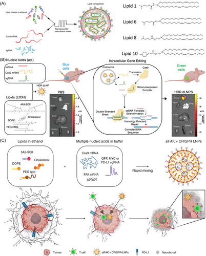
Despite highly efficient carriers exit, such as FDA-approved DLin-MC3-DMA LNPs, merely induce 1%–4% of RNA entering the cytoplasm, making endosomal escape the most challenging delivery stage. On this basis, Siegwart's group synthesized a series of novel phospholipids (iPhos) showing powerful endosome escape properties.213 In the acidic endosomal environment, tertiary amines were protonated, and phospholipids formed a smaller amphiphilic head and a larger triple hydrophobic chain tail, which formed a conical structure upon insertion into the endosomal membrane, facilitating the membrane transition to the hexagonal crystalline phase thereby enabling endosome escape. Organ-selective delivery of mRNA and CRISPR/Cas genome modification were made possible by iPhos-based LNPs (iPLNPs), which had a high in vivo effectiveness in delivering, indicating that the iPLNPs system had a high potential for clinical translation.
In another study, to deliver an all-nucleic acid CRISPR/Cas9 platform composed of Cas9 mRNA, sgRNA, and donor ssDNA for in vivo HDR correction, Siegwart et al. designed dendrimer-based LNPs (dLNPs) (Figure 5B).211 An array of ionizable dendrimer-based lipids was used to create the dLNPs, and these lipids showed the ability to be charged positive at low pH values for binding RNAs during self-assembly, uncharged at neutral pH to reduce cytotoxicity, and positively charged once more at endosome pH to allow endosomal liberation. Because of their all-in-one simplicity and great performance, the developed dLNPs edited more than 91% of all cells with 56% HDR efficacy in vitro and more than twenty percent HDR efficacy in xenograft tumors in vivo. It is necessary to remind here that HDR correction is significantly more complicated than NHEJ gene knockouts as it necessitates concurrent transport of the Cas9 mRNA, sgRNA and donor template ssDNA into one cell. Therefore, this study lays a good foundation for realizing gene editing and correction.
To facilitate tumor distribution and increase the effectiveness of gene editing, Siegwart et al. further used the above dLNPs to co-deliver siRNA, Cas9 mRNA, and sgRNA (siFAK + CRISPR-LNPs) targeting Focal adhesion kinase (FAK) (Figure 5C).212 It was discovered that this siFAK delivery-induced gene-editing improvement (10 times more in tumor spheroids) enhanced dLNPs intake by cells and tumor infiltration by lowering membrane stress regulated through the contracting strength of tumor cells. Four mouse cancer models showed significantly decreased tumor expansion and metastasis while siFAK + CRISPR-PD-L1-LNPs effectively disrupted PD-L1 production by CRISPR/Cas genome modification and decreased extracellular matrix stiffness. Wang's group constructed PBA-BADP, a cationic lipid coupled with PBA, to electrostatically assemble Cas9 mRNA into nanoparticles.214 Because of the PBA groups at the outer layer of the membrane, the nanoparticles demonstrated increased cellular absorption by cancerous cells through the PBA-SA interaction, leading to a 300-fold higher luciferase reporter expression of genes in cancerous cells than in noncancer cells. Additionally, PBA-BADP/Cas9 mRNA NPs selectively and powerfully suppressed the gene production in malignant cells.
Delivery of CRISPR systems in the form of mRNA is mainly based on LNPs, with a few studies also using polyplexes, extracellular vesicles. Kazunori et al. utilized a PEGylated polyplex micelle (PM) to complex both the Cas9 mRNA and the sgRNA in the core.215 PM was prepared by conjugating PEG with poly(N′-(N-(2-aminoethyl)-2-aminoethyl) aspartamide (PAsp(DET)). By compressing the negatively charged RNA through electrostatic contact, the amino-rich PAsp could aid endosomal escape. Also, for improved long-distance circulation and brain tissue diffusion, the spontaneously produced PEG corona might shield RNA molecules from enzymatic degradation and remove reticuloendothelial system (RES) components. It's interesting to note that compared to PM loading sgRNA alone, co-loading Cas9 mRNA and sgRNA significantly increased sgRNA stability. This is a good example of solving the problem of concurrent Cas9 mRNA and sgRNA delivery and ensuring their stability with the assistance of polyplexes.
With superior biostability and cellular internalization efficacy and little cytotoxicity, extracellular vehicles (EVs) offers promising inspiration in the delivery vector for RNA.216-220 Le and coworkers boosted the manufacturing of massive quantities of EVs for RNA therapeutics delivery using human red blood cells (RBCs).221 With no detectable toxicity, the mRNA-related CRISPR/Cas9 platform delivery with RBCEVs exhibited exceptionally potent genetic modification both in xenograft mice models and human cells. However, the loading efficiency is limited with the RNA encapsulation by electroporation. In Yuan and coworkers’ study, they developed a novel method for improved RNA payload encapsulating into modified exosomes.222 They combined the RNA-binding protein HuR, which has a really strong attraction for the AU-rich regions (AREs) in RNA molecules, with the exosome membrane protein CD9. It was revealed that the modified exosomes were remarkably effective for encapsulating AREs modified Cas9 RNA.
4.3 RNP-based CRISPR/Cas9
Transporting the RNP complex containing the Cas9 protein and sgRNA is the simplest method for genome editing. This method has various advantages over plasmids and other RNA delivery forms, including quick action, low off-target editing rates, and low cellular toxicity.223-226 However, in contrast to the transport of RNA or plasmid systems, RNPs provide unique hurdles because of their complex composition and charge characteristics.227 The sgRNA has approximately 100 negative charges, and the RNP complex has a net negative charge while the Cas9 protein has 22 positive charges, making it challenging to cross the cell membrane. For the delivery of RNP, numerous nanocarriers have been constructed over the past few years.228, 229 Nanocarriers encapsulated with Cas9 RNP can reach the nucleus to carry out CRISPR/Cas9-mediated genome editing after being internalized into targeted cells through endocytosis and endosomal escape. In this section, we divided the most often employed Cas9 RNP delivery nanotechnologies into four main groups: lipid-based nanotechnology, polymer-based nanotechnology, inorganic nanotechnology, and cell membrane-based nanotechnology.
4.3.1 Lipid-based nanotechnology
LNPs stand out as a promising choice among different nanotechnology-based delivery systems due to their well-established track record of effectively delivering genes and proteins in both laboratory settings (in vitro) and living organisms (in vivo). Up to now, three in vivo CRISPR-based gene therapies have obtained approval for clinical trials, one of which NTLA-2001 uses LNP delivery system.230 Leavitt et al. tested a LNP-RNP delivery platform to ex vivo transport template DNA and RNPs to mouse cortical neurons, has demonstrated exceptional genome editing efficiency.231 Harashima et al. presented the first report of the production of CRISPR/Cas RNP-loaded LNPs utilizing a clinically applicable microfluidic device.232 The optimized formulation showed outstanding base substitution (up to 23%) and gene disruption (up to 97%) with no discernible cytotoxicity. In another study, an FDA approved DLin-MC3-DMA LNPs were used as nano-vectors to encapsulate the RNP targeting Nuclear factor (erythroid-derived 2)-like 2 (NFE2L2) and a sonosensitizer hematoporphyrin monomethyl ether (HMME).233 Under ultrasound irradiation, the HMME produced a large amount of ROS, which led to lysosomal destruction, further resulting in RNP release. The knockout of NFE2L2 significantly enhanced the therapeutic efficacy of sonodynamic therapy (SDT).
Mastrobattista et al. improved the LNP formulation parameters for delivering the RNP in ready-to-use form.234 In the case of LNPs encapsulating Cas9 RNP alone for gene knockout or Cas9 RNP with an additional HDR template DNA for gene correction, modifications were made to the buffer composition during RNP and LNP complex formation, as well as adjustments in the concentrations of the cationic lipid DOTAP. They showed that LNP for gene knockout was not always dependent on DOTAP, whereas LNP for gene correction was only functional at low DOTAP concentrations. This result helped to clarify the requirement for ideal formulation parameters to produce LNP for direct in vivo transport of CRISPR/Cas9 components. When using the CRISPR/Cas9 RNP at nanomolar concentrations, gene knockout and gene correction efficiencies were obtained up to 80% and 20%, respectively. It's also worth mentioning that NHEJ-based gene knockout is more effective than HDR-based gene correction. Before HDR can be taken into consideration for clinical use, this ratio needs to be significantly improved.
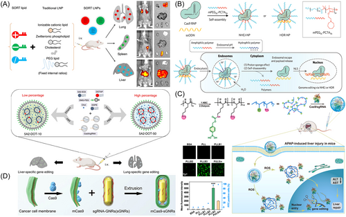
In another application of DOTAP-based LNPs, Siegwart's research team presented a broadly applicable method for achieving efficient delivery of RNPs into various cell types and enabling genome editing in tissues including muscle, brain, liver, and lungs. (Figure 6A).235 Based on the experience accumulated in the previous stage, the cationic lipid DOTAP was introduced into the original four core lipids of LNPs, which not only enhanced the encapsulation of Cas9/sgRNA RNPs under neutral conditions and preserved Cas9 function, but also retained the efficient delivery capability of conventional LNPs.239 Using this approach, they achieved the successful delivery of Cas9/sgRNA RNPs into cells, resulting in a gene editing efficiency exceeding 80%. The developed delivery system for RNPs not only simplifies the construction of in situ tumor models in mice, but also demonstrates the value of gene therapy in genetic diseases.
To ablate leukemia stem cells (LSCs), Leong et al. targeted the crucial gene Interleukin-1 receptor accessory protein (IL1RAP) in human LSCs using a bioreducible lipidoid-encapsulated RNP.240 Eight bioreducible lipids were created and evaluated to improve the LNP system that targets LSCs. To further improve the tissue specificity, the chemoattractant CXCL12α was loaded together onto mesenchymal stem cell membrane-coated nanofibril (MSCM-NF), imitating the bone marrow environment. The CXCL12 release promotes LSC migration and homing to the bone marrow niche, while the release of LNP-encapsulated Cas9 RNPs leads to efficient gene editing. The in vivo persistence duration of Cas9 RNP at the injection site was notably prolonged when Cas9 RNP was immobilized on the surface of nanofibers (NF), in contrast to the free Cas9 RNP. Besides, the enzymatic degradation of the RNP was effectively avoided and the cytotoxicity was reduced. Altogether, this nanofibril scaffold offers a secure and effective Cas9 delivery platform to target LSCs for enhanced acute myeloid leukemia therapy.
In a recent investigation, Wang and colleagues utilized a biodegradable LNP to transport an enhanced inactivated CRISPR (eiCRISPR) system within a xenograft tumor model. This eiCRISPR system comprises the Cas9 protein, a self-contained inactive sgRNA (bsgRNA), and a chemically caged deoxyribonuclease (DNAzyme), allowing for controlled activation of eiCRISPR and bsgRNA within the tumor environment.241 The bsgRNA contained an extension sequence that prevented the base-pairing contact between bsgRNA and the target gene, shutting down gene editing by eiCRISPR. The chemically caged DNAzyme exclusively broke the blocking extension of bsgRNA following stimulation by the overexpressed NAD(P)H:quinone oxidoreductase (NQO1) in tumor cells. Therefore, this study is expected to realize the cell selectivity and spatiotemporal controllability of gene editing.
4.3.2 Polymer-based nanotechnology
With better colloidal stability, excellent biocompatibility, and appropriate surface manipulation that enabled targeted transportation and cell membrane penetrating, polymer-based nanocarriers were constructed to transport Cas9 RNP. In addition to using PEI for Cas9 encapsulation, Chung et al. chemically tailored the Cas9 protein with branched PEI to obtain a Cas9 derivate (Cas9-P).242 RNP based on the Cas9-P could incorporate well with phosphorothioate-modified DNA oligonucleotides (NanoRNP), showing enhanced colloidal stability and prolonged activity. The NanoRNP could be internalized into B16 melanoma cells efficiently, resulting in effective PD-L1 gene disruption and tumor growth suppression.
Biocompatible DNA nanostructure-based nano-vectors are developing among the established polymer techniques because of their superior encapsulation ability through molecular recognition instead of charge interactions.243 A DNA nanoflowers (DNFs)-based nanoplatform was developed by Liu et al. to effectively load RNP by sequence hybridization.244 The DNFs were engineered with multiple copies of MUC1 aptamers for precise tumor cell targeting and were incorporated with miR-21 binding sequences to enable cell-type-specific release of RNPs. Different from the PEI or LNP for encapsulation, this strategy provided a versatile way to protect RNP activity as well as realizing responsive release in specific cells. Besides, various DNA aptamers could be encoded to accomplish the targeting of different cells. In another study, ultra-long ssDNA chains were utilized to co-deliver RNP and DNAzyme for combined gene cancer treatment.245 The ultra-long DNA chain was engineered using repeating sgRNA recognition sequences, DNAzyme, and HhaI cleavage sites through the rolling circle amplification (RCA) technique. In this system, Mn2+ in the DNAzyme as the cofactors was also served to compact the DNA chains to form nanoparticles.
Lächelt et al. described a FA-PEG decorated oligoamino amides delivery system to encapsulate RNP targeting PD-L1 and PVR immune checkpoint genes.246 After being injected into CT26 tumors in vivo, FA-modified nanocarriers significantly outperform nontargeted nanoparticles in terms of gene editing, including dual PD-L1/PVR gene disruption. Gong's team developed a pH-sensitive amphiphilic polymer, mPEG-PC7A, for the encapsulation of Cas9 RNP or Cas9 RNP + ssODN (Figure 6B).236 This polymer facilitates the delivery of Cas9 RNPs for NHEJ or Cas9 RNPs with ssODNs for HDR by modifying its hydrophobicity and cationic charge density. In Gong's another study, a customizable polymer called nanocapsule (NC) was synthesized to deliver RNP complex.247 The NC consisted of a mixture of cationic and anionic monomers to interact with the diverse surface charges of the RNP. In addition, the NC contained imidazole monomers to contribute to proton sponge effect-induced endosomal escape. Besides, the GSH-degradable crosslinkers in the NC were the key to ensure the efficiency of gene editing. Furthermore, PEG conjugated with ligands could be modified on the surface, which demonstrated the versatility and enhanced the cellular uptake. This biodegradable NC delivers RNP efficiently and showed robust gene editing outcomes both in vitro and in vivo without any obvious cytotoxicity.
Zheng et al. synthesized an angiopep-2 modified, guanidinium and fluorine decorated polymer to encapsulate RNP.248 Angiopep-2 peptide on the nanoparticles enabled the brain targeting and blood-brain barrier (BBB) penetrating for the treatment of glioblastoma multiforme (GBM). The guanidinium and fluorine components improved the interaction with RNP and its stability in the blood circulation. Furthermore, by targeting PLK1, the RNP nanoparticles achieved approximately 32% gene knockout and 67% protein reduction in vitro. In vivo studies demonstrated an extension of the median survival time of glioblastoma-bearing mice to 40 days.
Chen's group reported a boronate moieties decorated poly-l-lysine (PLL) to load Cas9 RNP through nitrogen/boronate coordination (Figure 6C).237 Diverse cargo proteins were effectively transported by the lead polymer into living cells while maintaining their protein activity. Under ROS condition, proteins could be efficiently released via the oxidation of boronate ligands for effective CRISPR/Cas9 gene editing. Similarly, Ping and Chen collaboratively developed a boronic acid-rich dendrimer as a versatile approach for protein delivery, including Cas9 RNP. This polymer can interact with both the anionic and cationic groups on the protein surface.249 On the one hand, through the process of nitrogen-boronate complexation, PBA, an electron-deficient group, could bind with cationic groups like imidazole and amine on proteins. On the other hand, the cationic dendrimer could coordinate with anionic carboxylates on protein. The dendrimer ensured efficient intracellular transport of Cas9 protein into various cell lines and exhibited strong efficacy in CRISPR/Cas9 genome editing. For human colorectal cancer (CRC) gene therapy, Ping's group utilized this phenylboronic dendrimer (PD) strategy to deliver Cas9 RNP with two sgRNAs that targeted both APC and KRAS mutations.250 Further, the PD/RNP nanoparticle was coated with anionic HA (HAPD) for positive charge shielding and CRC cells targeting. In xenografted and orthotopic mouse models of CRC, systemic administration of Cas9 RNP targeting both APC and KRAS via HAPD significantly halted cancer advancement and substantially decreased lung and liver metastases.
4.3.3 Inorganic materials-based nanotechnology
Inorganic materials have displayed remarkable promise for improving the loading and delivery abilities of Cas9 RNP. Inorganic materials have the advantages of easy preparation, structural stability, and compositional functionality, which could bind the Cas9 RNP and prevent the Cas9 RNP from rapid degradation.
MOFs with appropriate biocompatibility and biodegradability and adjustable architectures have been constructed for an efficient RNP delivery method. For example, Khashab et al. encapsulated Cas9 RNP into ZIF-8 and further coated it with a biomimetic cancer cell membrane.251 In vitro cellular uptake and transfecting efficiency as well as in vivo genome editing revealed the cell-specific targeting property corresponding to the cell membrane type owing to the homotypic binding ability. In another study, ZIF-8 was used to co-deliver Cas9/sgRNA targeting Indoleamine-2,3-dioxygenase-1 (IDO1) and a sonosensitizer hematoporphyrin monomethyl ether, and further electrostatically adsorbed onto the surface of Lactobacillus rhamnosus GG (LGG).252 Owing to the hypoxia homing capability of LGG, this CRISPR/Cas9 nanosystem (MHS) was able to penetrate the hypoxia tumor center, effectively boosting the accumulation of MHS in the tumor region. Upon US stimulation, the generated ROS damaged the endosomal/lysosomal membrane, leading to Cas9/sgRNA escaping and further efficient IDO knockout, thus lifting the immunosuppression of the TME.
To achieve on-demand release, Xu et al. developed a sono-controllable and ROS-responsive MOF to anchor with Cas9 RNP complex targeting MTH1 (P/M@CasMTH1).253 P/M@CasMTH1 was fabricated by covalently attaching RNP to porphyrin-integrated nMOFs through a ROS-sensitive thioketal linker, and then coated with PEI. The constructed P/M@CasMTH1 not only played the role of RNP delivery vector, but also serve as a sonoregulator to respond to ultrasound (US) energy, thus spatiotemporally activating the genome editing. This research established the first concept on the appropriate integration of SDT and gene editing for intelligent, effective, and combinatorial cancer treatment.
Owing to the diverse structural modification capabilities and beneficial biological activity, Au-based nanoparticles for RNP delivery are gaining popularity.254, 255 Besides, Au nanoparticles also showed outstanding photothermal conversion efficiency in the near-infrared region (NIR).256 For example, Ding's group functionalized gold nanorods (GNRs) with the TAT peptide and a thiolated DNA linker with the cell-type-specific aptamer to effectively load the RNP complex. With the assistance of both tumor cell targeting aptamer and TAT, this multifunctional nanoplatform caused efficient RNP transportation to tumor cells and PLK1 gene editing. With mild photothermal treatment, this nanoplatform realized excellent tumor cell proliferation inhibition.257 Zhao et al. developed a multi-branched Au nanooctopus (AuNO) as the core of the nanoparticle, and coated with the shell mesoporous polydopamine (mPDA) to encapsulate CRISPR/Cas9 RNP targeting the heat shock protein (HSP) gene and finally coated with a PEG-FA (AP).258 After entering the cell, both acid and NIR irradiation can accurately induce the release of RNP into the nucleus. Following knockout of HSP90, tumor cells become vulnerable to the moderate hyperthermia produced by AP under NIR-II radiation, which caused tumor cell death while the adjacent normal cells survived. In a similar study, to overcome tumor thermos-resistance and prevent thermal harm to adjacent normal tissues, Song et al. constructed a hypoxia responsive AuNRs for the delivery of CRISPR/Cas9 RNP targeting HSP90.259 To trigger hypoxia-responsive, controlled release of RNP, gold nanorods (AuNRs) are functionalized with azobenzene-4,4'-dicarboxylic acid (p-AZO). Therefore, the RNP complex could be released in the hypoxic condition of tumor cells to specifically deplete HSP90α gene, resulting in reduced thermal tolerance. In another study, Wang et al. used cancer cell membranes-derived nanovesicles to encapsulate the GNRs and RNP complex targeting oncogene Survivin (BIRC5) (Figure 6D).238 Through downregulating HSP70, the reduction of survivin expression enabled tumor cells more sensitive to heat, resulting in improved photothermal/gene therapy.
NIR-triggered photothermal controlled release of Cas9 RNP to suppress the generation of HSPs presents a prospective method to realize synergistic mild-PTT effects. Han's group introduced another PTT nanomaterial semiconductor copper sulfide (CuS) NPs to deliver RNP targeting HSP90α and DOX for combined gene therapy, chemotherapy, and PTT.260 In response to photothermal activation, the double-strand formed between CuS NPs-linked DNA fragments and sgRNA served as a regulatory element for controlled genome editing and the release of DOX. Additionally, the cationic PEI was applied to coat the surface to facilitate endosomal escape. The reduced tumor thermal resistance was achieved for improved mild-PTT benefits through photothermally controlled genome editing to alter HSP90. Two rounds of NIR light irradiation triggered the release of Cas9 RNP and DOX, inducing mild PTT effects both in laboratory settings and in live subjects. This, in turn, resulted in a significant combined impact on inhibiting tumor growth. In their another study, CuS NPs were also served as a basic carrier to modify Cas9 RNP targeting Protein tyrosine phosphatase non-receptor type 2 (PTPN2).261 Efficacious PTPN2 reduction sensitized the immunotherapy, embodying in the increased infiltration of CD8+ T cells, and upregulated IFN-γ and TNF-α in the tumor site. In addition, mild PTT induced ICD, which amplified the effects of tumor immunotherapy.
Silica nanostructures with advantages such as structural adjustability, stability in harsh biological conditions and good safety, are gaining more and more attention in therapeutic and diagnostic applications.262-265 Lee and colleagues designed a sponge-like silica nanoconstruct (SN) to deliver therapeutic RNPs, including Cas9 nuclease RNP (Cas9-RNP) and base editor RNP (BE-RNP).266 Targeting distinct cell types and genes led to a 50-fold increase in gene deletion efficiency and a 10-fold increase in base substitution efficiency, while maintaining low off-target effects, as compared to lipid-based methods in vitro. Besides, in vivo intravenous injection in a solid tumor model induced the successful gene correction. Song et al. coated silica spheres with CaCO3 and integrated an H2O2-responsive manganese carbonyl (MnCO), glucose oxidase (GOx), and GSH-sensitive Cas9 RNP targeting Nuclear factor E2-related factor 2 (Nrf2) gene to obtain a cascade nanoreactor.267 CaCO3 degradation was activated in the acidic TME to cause carbon monoxide (CO) generation and Ca2+ overloading. Elevated levels of GSH within tumor cells triggered the release of CRISPR/Cas9, leading to Nrf2 gene editing and increased susceptibility of tumor cells to damage induced by ROS. Liu's group developed a surface-thiolated MSN to simultaneously encapsulate the RNP designed to target PD-L1, and a small molecule drug called axitinib. Additionally, they coated the nanoparticle with a lipid layer to improve its circulation in the bloodstream and safeguard the RNP from enzymatic degradation.268 The CRISPR/Cas9 system and axitinib were released as the nanoparticle entered the reductive tumor cell microenvironment, which led to the combined modulation of several cancer-related pathways.
4.3.4 Other materials-based nanotechnology
EVs are membrane vesicles generated from cells that act as messengers for cell-to-cell communication.216, 269, 270 EVs are mainly composed of exosomes and microvesicles, which transport materials and information from donor cells to recipient cells, thus making them potential drug carriers and show considerable promise for systemic delivery of RNP.271-274 RNP loading can be achieved by fusing Cas9 with either the intracellular or extracellular domain of the EVs scaffolding proteins.275 For instance, Cas9 has been fused with a WW domain, resulting in WW-Cas9, which along with sgRNA targeting GFP, was enclosed within the lipid bilayer of EVs due to molecular recognition between WW and ARRDC1.276 After entering recipient cells, the GFP fluorescence signal disappeared, and the GFP gene knockout was confirmed by direct DNA sequencing and T7E1 assay. This study demonstrates that EVs are an effective delivery technology for Cas9 RNP. To enhance the loading efficiency of Cas9 protein into exosomes, Li and colleagues created a modified exosome by combining an exosome membrane protein with GFP. They then fused the Cas9 protein with a GFP nanobody for effective encapsulation.277 Afterwards, the affinity of the GFP-GFP nanobody allowed Cas9 proteins to be effectively and precisely packed into exosomes instead of random loading. Utilizing A549stop-DsRed reporter cells, the enhanced loading of Cas9 RNP demonstrated an improved gene knockout efficiency, which provide an alternative method for Cas9 RNP delivery. In addition, studies have been conducted to increase the targeting of EVs and reduce off-target effects. For specific cell targeting, Zhang and coworkers modified EVs with DNA aptamers conjugated with valency-controlled tetrahedral DNA nanostructures (TDNs) through cholesterol anchoring.278 TDNs applied in this study were to accurately bind DNA aptamer to the membrane of EVs. In cultured HepG2 cells, as well as in human primary liver cancer-derived organoids and xenograft tumor models, TDNs containing one aptamer and three cholesterol anchors significantly improved the tumor-specific aggregation of EVs. The delivery of RNP targeting WNT10B displayed significant tumor growth inhibition, providing a promising platform for EVs-based cell-selective Cas9 delivering.
Polymeric micelles are advantageous in that they are simple to functionalize, very stable, and biocompatible, leading to an increased delivery efficiency of RNP systems. Zhang et al. constructed a new mesoporous nanoflower nanoplatform termed Cas9-NF.279 They crosslinked Cas9 RNP with Pluronic 127 (F127) micelles via a reduction-responsive and self-immolative linker that allowed for effective intracellular transport and regulated release of Cas9. Due to its encapsulation within the network of PEG-enriched Pluronic micelles, the Cas9 protein remained stable even in highly acidic or alkaline environments. In a mouse tumor model, this crosslinked nanoparticle showed great capacity to suppress oncogene expression and prevent tumor progression. In this research, Cas9 itself was utilized as an integral component of a vector, facilitating efficient intracellular delivery and gene editing by Cas9. Furthermore, Cas9-NF nanoflowers can serve as a drug delivery platform capable of transporting both hydrophobic and hydrophilic drugs. This enables the integration of gene therapy with other therapeutic approaches.
5 THE CLINICAL APPLICATIONS OF CRISPR/CAS9 SYSTEM
CRISPR/Cas9-based gene editing delivered via nanotechnology represents a new era in the fight against cancer. However, there remain barriers between studies and clinical applications that need to be addressed, despite the fact that so many outstanding studies have been published to actively deploy the CRISPR system.280 At present, clinical trials have not yet made extensive use of the CRISPR/Cas9 technology, which is mostly limited in ex vivo level, such as manipulating T cells or stem cells before re-injecting them into patients (Table 1). However, compared with ex vivo gene editing, in vivo gene editing offers a number of benefits, including the ability to simultaneously alter the genes in numerous tissue types and time and money savings.281 Crucially, current Cas9 delivery techniques either have efficiency limitations or safety concerns that prevent clinical usage in patients. High gene editing efficiency can be achieved by using nanomaterials capable of rapid lysosomal escape and nuclear localization. In addition, the development of smaller size Cas proteins has been under exploration to achieve more efficient encapsulating.282-285 The primary requirements for safety are transient CRISPR/Cas expression and nanomaterials with acceptable biocompatibility. Second, the off-target effect of CRISPR/Cas9 system is also worth considering. To solve this problem, there are two ways to start. On the one hand, tissue-specific promoters could be utilized to activate CRISPR/Cas9 specifically and accurately. On the other hand, nanoparticles can be constructed capable of spatiotemporal control and ligand-mediated active targeting. The last but not the least, nanocarriers must adhere to current good manufacturing practice (CGMP) rules for the intended clinical translation.
| Targets | Condition or disease | Intervention/treatment | Status | Delivery method | Phase | NCT identifier |
|---|---|---|---|---|---|---|
| PD-1, TCR | Solid tumor, adult | Anti-mesothelin CAR-T cells | Unknown | Electroporation | 1 | NCT03545815 |
| CISH | GEC | TIL | Recruiting | Unknown (ex vivo) | 1/2 | NCT04426669 |
| E6/E7 | Human papillomavirus-related malignant neoplasm | TALEN + CRISPR/Cas9 | Unknown | Administration (in vivo) | 1 | NCT03057912 |
| PD-1 | Solid tumor, adult | Mesothelin-directed CAR-T cells | Unknown | Unknown (ex vivo) | 1 | NCT03747965 |
| TRAC, β2M | B-cell malignancy | CTX110 | Recruiting | Unknown (ex vivo) | 1 | NCT04035434 |
| PD-1 | EC | PD-1 knockout T cells | Completed | Unknown (ex vivo) | 2 | NCT03081715 |
| CISH | NSCLC | TIL | Not yet recruiting | Unknown (ex vivo) | 1/2 | NCT05566223 |
| CD19 | B Cell lymphoma | CTX112 | Not yet recruiting | Unknown (ex vivo) | 1/2 | NCT05643742 |
| PD-1 | Metastatic NSCLC | PD-1 knockout T cells | Completed Has Results | Electroporation | 1 | NCT02793856 |
| NF1 | Tumors of the central nervous system | Collection of stem cells | Suspended | Unknown | Not disclosed | NCT03332030 |
| CD70 | T cell lymphoma | CTX130 | Recruiting | Electroporation | 1 | NCT04502446 |
| TGF-β receptor II | Solid tumor, adult EGFR overexpression | TGFβR-KO CAR-EGFR T cells | Recruiting | Unknown (ex vivo) | 1 | NCT04976218 |
| PD-1 | HRPC | PD-1 knockout T cells | Withdrawn | Unknown (ex vivo) | Not disclosed | NCT02867345 |
| PD-1 | IBC stage IV | PD-1 knockout T cells | Withdrawn | Unknown (ex vivo) | 1 | CT02863913 |
| HPK1 | ALL | XYF19 CAR-T cell | Recruiting | Electroporation | 1 | NCT04037566 |
| CD5 | Hematopoietic malignancies | CT125A cells | Not yet recruiting | Unknown (ex vivo) | 1 | NCT04767308 |
| TRAC, β2M | Multiple myeloma | CTX120 | Active, not recruiting | Electroporation | 1 | NCT04244656 |
| BCMA | Relapsed/refractory multiple myeloma | CB-011 | Recruiting | Unknown (ex vivo) | 1 | NCT05722418 |
| TRBC, CD52, CD7 | Relapsed/refractory T-cell ALL | Cryopreserved BE CAR7 T cells (BE752TBCCLCAR7PBL) | Recruiting | Cas9 mRNA, electroporation | 1 | NCT05397184 |
| PD-1 | EBV associated malignancies | PD-1 knockout EBV-CTL cells | Unknown | Cas9 plasmids, electroporation | 1/2 | NCT03044743 |
| CD33 | Relapsed/refractory T-cell ALL | CD33-deleted CD34 + HSC | Not yet recruiting | Unknown (ex vivo) | 1 | NCT05662904 |
| WT1 | AML | NTLA-5001 | Terminated | Cas9 RNP, electroporation | 1/2 | NCT05066165 |
| TCR, B2M | B cell leukemia B Cell Lymphoma | CD19-specific CAR-T cell | Unknown | Cas9 mRNA, electroporation | 1/2 | NCT03166878 |
| CD33 | Leukemia, myeloid, acute | CD33-deleted hematopoietic stem and progenitor cell | Recruiting | Unknown (ex vivo) | Not disclosed | NCT05309733 |
| TCR, PD-1 | Multiple myeloma, melanoma, synovial sarcoma, myxoid/round cell liposarcoma | TCR and PD-1 knockout T cells | Terminated | Cas9 RNP, electroporation | 1 | NCT03399448 |
| TCR, MHC I | RCC | Universal anti-CD70 CAR-T-cells (CTX130) | Recruiting | Electroporation | 1 | NCT04438083 |
| PD-1 | Relapsed/refractory B Cell NHL | Anti-CD19 CAR-T-cells (CB010) | Recruiting | Unknown (ex vivo) | 1 | NCT04637763 |
| B2M, CIITA, TRAC | ALL CLL NHL | PACE CART19 | Withdrawn | Cas9 mRNA, electroporation | 1 | NCT05037669 |
| TRAC, CD52 | B Cell ALL | TRAC and CD52 knockout anti-CD19 CAR-T-cells (PBLTT52CAR19) | Recruiting | Cas9 mRNA, electroporation | 1 | NCT04557436 |
| CD19, CD20 or CD22 | B cell leukemia B cell lymphoma | Universal dual specificity CD19 and CD20 or CD22 CAR-T cells | Unknown | Cas9 RNP, electroporation | 1/2 | NCT03398967 |
| TRAC | B cell NHL | Autologous CD19-STAR-T cell | Recruiting | Unknown (ex vivo) | 1/2 | NCT05631912 |
| PD-1 | AHC | PD-1 knockout engineered T cells | Recruiting | Unknown (ex vivo) | 1 | NCT04417764 |
| PD-1 | Metastatic RCC | PD-1 knockout T cells | Withdrawn | Unknown (ex vivo) | 1 | NCT02867332 |
- Abbreviations: AHC, advanced hepatocellular carcinoma; ALL, acute lymphoblastic leukemia; AML, acute myeloid leukemia; B2M, beta-2 microglobulin; CIITA, Class II major histocompatibility complex transactivator; CLL, chronic lymphocytic leukemia; EC, esophageal cancer; EBV, Epstein-Barr virus; EGFR, epidermal growth factor receptor; GEC, gastrointestinal epithelial cancer; HRPC, hormone refractory prostate cancer; IBC, invasive bladder cancer; NHL, non-Hodgkin lymphoma; NSCLC, non-small cell lung cancer; RCC, renal cell carcinoma; TIL, tumor infiltrating lymphocytes; TRAC, TCR-α chain; WT1, Wilms’ tumor 1.
6 CONCLUSIONS AND PERSPECTIVES
The CRISPR/Cas9 technique has revolutionized genome editing, which brought ground-breaking innovations to life sciences.65, 286, 287 The establishment of secure and efficient deliver strategies is still crucial for the introduction of this technology into clinical settings. The main issues using the CRISPR/Cas9 system for therapeutic applications are off-target consequences, toxicity, systemic delivery, and editing effectiveness.288-291 Although viral vectors are employed in the majority of current clinical research, obstacles like tumorigenic risks, inability to specific targeting, toxicity, and immunogenicity remain to be overcome.292 Rapid advancements in nanotechnology encourage the promise of in vivo CRISPR/Cas9 system delivery. There are three forms of CRISPR/Cas9 payloads namely DNA plasmids, mRNA/sgRNA, and RNP complex that could be delivered by nanotechnology. In the use of CRISPR/Cas9 for gene editing, attention should be taken in selecting the form of CRISPR/Cas9 payloads.
Among the three, Cas9 plasmids are the most stable, economical, and simple-to-fabricate form.293 Moreover, plasmids can be engineered with tissue- or cell-specific promoters to facilitate site-specific expression of Cas9 nuclease and sgRNA.294, 295 Additionally, drug-inducible, light-sensitive, or other responsive promoters can be incorporated into plasmids for regulating the expression of Cas9 nuclease and sgRNA,203, 296-299 thus there is an opportunity for controlled gene editing. However, Cas9 proteins need to be produced by transcriptional and translational pathways from Cas9 plasmids, the gene editing timeline will be delayed.300 Furthermore, sustained Cas9 protein expression mediated by plasmid DNA can potentially elevate off-target effects and pose a risk of random integration of plasmid DNA into the host genome. Compared to Cas9 plasmids, Cas9 mRNAs translate more rapidly in the cell, limiting protein persistence and thus reducing potential off-target effects.301, 302 Nevertheless, poor stability of mRNA and transient expression of mRNA in the cell limit the duration and the effectiveness of gene editing.303 Therefore, it is difficult to apply these two delivery forms to the in vivo treatment of diseases.
Thirdly, Cas9 RNP offers the advantage of swiftly performing genome editing without the need for transcription and translation processes. This results in higher gene-editing efficiency and a reduced risk of off-target effects, making direct delivery of CRISPR/Cas9 through RNP the preferred method.117, 227, 304, 305 Furthermore, the Cas9-sgRNA RNP complex can effectively target a wide range of cell types, including those that are typically challenging to transfect, such as immune cells and stem cells. This versatility is a crucial factor in determining the clinical therapeutic potential of Cas9-sgRNA RNP.110, 306 The challenges of RNP-mediated CRIPR/Cas9 gene editing are the large size of the RNP molecule, which limits its penetration into the cell membrane. This also involves the requirement to maintain the purity and functional activity of the Cas9 protein throughout the manufacturing process.307-309 To summarize, the choice of the CRISPR/Cas9 tool should be considered from a variety of perspectives and in the context of specific needs.
Numerous nanotechnology-based delivery systems have demonstrated their effectiveness in introducing various forms of CRISPR/Cas9 into target cells, both in ex vivo and in vivo settings.41 These nanocarriers encompass cationic lipids, cationic polymers, DNA nanostructures, MOFs, AuNPs, cell-derived vesicles, and other inorganic materials, each offering unique advantages in CRISPR/Cas9 delivery.42, 310 As shown by the numerous instances provided in this review, these delivery systems have significant therapeutic potential for treating cancer. First, nanotechnology-based delivery systems are ideal to effectively encapsulate and protect various types of CRISPR/Cas9 components from being degraded by the nucleases and proteases in blood. Second, as the various CRISPR/Cas9 components are biomolecules and are negatively charged, they are difficult to enter cells. The ability of the nanocarriers to accommodate or modify a variety of highly reactive chemical groups can be employed for improved accumulation in the tumor site and intracellular uptake. Third, nanocarriers can also be engineered to respond to internal biochemical signals such as pH, ATP, and intracellular reducing environment, as well as external physical signals such as light, magnet, ultrasound, and heat.311-318 Thus, the CRISPR/Cas9 system may selectively exercise the function of gene editing at predetermined site and time, thereby decreasing side effects and off-target phenomena.
Even though the majority of CRISPR/Cas9 nanocarriers available today fall short of all clinical trial standards, the outlook is unquestionably favorable. Researchers from several domains such as biomedicine, bioengineering, and biomaterials will eventually address and resolve any constraints with their consistent focus. We anticipate that advancements in nanotechnology-based vectors will expand manufacturing capabilities and broaden the applications of CRISPR/Cas9-based therapeutic genome editing.
AUTHOR CONTRIBUTIONS
Shiyao Zhou planned, drafted the manuscript and designed the diagrams. Yingjie Li collected information. Qinjie Wu and Changyang Gong revised the manuscript. All the authors read and approved the final manuscript.
ACKNOWLEDGEMENTS
Graphical abstract image, Figures 1-3 in this work were drawn by Biorender.com. This study was funded by Key Research and Development Program of Sichuan Province (2023YFS0153), the National Natural Science Foundation of China (82172094), and Sichuan Province for Distinguished Young Scholar (2021JDJQ0037).
CONFLICT OF INTEREST STATEMENT
The authors declare no conflict of interest.
ETHICS STATEMENT
The authors declare that human ethics approval was not needed for this study.
Open Research
DATA AVAILABILITY STATEMENT
No data sets were generated or analyzed during the current study. Thus, data sharing is not applicable to this article.


