

Journal list menu

 Issue
IssueJournal of Medical Radiation Sciences: Volume 62, Issue 1
Special Issue:Themed Issue: Breast
1-88March 2015Guest Editor(s):
Export Citations
Download PDFs
Editorials
Rolling out radioguided occult lesion localisation for breast tumours
- Pages: 1-2
- First Published: 10 January 2015

Radioguided occult lesion localisation (ROLL) is a simple but effective method of localising impalpable breast tumours for surgical excision. It utilises radiotracers and equipment already available in surgical units performing sentinel lymph node biopsy. Once the operators familiarise themselves with the technique, ROLL can substitute for needle wire localisation.
What is the best way to support patients undergoing radiation therapy?
- Pages: 3-5
- First Published: 12 February 2015

This editorial explores ways of providing support to patients undergoing radiation therapy. We highlight the importance of preparing patients and their families for radiation therapy. We also emphasise that patients with children need to be supported and be given advice on communicating with their children. Survivorship and treatment follow-up are also discussed.
Original Articles
Radioguided localisation of impalpable breast lesions using 99m-Technetium macroaggregated albumin: Lessons learnt during introduction of a new technique to guide preoperative localisation
- Pages: 6-14
- First Published: 25 December 2013
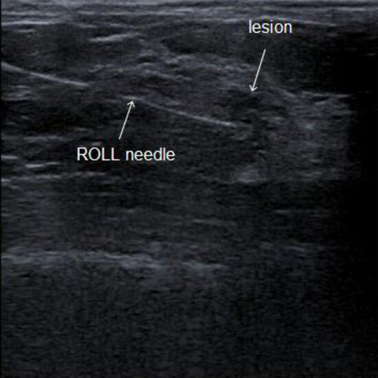
Radioguided localisation of impalpable breast lesions is routinely used in many centres around the world. Advantages over other localisation methods are well described in published literature, but there is little documentation of its use in Australia. This article reports our experience during an audited introduction of the technique.
Evaluation of radiographers’ mammography screen-reading accuracy in Australia
- Pages: 15-22
- First Published: 06 August 2014
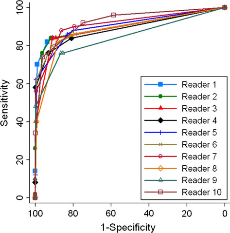
This research provides evidence of the accuracy of radiographers’ screen-reading mammograms. Radiologist workforce shortages may contribute to delays in women receiving their results in the BreastScreen Australia program. This research provides evidence to support a similar strategy being implemented within the BreastScreen Australia program, as has been successfully implemented in the United Kingdom, of radiographers screen-reading to contribute to the timeliness and accuracy of program outcomes.
Evaluating radiographers' diagnostic accuracy in screen-reading mammograms: what constitutes a quality study?
- Pages: 23-31
- First Published: 14 August 2014
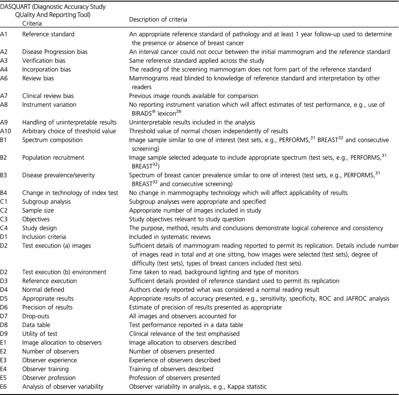
This paper has been written firstly to evaluate the quality of studies that evaluate the accuracy of radiographers' screen-reading mammograms. Secondly, an appropriated tool has been developed to determine the essential issues of quality when evaluating these studies. We believe that this tool, with further refinement, and application can contribute to future quality studies in this important area of research and practice.
Imaging dose in breast radiotherapy: does breast size affect the dose to the organs at risk and the risk of secondary cancer to the contralateral breast?
- Pages: 32-39
- First Published: 07 January 2015
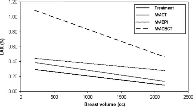
This study investigates the imaging doses related to breast size for three imaging modalities used in breast radiotherapy. The study's main finding was that the primary breast volume inversely correlated with the contralateral breast dose for all three imaging modalities. As the primary breast volume increases, the likelihood of a patient developing a radiation-induced secondary cancer to the contralateral breast decreases.
Quantifying intra- and inter-fractional motion in breast radiotherapy
- Pages: 40-46
- First Published: 13 July 2014
An investigation into the magnitude of intra- and inter-fractional motion variation present in the set up of breast cancer patients receiving radiotherapy. Variation was greatest inter-fractionally but still within tolerance. This review of the breast technique provides confidence in our set up and the ability to move towards advanced treatment techniques.
Review Articles
Expanding the indications for MRI in the diagnosis and treatment of breast cancer: what is best practice?
- Pages: 47-53
- First Published: 31 January 2015
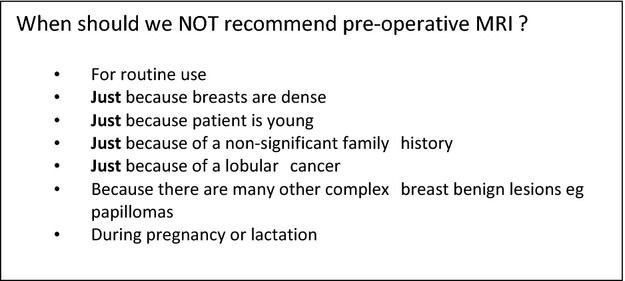
Breast magnetic resonance imaging (MRI) now has an accepted place in screening younger women at high risk of breast cancer, and is increasingly used in a number of other settings including assessment of response to neo-adjuvant therapy and local staging of cancer. Although the evidence for its general use in these settings is very limited, in highly selected patients, especially where discordance with conventional measurements occurs, MRI can have a place in assessing the extent of disease, both whether operable and how operable, and guiding surgery. These scenarios and future indications and alternative technologies are explored in this paper.
The role of general nuclear medicine in breast cancer
- Pages: 54-65
- First Published: 12 February 2015
Routine nuclear medicine is a major contributor to a full gamut of clinical studies. Developments in instrumentation such as the high-resolution dedicated breast device coupled with the diagnostic versatility of conventional cameras have reinserted nuclear medicine as a valuable tool in the broader clinical setting.
The cardiac dose-sparing benefits of deep inspiration breath-hold in left breast irradiation: a systematic review
- Pages: 66-73
- First Published: 07 January 2015
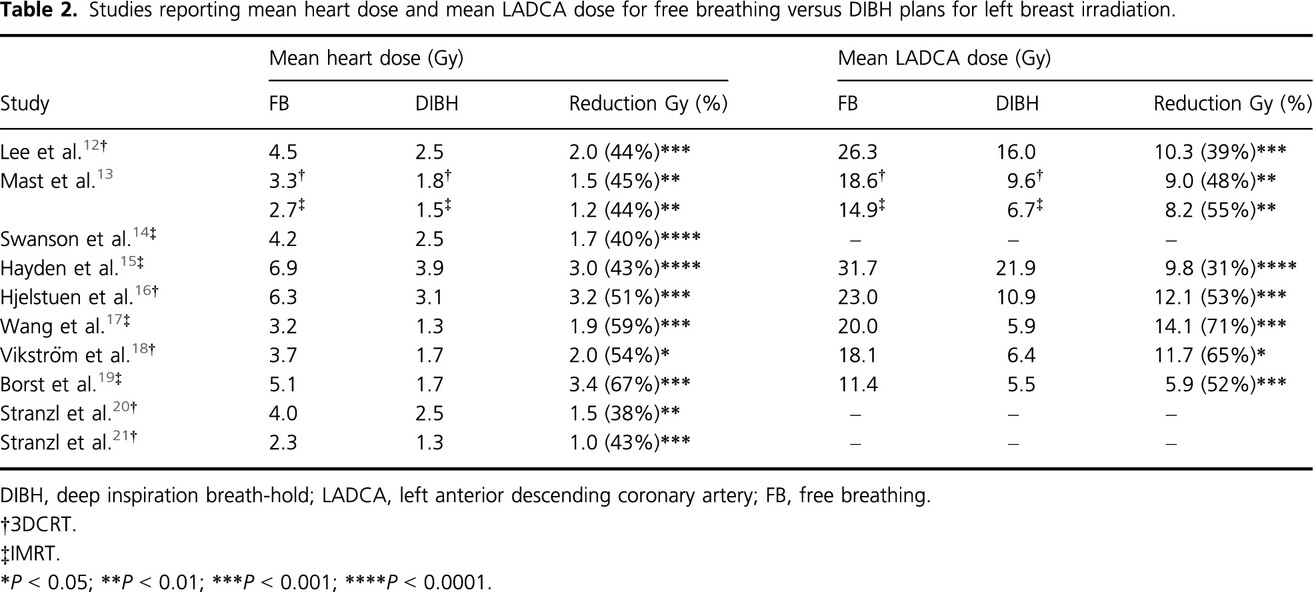
Despite the technical advancements in breast radiation therapy, cardiac structures are still subjected to significant levels of irradiation. The primary aim of this review was to evaluate the cardiac dose-sparing benefits of the deep inspiration breath-hold (DIBH) technique. Based on a review of the literature, DIBH is a reproducible and stable technique for left breast irradiation showing significant promise in reducing the late cardiac toxicities associated with radiation therapy.
Review of deep inspiration breath-hold techniques for the treatment of breast cancer
- Pages: 74-81
- First Published: 16 February 2015
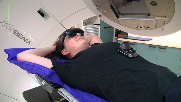
Despite variation in the literature regarding the deep inspiration breath-hold (DIBH) delivery method, patient coaching, visual feedback mechanisms and treatment verification, all methods of DIBH delivery reduce radiation dose to the heart. Further research is required to determine optimum protocols for patient training and treatment verification to ensure that the technique is delivered successfully.
Reflections
Finding my own voice through the breast cancer journey: humour, sadness and smurfs
- Pages: 82-85
- First Published: 14 January 2015

Finding your voice as a cancer patient can be hard, especially when you do not know what to say yourself. This article reveals a personal story of a diagnostic radiographer who thought she knew it all until she was diagnosed with breast cancer. Using humour and sadness as a scaffold for support, the voice of a young mother with cancer is heard within this paper.
Book Reviews
Diagnostic Breast Imaging: Mammography, Sonography, Magnetic Resonance Imaging, and Interventional Procedures
- Pages: 86-87
- First Published: 07 January 2015

Diagnostic Breast Imaging is the third edition of a highly acclaimed text which focuses on imaging modalities used in the diagnosis of breast cancer. This edition has been updated to include advances in current breast imaging modalities such as mammography, ultrasound, magnetic resonance imaging and percutaneous interventions. Significant emerging technologies for example digital breast tomosynthesis, are presented using evidence-based methods. This is an essential text for health professional students and practitioners involved in the diagnosis and treatment of breast cancer.
RadCases Breast Imaging
- Page: 88
- First Published: 04 December 2014

RadCases Breast Imaging is a new addition to the successful and proven RadCases series. With the 100 cases presented in the hardcopy and many more in the online version, the book not only covers the essentials of breast pathology but also provides pearls and pitfalls for each case. RadCases Breast Imaging is suitable for radiology trainees, preparing for their exams, for junior radiologists needing to fill knowledge gaps in a quick and efficient way, and general radiologists involved in breast imaging, providing that extra reassurance needed at times.
Sign up for email alerts
Tools


 invites authors to submit articles to a special themed issue on paediatric medical imaging and radiation therapy")

More from this journal

-1693813706.png)