Journal list menu

 Issue
IssueJournal of Biophotonics: Volume 8, Issue 4
In Honor of Valery V. Tuchin's 70th Birthday
273-356April 2015
Export Citations
Download PDFs
Cover Picture
Cover Picture: Optical coherence elastography for tissue characterization: a review (J. Biophotonics 4/2015)
- First Published: 02 April 2015
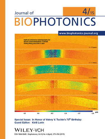
Optical coherence elastography (OCE) is an emerging 3D nondestructive biomechanical imaging technique that has experienced a rapid development. S. Wang and K. Larin review the state-of-the-art OCE techniques from the application point of view and discuss current challenges and potential solutions.
(Picture: S. Wang and K. Larin, pp. 279–302 in this issue)
Issue Information
Contents
Editorial
Celebrating the 70th birthday of Valery V. Tuchin
- Pages: 277-278
- First Published: 02 April 2015
Review Article
Optical coherence elastography for tissue characterization: a review
- Pages: 279-302
- First Published: 20 November 2014
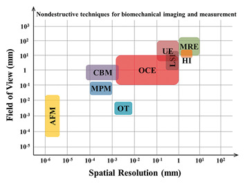
Optical coherence elastography (OCE) is an emerging 3D nondestructive biomechanical imaging technique that has recently experienced a rapid development. In this paper, the mechanical contrast employed by OCE is described, the state-of-the-art OCE techniques are reviewed from the application point of view, and the current challenges and potential solutions are discussed. Throughout this review, the unique role of OCE for the mechanical characterization of tissue are emphasized and highlighted.
Letter
Three-dimensional arbitrary trajectory scanning photoacoustic microscopy
- Pages: 303-308
- First Published: 30 July 2014
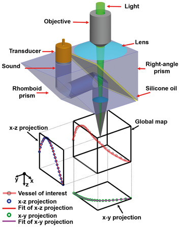
Photoacoustic microscopy was enhanced with three-dimensional arbitrary trajectory (3-DAT) scanning, which can rapidly image selected vessels over a large field of view (FOV) and maintain a high signal-tonoise ratio (SNR) despite the depth variation of the vessels. It is shown that hemoglobin oxygen saturation (sO2) and blood flow can be measured simultaneously in a mouse ear in vivo at a frame rate 67 times greater than that of a traditional two-dimensional raster scan. sO2 dynamics were also observed in response to switching from systemic hypoxia to hyperoxia.
Editor's Choice
Quick analysis of optical spectra to quantify epidermal melanin and papillary dermal blood content of skin
- Pages: 309-316
- First Published: 09 December 2014
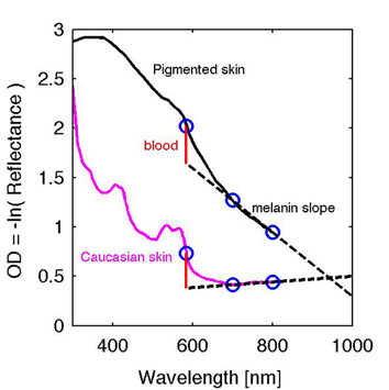
A practical approach for assessing the melanin and blood content of the skin from total diffuse reflectance spectra is presented. The paper describes the non-rectilinear character of a quick analysis, which uses just three wavelengths, and shows that most any choice of two wavelengths in the 600–900 range can achieve the characterization of melanin. Monte Carlo simulations created spectral data for a skin model with epidermis, papillary dermis and reticular dermis to illustrate the analysis.
Full Articles
Application of circularly polarized light for non-invasive diagnosis of cancerous tissues and turbid tissue-like scattering media
- Pages: 317-323
- First Published: 18 October 2014
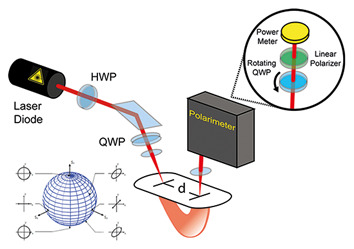
The directional awareness of circularly and elliptically polarized light backscattered from turbid tissue-like scattering media is exploited. Circularly and elliptically polarized laser light is applied which illuminates the tissue samples of interest. It is demonstrated that the Stokes vector of backscattered light depicted on a Poincaré sphere can be used for characterization of cancerous and non-cancerous tissue samples in vitro.
Wavelength optimized cross-polarized wide-field imaging for noninvasive and rapid evaluation of dermal structures
- Pages: 324-331
- First Published: 19 January 2015
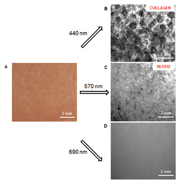
Polarization optical imaging was used for noninvasive quantitative evaluation of dermal collagen. Collagen content and intensity histogram were computed from the optical images of 17 human subjects. Quantitative analysis showed a decrease in collagen content and average pixel value as well as an increase in the full width at half maximum of collagen image histogram with age. The results indicate that polarization optical imaging has potential for noninvasive in vivo evaluation of human dermis.
Ex vivo optical measurements of glucose diffusion kinetics in native and diabetic mouse skin
- Pages: 332-346
- First Published: 11 March 2015
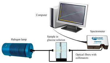
The motivation of the study is to quantify diffusion processes of glucose in skin in order to understand tissue structural alterations due to diabetes mellitusdevelopment. Optical clearing method for examination of skin samples taken from healthy and alloxan induced diabetic mice was used. Diffusion coefficients for glucose transport in normal and diabetic skin were measured ex vivo.
Non-linear imaging and characterization of atherosclerotic arterial tissue using combined SHG and FLIM microscopy
- Pages: 347-356
- First Published: 11 March 2015
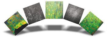
Atherosclerosis is among the most widespread cardiovascular diseases and its early diagnosis is crucial for avoiding life threatening consequences. Non-linear microscopy can diagnose tissues and atherosclerosis in a label-free modality, opening the way for a clinical use of these optical techniques. Combined SHG-FLIM microscopy is demonstrated to be extremely powerful for diagnosing and characterizing atherosclerosis.
Sign up for email alerts
Tools
More from this journal
Related Titles
 Volume 19, Issue 14July 22, 2025
 Volume 5, Issue 3-4August-November 2023
 Volume 13, Issue 21July 25, 2025